

Thorax Case 3
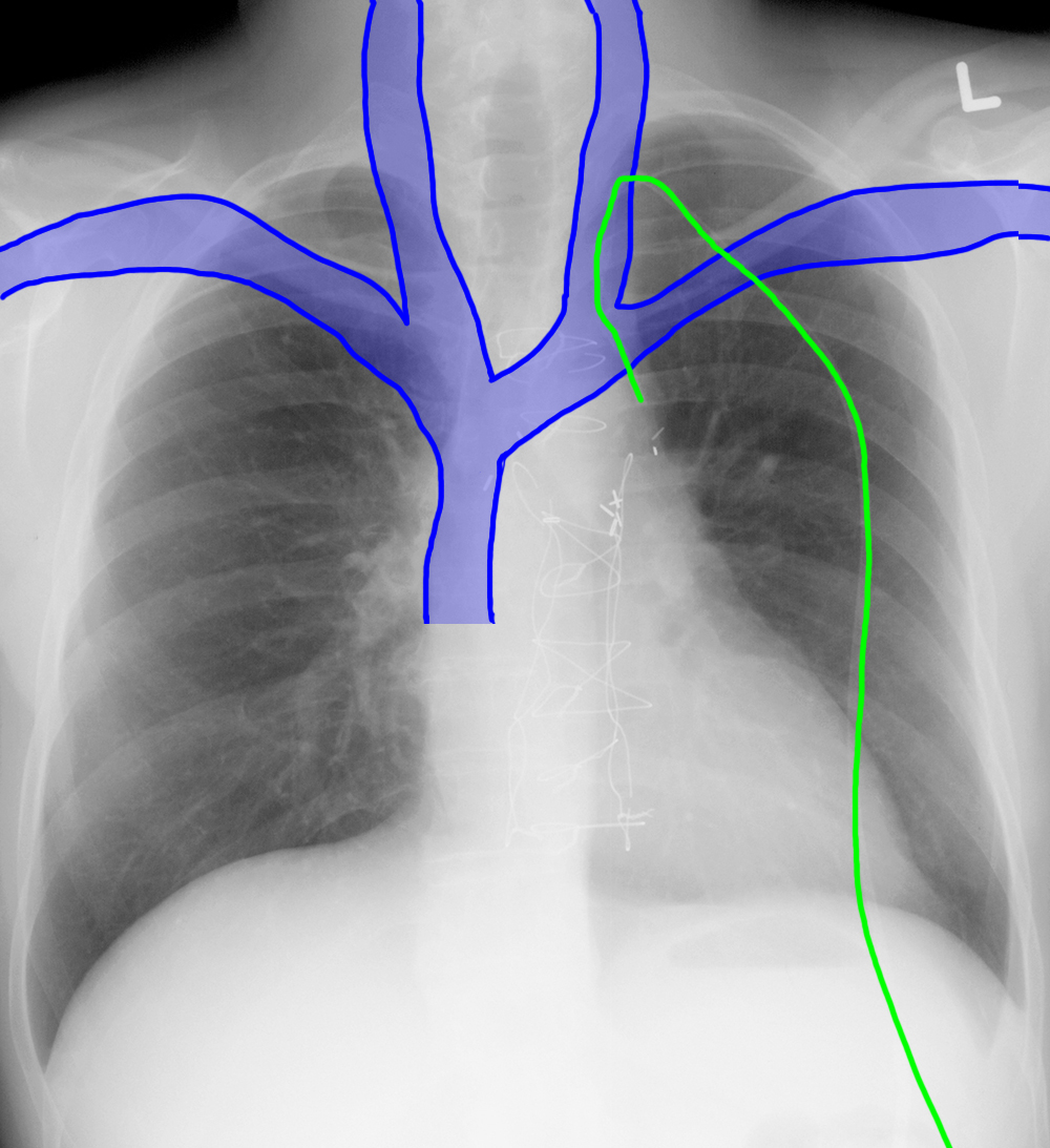
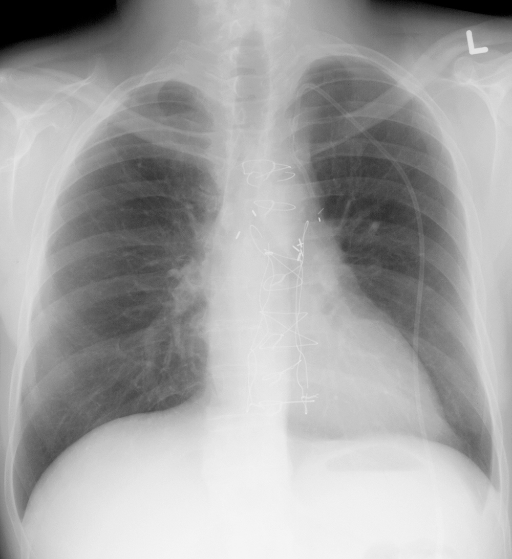
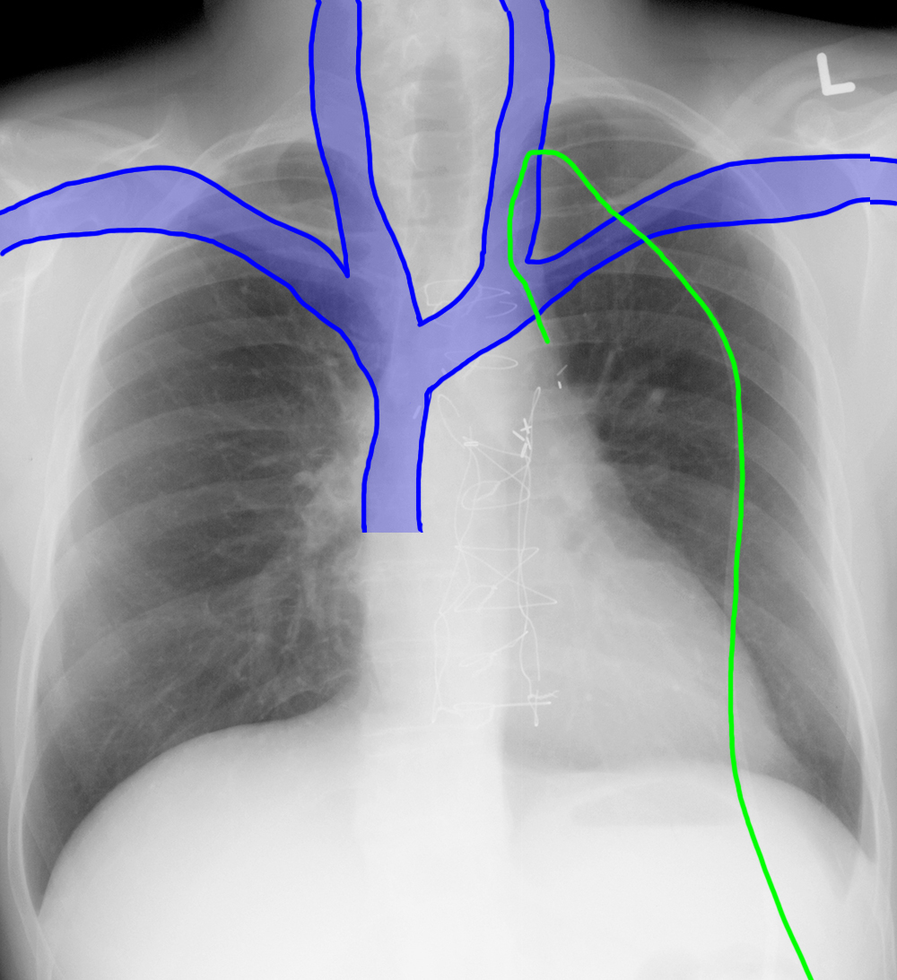
This is a patient who had a central line placed.
Question 1:
a) What vessel was used to access the central venous system?
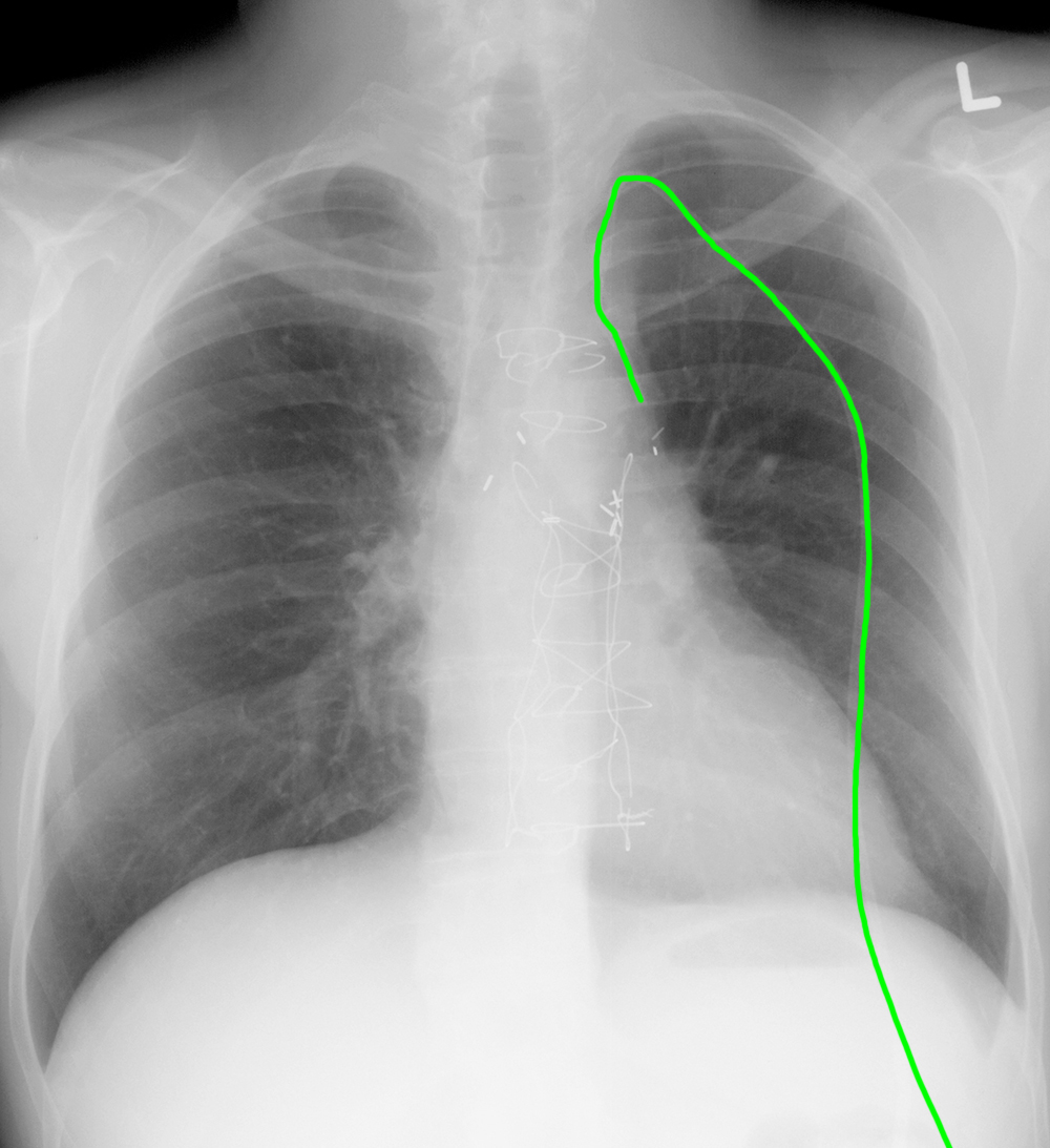
Much of the catheter on the image is OUTSIDE the patient. The supraclavicular region is the likely site of skin entry, and from that point, the catheter passes relatively vertically along the left upper mediastinum, consistent with a left internal jugular approach.
b) Check the label for veins. What are possible explanations for the course of this catheter?
When a catheter does not appear to be following the expected course of any of the large mediastinal veins, there are three things to consider. The catheter could be extra-vascular (not in a vein at all, often accompanied by marked mediastinal widening and pleural effusion, NOT see in this case). The catheter could be in anomalous vessel (in this location, a left-sided SVC is a possibility). OR the catheter could be in a small tributary vein.
Thorax Case 3
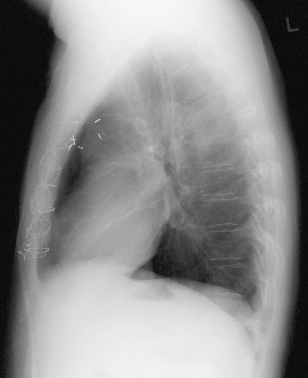
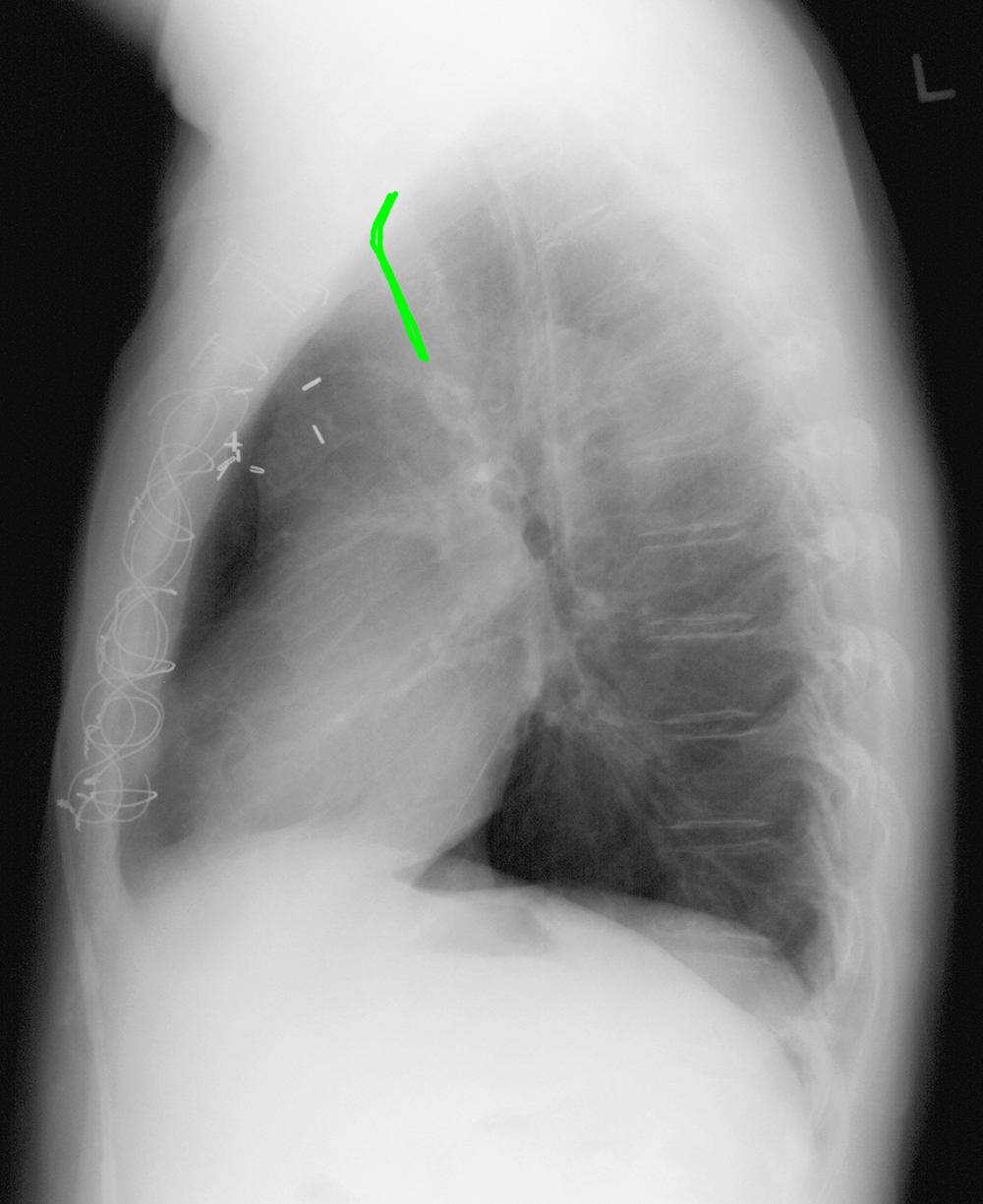
This is the lateral view of this same patient.
Question 2:
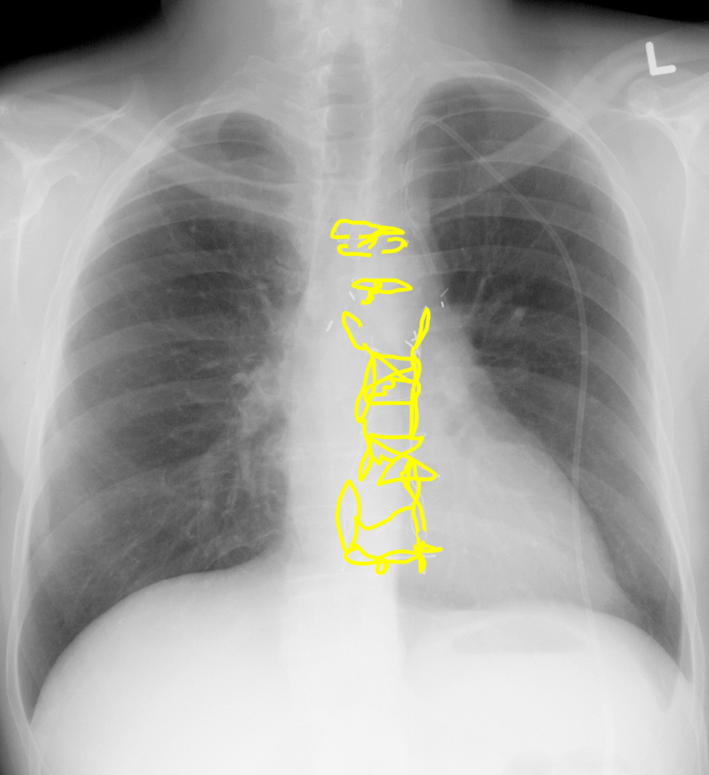
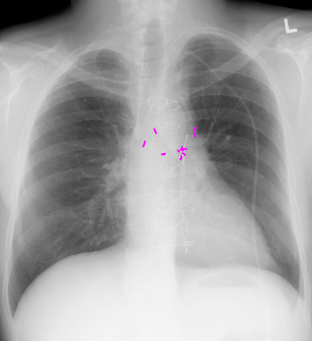
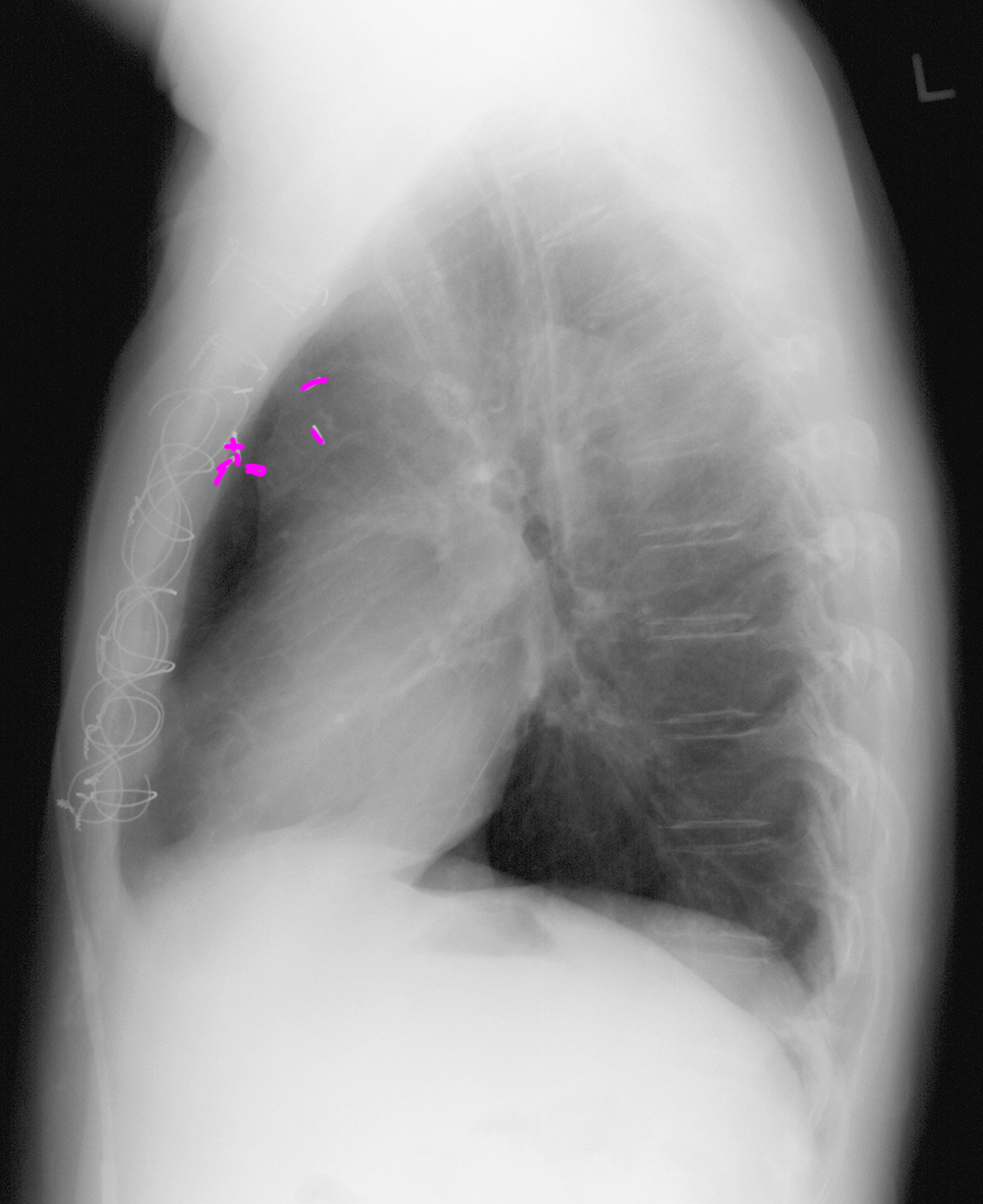
a) What do the wires and surgical clips indicate was likely done in this patient in the past?
Sternal wires like this patient has, with surgical clips in the mediastinum often indicate prior cardiac surgery, particularly involving the coronary arteries. At the time of this patient's surgery, no left SVC was seen.
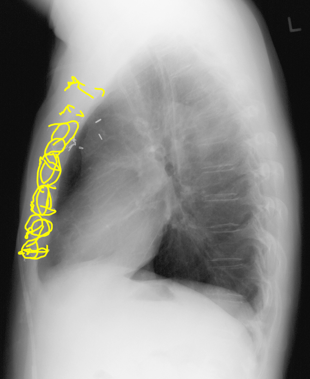
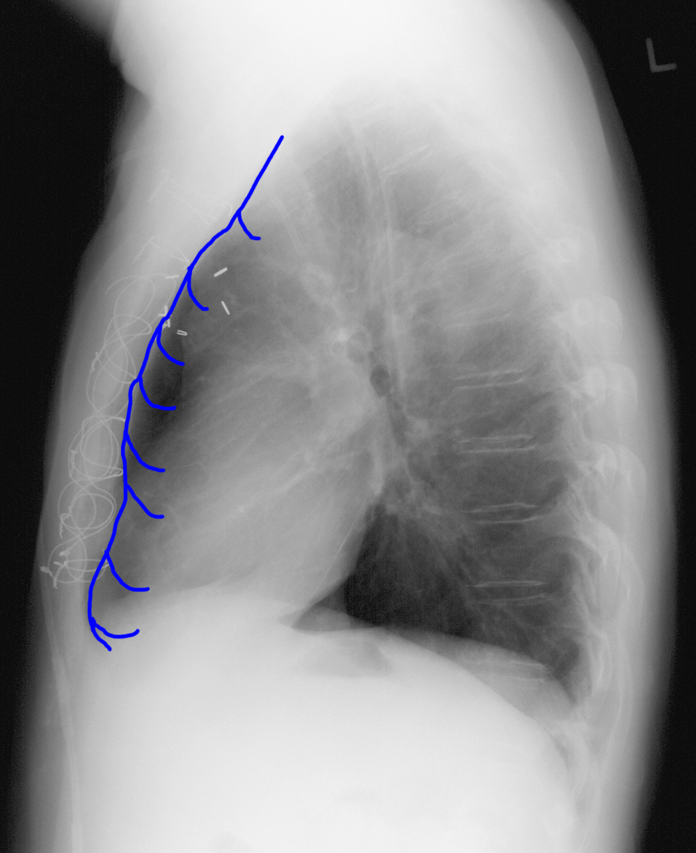
b) What small tributary vein is indicated on this image?
The label of the small vein shows a vessel running just along the anterior deep surface of the sternum. This is the expected course of the internal mammary vein. This is one small tributary that could be considered for this CVL on the frontal view, but the course of the CVL does not match the internal mammary vein on the lateral view.
Thorax Case 3
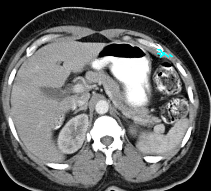
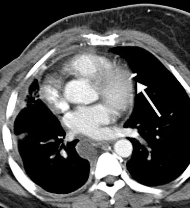
This is a chest CT done on a different patient. This patient had a history of a right mastectomy for breast cancer, and had many central lines during her treatment. She now has swollen veins, particularly on the left side of her neck and chest wall. She currently has a left subclavian CVL with subcutaneous injection port in place.
Question 3:
a) Was IV contrast given for this study?
Yes. The chambers of the heart and the large mediastinal vessels are all whiter than muscle.
b) Given this patient's history, what possible explanation could you give for the very bright contrast seen in small vessels in the chest wall and mediastinum?
She might have developed a stenosis or clot in one or more of her large mediastinal veins, due to having multiple prior CVLs. This would cause the IV contrast, which is injected via an arm vein, to be diverted from the main path back to the heart and into small tributary vessels.
c) What is the vessel indicated by the arrow on this CT scan?
This is a small tributary that usually accompanies the phrenic nerve, the pericardiacophrenic vein. It drains the diaphragm and passes anterior to the hilum to drain into the left brachiocephalic vein, in most cases.
Thorax Case 3
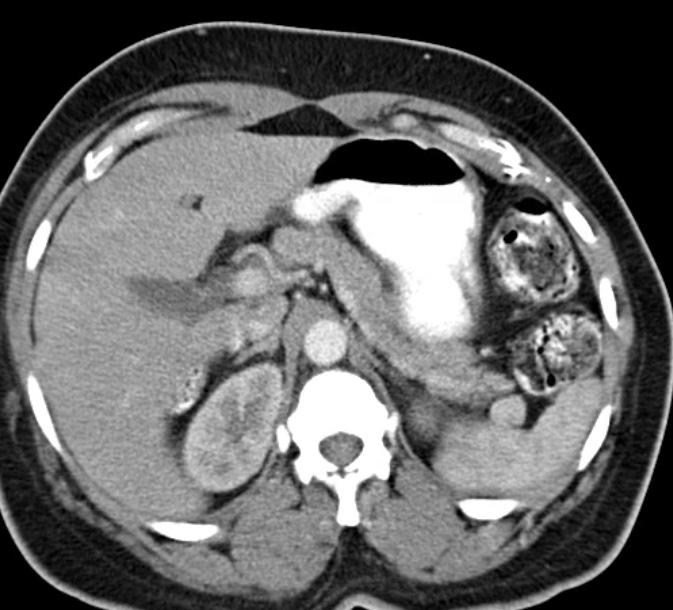
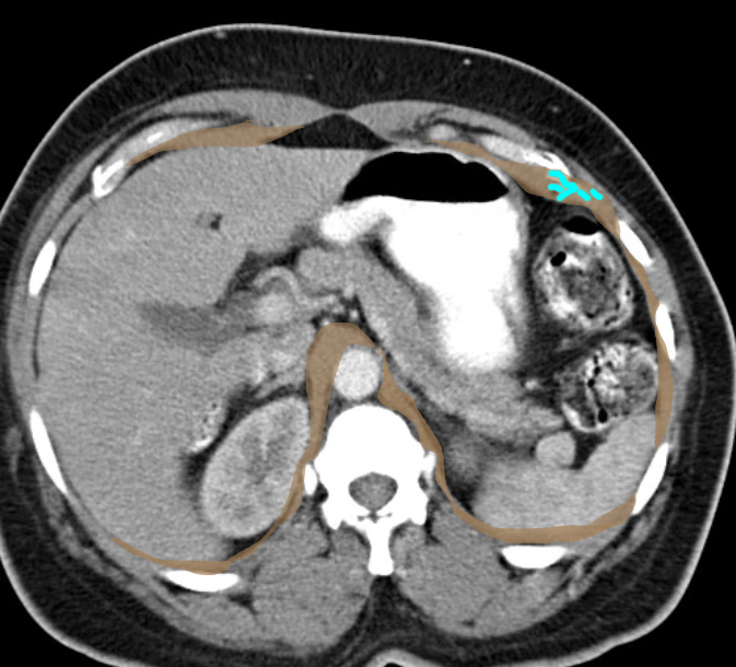
This is the bottom slice of the CT shown on the previous page.
Question 4:
a) The label of 'veins' below shows the small vessels that make up the inferior-most portion of the left pericardiacophrenic vein. What is the structure labeled in brown?
The structure is the diaphragm, which appears in many pieces at this level as we are slicing it near the bottom of its extent, there it is attaching in many places around the inside of the ribcage.
b) The pericardiacophrenic vein is normally very small. Do you think it would be likely for a CVL to enter this vein in a normal person?
No. In most cases where a CVL enters a tributary like this, it can only happen if the vessel is LARGER than usual. So when this happens, one should consider whether the patient has a reason for collateral veins to be abnormally large (such as stenosis or clot, as in the patient whose CT we reviewed).
