

Thorax Case 4
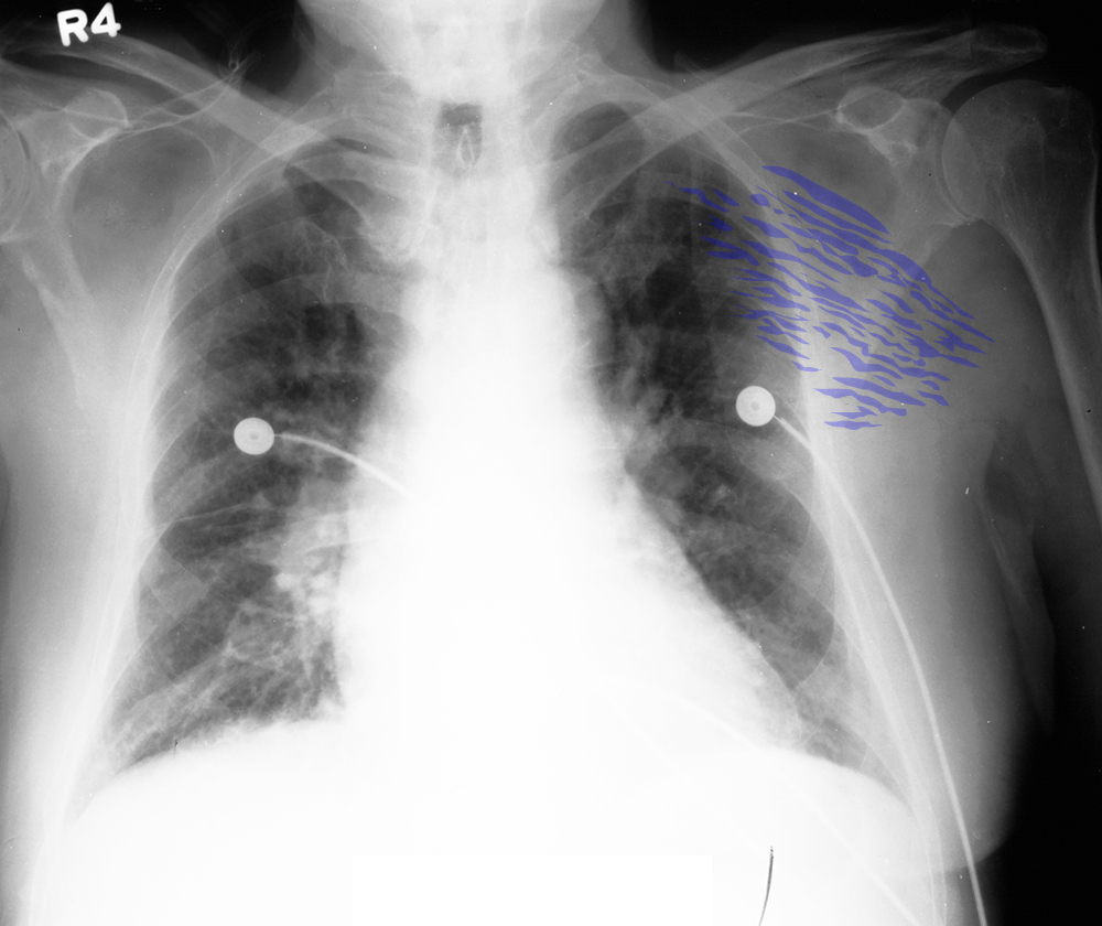
This patient has pain and swelling in their left anterior chest wall, with fever and elevated white blood cell count (suggesting infection).
Question 1:
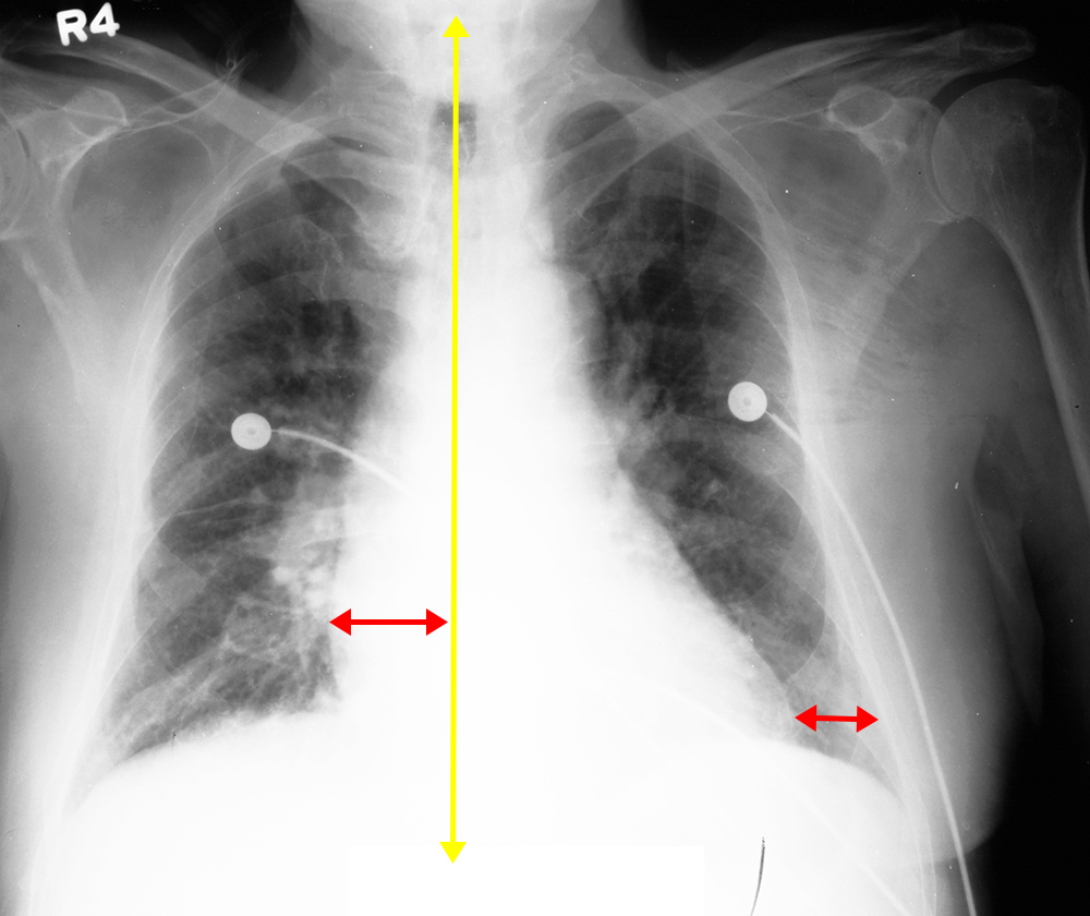
a) What do you think of this patient's heart size?
Using our line segments, we can see that the one from the patient's right heart border to the midline is LONGER than the line segment from the patient's left heart border to the inside of the ribs. This means that the cardiac contour is enlarged, which could be due to actual cardiac dilatation or pericardial disease, such as an effusion.
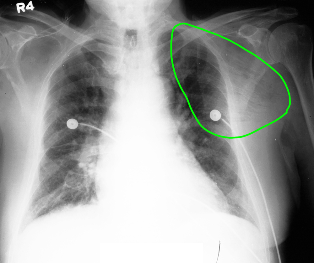
b) If you click on the 'question' label below, try to determine what is abnormal in the circled region before checking the answers. What density is the abnormality in this region?
The abnormality consists of dark streaks visible within the midrange grey of soft tissue. Despite being quite small, each little streak is visibly darker than surrounding tissue, indicating that they are likely gas. On a chest radiograph, fat and muscle/fascia are much more similar in appearance, and it would be unlikely that tiny pockets of fat would be dark enough to see.
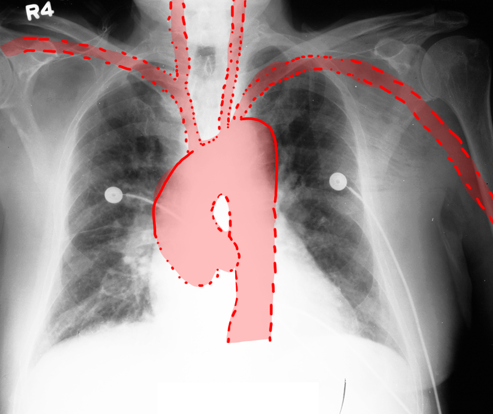
c) What do you think of the contour of the ascending aorta?
The outward convexity of the ascending aorta (shown on the label below 'aorta') is a bit too large, suggesting that the ascending aorta is likely dilated (as shown in the annotated image). Granted, we cannot actually see the medial border of the vessel, so we are making an estimate based on the lateral border, which we can see.
Thorax Case 4
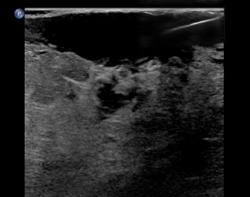
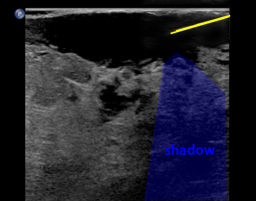
This is the next study that was done, as there was a painful red lump in the patient's upper arm. The patient reported that they had recently been fishing and a fishhook had lodged in their arm and had been removed.
Question 2:
a) What type of study is this?
This is an ultrasound, which uses sound waves to generate an image of tissues.
b) What type of tissue appears black on ultrasound?
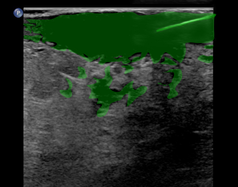
Fluid appears black on ultrasound. This is one of the things that ultrasound is most useful for--detection of fluid collections, which could include pleural fluid, pericardial fluid, or fluid in an abscess, as in this case.
c) Why is ultrasound a good method for guiding interventional procedures?
Unlike radiographs or CT images, which are still images, ultrasound is a real-time imaging modality. We can watch things like the heart beating, bowel loops contracting, or a needle being guided into a fluid collection, as in this case. Ultrasound is also very useful for placement of CVLs as you can see that you are entering the target vessel.
d) What is the significance of gas in the soft tissues in this patient?
Gas forming organisms can produce the appearance of streaks of gas in the soft tissues, and often indicate a severe infection. After draining the upper extremity abscess in this patient, cultures yielded Streptococcus and prompt treatment with IV antibiotics was ultimately successful in treating this serious infection. When gas forms in an infection of this type, it is called 'necrotizing fasciitis' and can be rapidly fatal if not treated promptly.
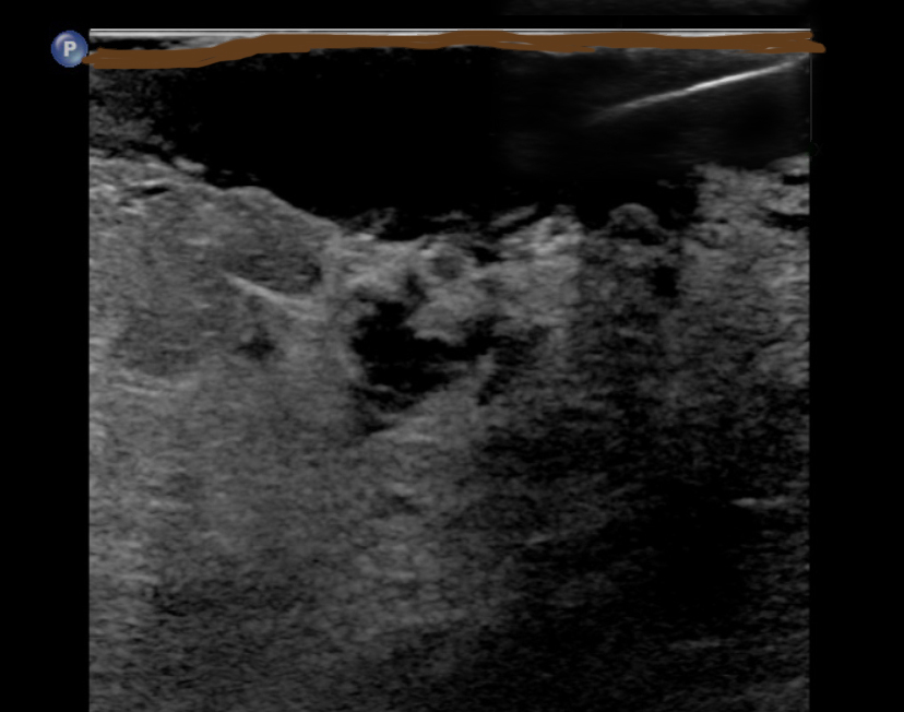
For ultrasound imaging, because the field of view is quite small, it is often difficult to know what tissue is being imaged without labels. The orientation of the transducer should also be indicated on the image, since the image is a slice and could be done in sagittal, coronal, or axial planes, depending on how the transducer is held against the patient's skin. By convention, for axial and coronal imaging, the patient's right is on the LEFT side of the image (just like for CT or MR). For sagittal imaging, the patient's head is on the LEFT and feet on the RIGHT.
Thorax Case 4
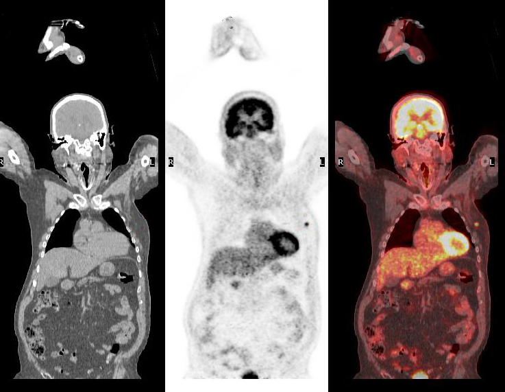
When infection is a consideration, particularly if the source of the infection is not clear, this is another type of study that can be helpful. Since it was clear in our patient that the source of infection was an upper extremity abscess, no further imaging was needed.
Question 3:
a) What type of study is this?
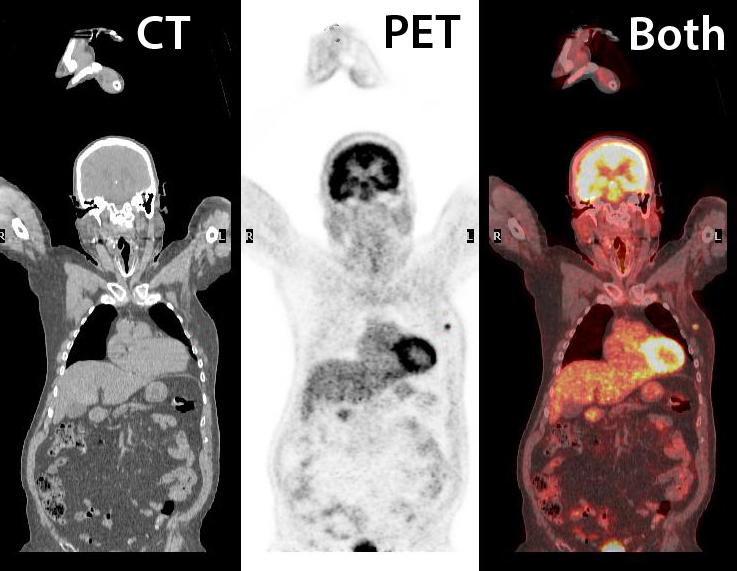
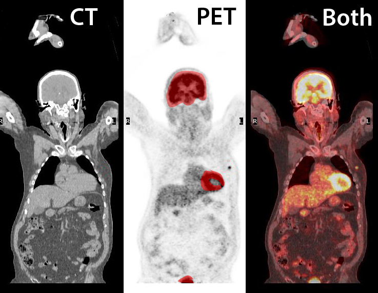
This is a PET/CT study. In this type of exam, a radioactive tracer (specific for cells with active uptake of glucose) is injected intravenously and imaging is done with a sensitive type of camera that can detect radiation emitted by the body. A CT scan is done simultaneously so that the two imaging modalities can be superimposed.
b) What tissues normally take up the PET tracer?
Brain and myocardium both rely on glucose for energy and therefore can show high uptake normally. The tracer is eliminated by the kidneys so uptake in the collecting systems and bladder is also normal.
In this case, a focal area of abnormal uptake is seen in the left chest wall anteriorly which was a tumor. Both tumors and infection can give abnormal uptake of the PET tracer.

Thorax Case 4
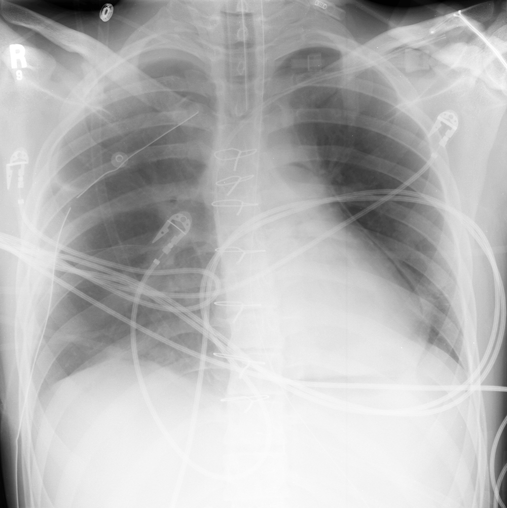
This is a portable chest radiograph of a different patient on the day of cardiac surgery.
Question 4:
a) What support devices can you identify?
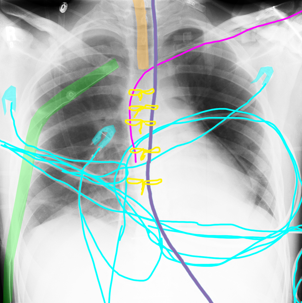
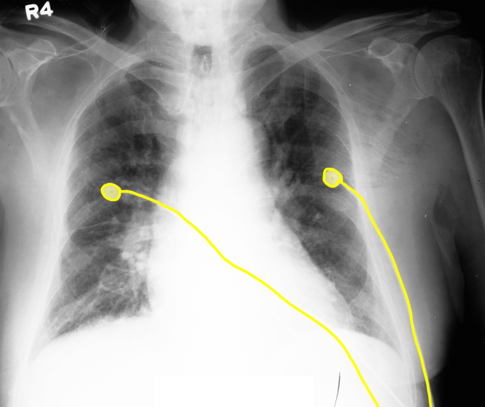
This image demonstrates the difficulty in many portable radiographs, with overlying material such as telemetry leads (shown in blue). There is an endotracheal tube (orange), a right chest tube (green), a left subclavian CVL (pink) and an NG tube (purple).
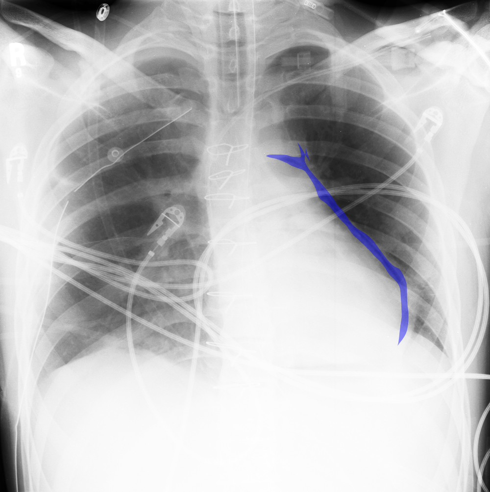
b) Where is the abnormal air located?
The crescent of air is wrapping around the left heart border and extending up along the margin of the main pulmonary artery, consistent with air in the pericardial space. This is not uncommon on the day of cardiac surgery, as air is expected in the immediate post-operative period if the pericardial space was entered. The air usually resolves in a few hours.
