

Case 7
70 year old female with painful lump in the left upper outer breast, poor historian
Question 1:
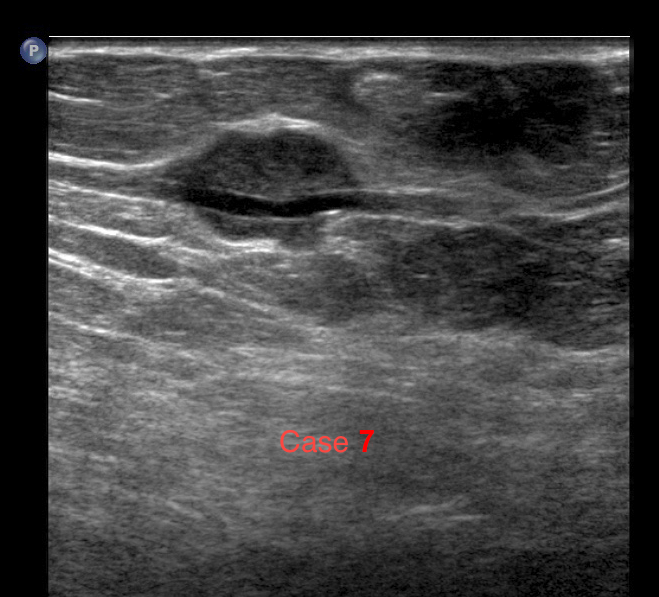
How would you describe the ultrasound findings? Why might history be particularly important in this case?
×
Answer:
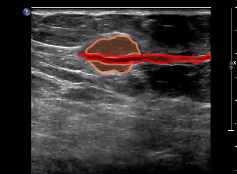
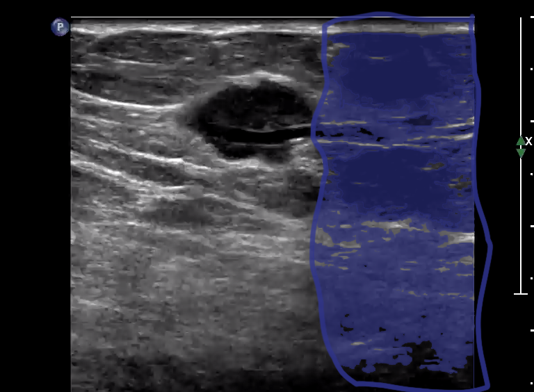
There is a small hypo echoic mass that seems to be associated with an anechoic tubular structure that could be a vessel. A history of trauma to the area might suggest vascular injury and formation of an AVM.
There is a small hypo echoic mass that seems to be associated with an anechoic tubular structure that could be a vessel. A history of trauma to the area might suggest vascular injury and formation of an AVM.
Further Explanation:
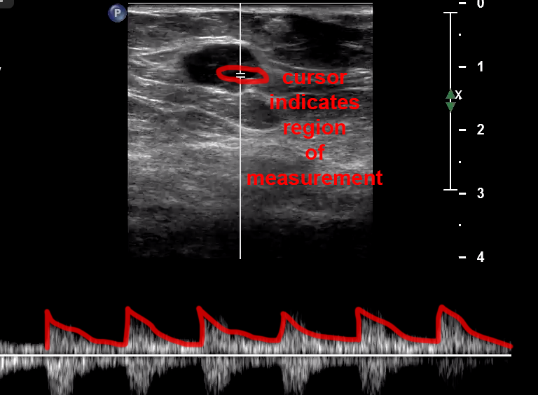
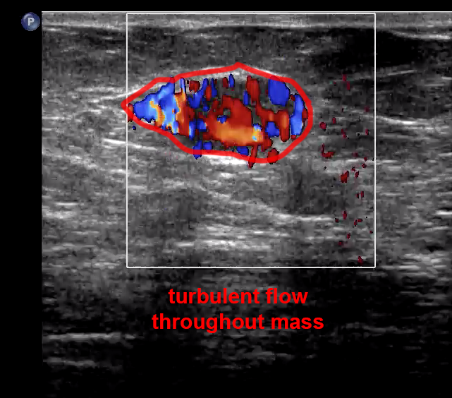
Case 7
(click on 'X' to return to Case List)
Question 2:
a) In what part of the breast is this lesion located?
×
Answer:
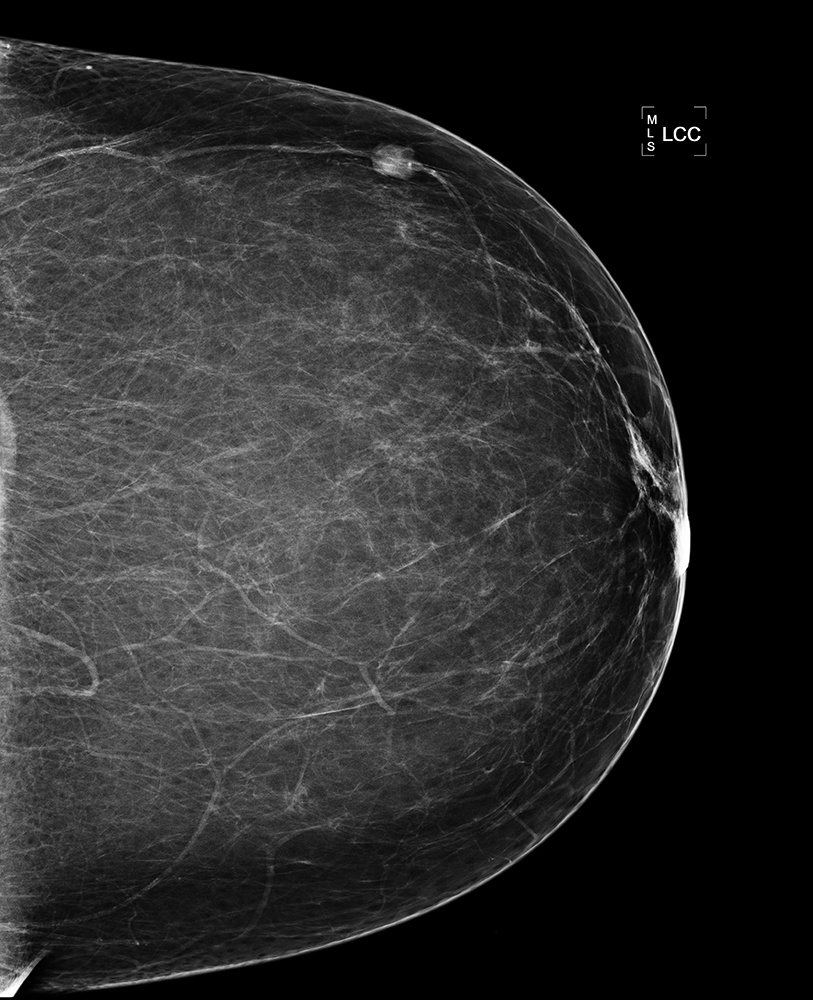
The image is labeled LCC, so this is a left CC view. On the CC view, the axilla is always shown at the top of the image, so this lesion is in the outer breast. Again, you cannot tell on this single view if it is upper or lower in the breast, but from the US you know its exact location (upper outer, so 1:00-2:00 position on the left).
The image is labeled LCC, so this is a left CC view. On the CC view, the axilla is always shown at the top of the image, so this lesion is in the outer breast. Again, you cannot tell on this single view if it is upper or lower in the breast, but from the US you know its exact location (upper outer, so 1:00-2:00 position on the left).
b) What is your top diagnosis?
×
Answer:
Given the ultrasound features, and the fact that it is associated with a vessel on both the US and mammogram, this is most likely a traumatic AVM. Upon further questioning, the patient did recall a recent fall and bruising in this part of her breast about two weeks ago.
Given the ultrasound features, and the fact that it is associated with a vessel on both the US and mammogram, this is most likely a traumatic AVM. Upon further questioning, the patient did recall a recent fall and bruising in this part of her breast about two weeks ago.
