

Case 2-PRE CLASS CASE
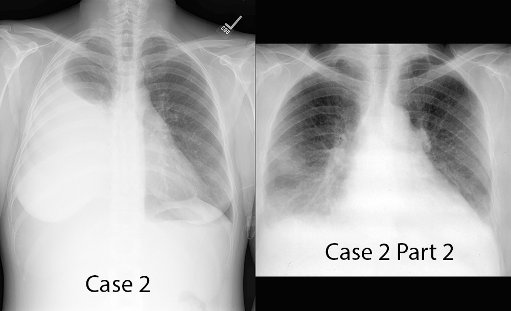
48 year old female with shortness of breath
Further Explanation:
What is the most significant finding?
Please ENTER your response before moving to the next page!
Case 2-PRE CLASS CASE
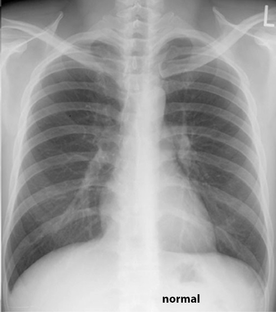
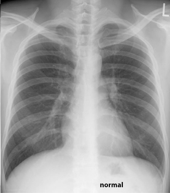
How is this case different?
Further Explanation:
Please enter your responses before going on to the next page!
Case 2-PRE CLASS CASE
Case 2 and Comparison--to view HINTS below, go back to the case page
Further Explanation:
Case 2--
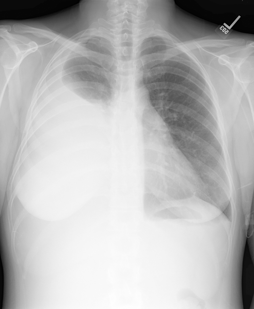
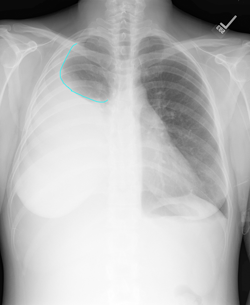
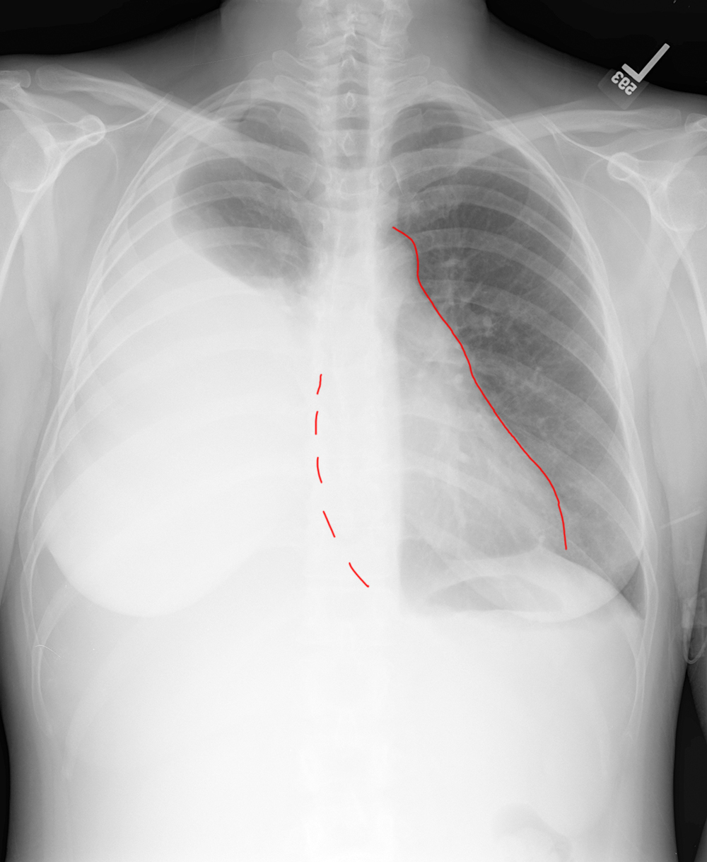
Hint 1: There is an obvious difference in density on the two sides of this chest radiograph, with the right side almost completely white. The upper surface of the right opacity is smooth and sharp, with a curve up the inside of the right chest wall (a meniscus) which suggests fluid.
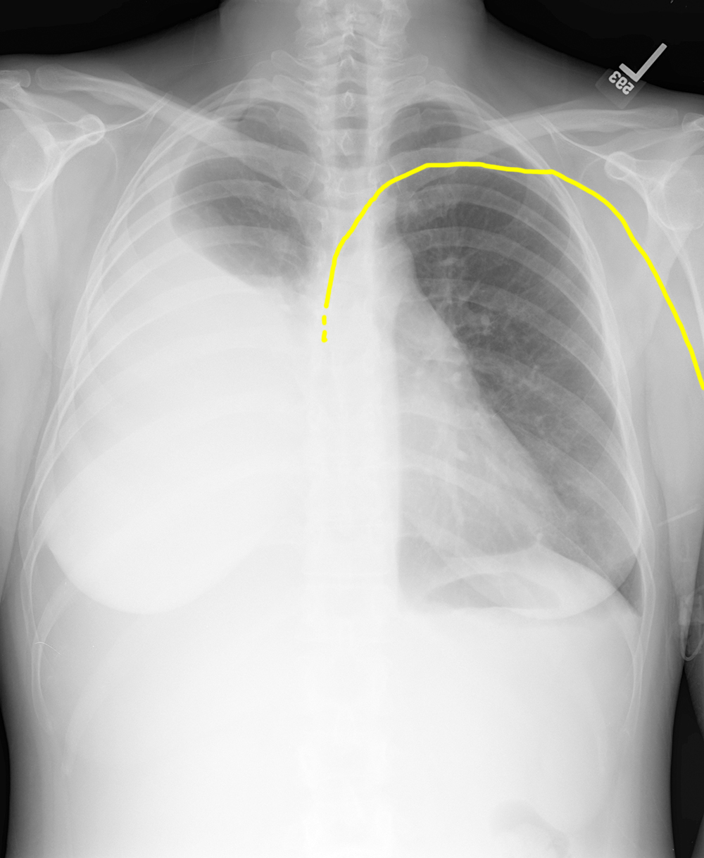
Hint 2: There is a left PICC (peripherally inserted central catheter) that passes through the left subclavian vein into the left brachiocephalic vein and shows the likely position of the SVC and right heart margin, which are otherwise obscured.
Hint 3: Using the PICC position, you can see that the heart is shifted to the LEFT. Without the PICC, this might just represent an enlarged heart, but given the approximate position of the right heart border, the heart is actually too far over, suggesting mass effect from the white opacity in the right hemithorax. The combination of these findings (right opacity with smooth upper margin and meniscus, shift of heart to the left) is consistent with a large right pleural effusion.
Comparison Case 2--
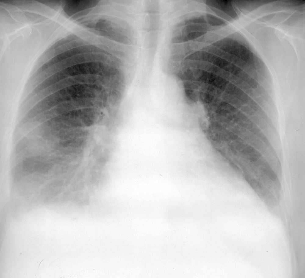
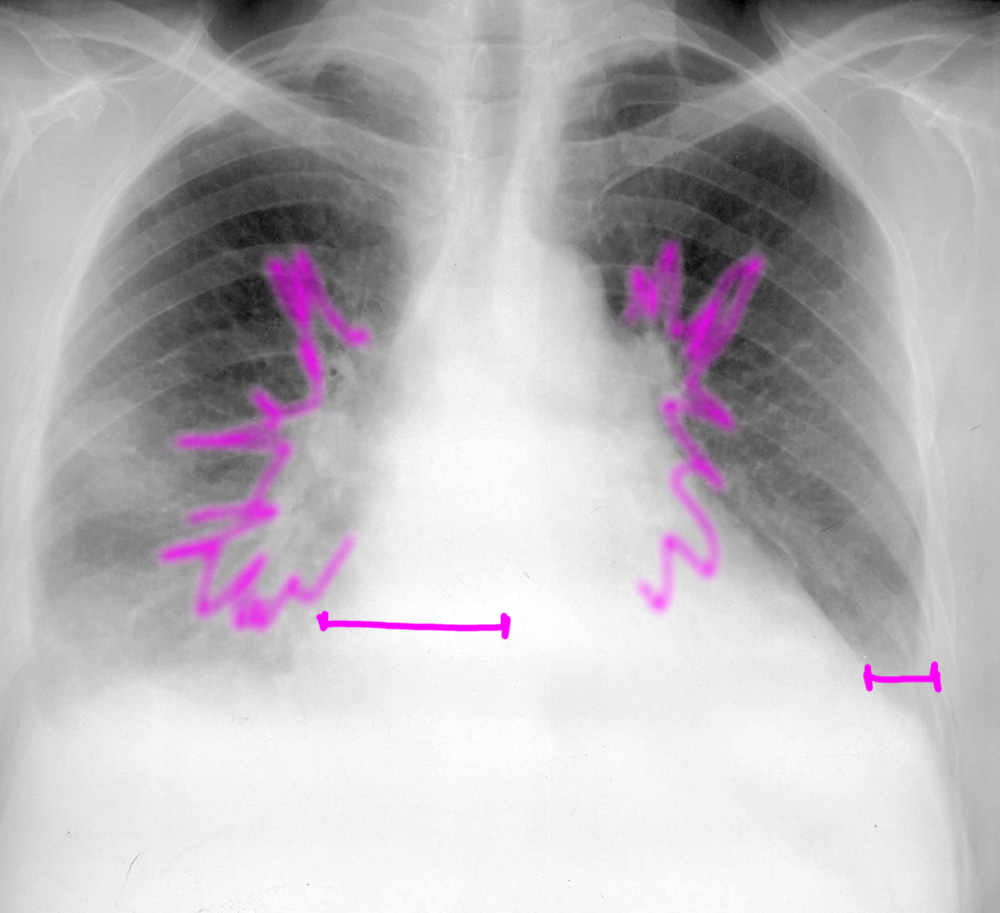
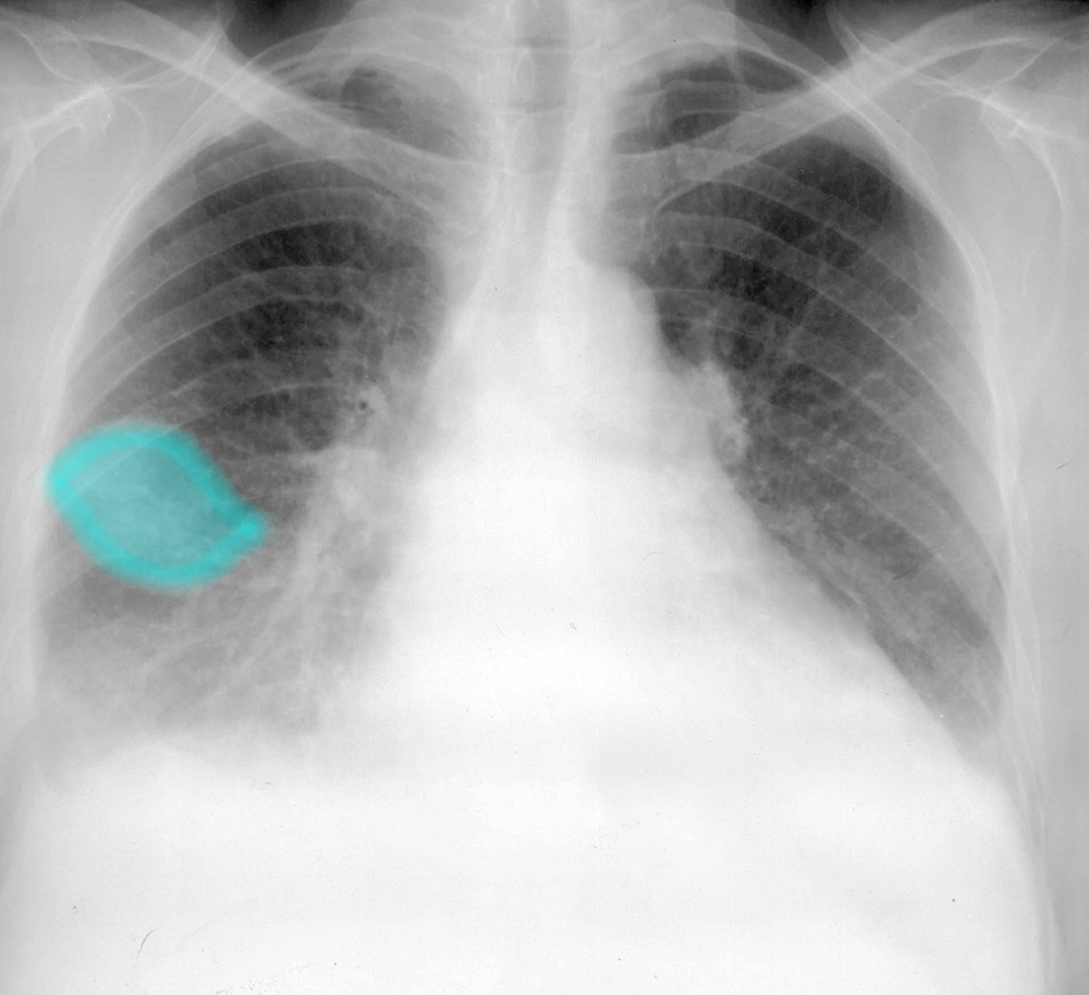
Hint 1: The line segments indicate enlargement of the heart/pericardium and the pulmonary vasculature is also engorged and hazy, suggesting fluid in the lung parenchyma.
Hint 2: There is a vague somewhat oval or lens-shaped opacity projecting in the mid portion of the right hemithorax.
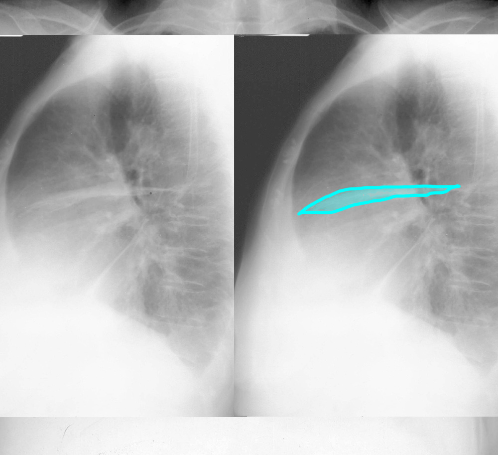
Hint 3: The lateral view for this patient shows opacity or thickening of the minor fissure. The combination of these findings (enlarged heart, engorged and hazy pulmonary vessels, and pleural fluid ) is most consistent with congestive heart failure. Pleural fluid does not always look the same on a frontal view, and its appearance depends on where it is located. When it is in the minor fissure, it can simulate a pneumonia or mass. You need to look at a lateral view to figure this out.
