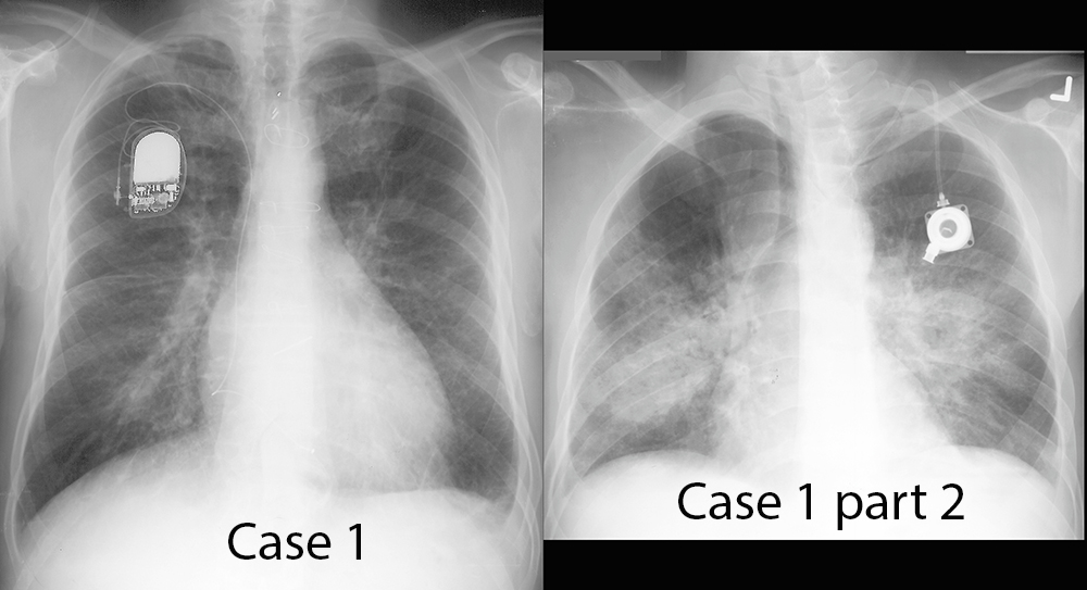


Case 1-PRE CLASS CASE
56 year old male with shortness of breath. Decide what you think of the image. You are given hints below to help in your analysis. Click on each 'Hint' to bring up annotations. To return to the unannotated image, just click on the 'Hint' button again.
Further Explanation:
Decide what is the most likely cause of this patients symptoms, based on the chest imaging findings. You can enter your response at the following website (which you can keep open in a separate tab so you can go back and forth from this site to the answer site). Your answers are anonymous and will be used to assess the utility of these case reviews.
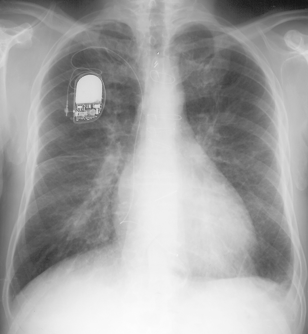
Case 1-PRE CLASS CASE
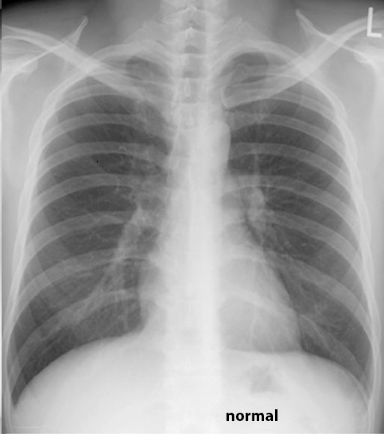
How is this case different from the previous case?
Further Explanation:
Return to the tab for the ANSWER SITE to enter your answer for this case, and all following cases. The next page will give answers and explanations for the cases shown. Please ENTER your responses before going on to the next page!
Case 1-PRE CLASS CASE
Case 1 and Comparison--to view HINTS listed below, go back to the case page
Further Explanation:
Case 1--
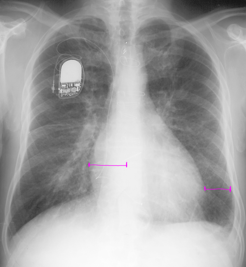
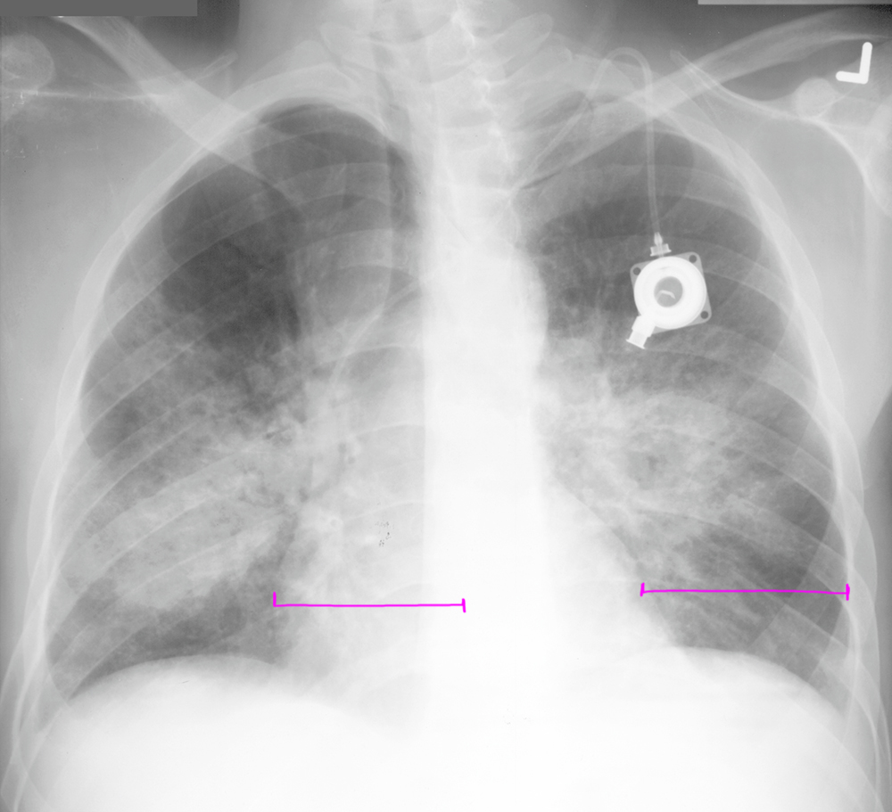
Hint 1: The line segments show a simple way to assess for heart size on chest radiographs. If the line from the right heart border to the middle of the spine is SHORTER than the line from the left heart border to the inside of the ribs, the heart is normal. In this case, the right heart-to-middle line is LONGER, so the heart contour is enlarged.
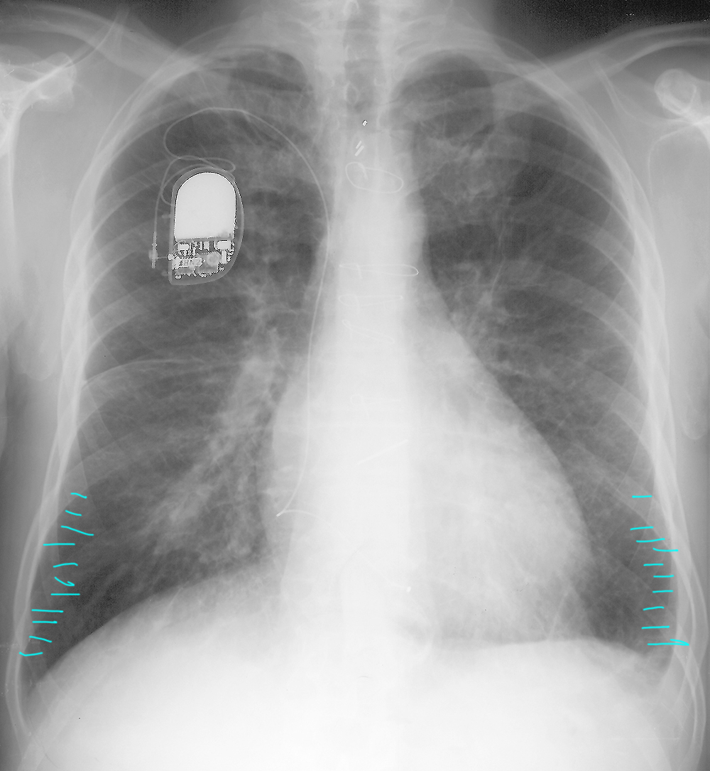
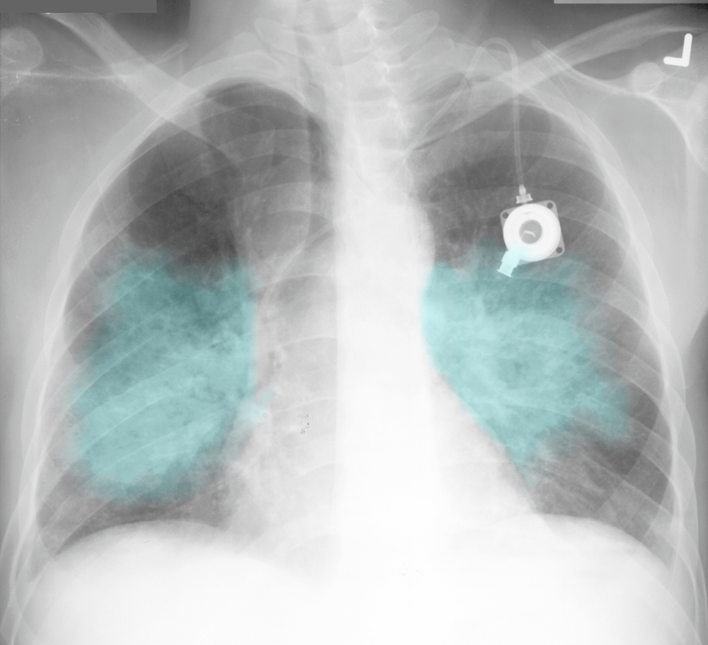
Hint 2: The aqua lines indicate characteristic appearance of Kerley B lines--short horizontal markings in the peripheral lung base that represent some dense material in the interlobular interstitium, most often seen in lung edema.
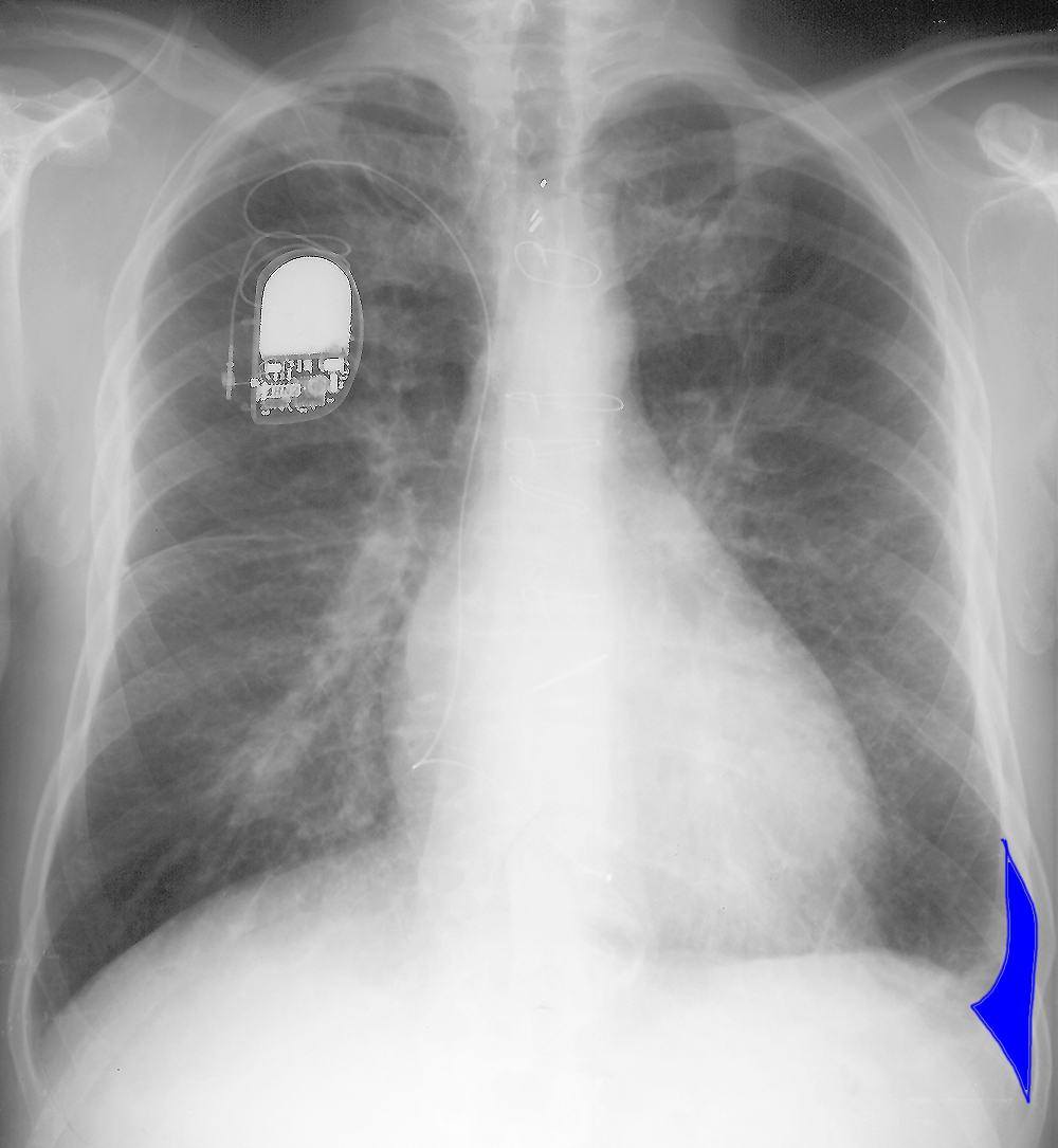
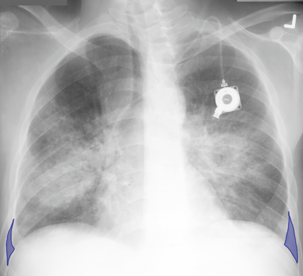
Hint 3: The blue area indicates a small left pleural effusion, with a meniscus creeping up the lateral pleural margin due to surface tension.
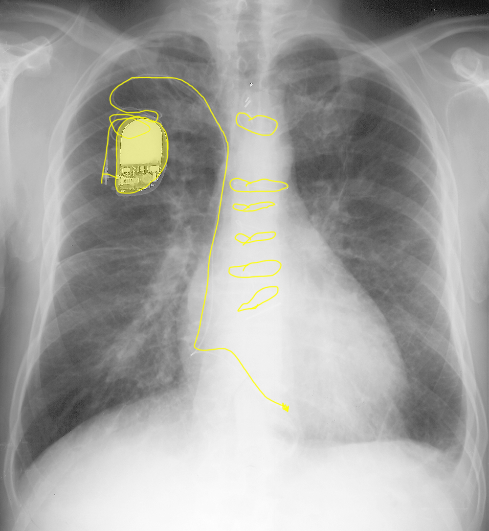
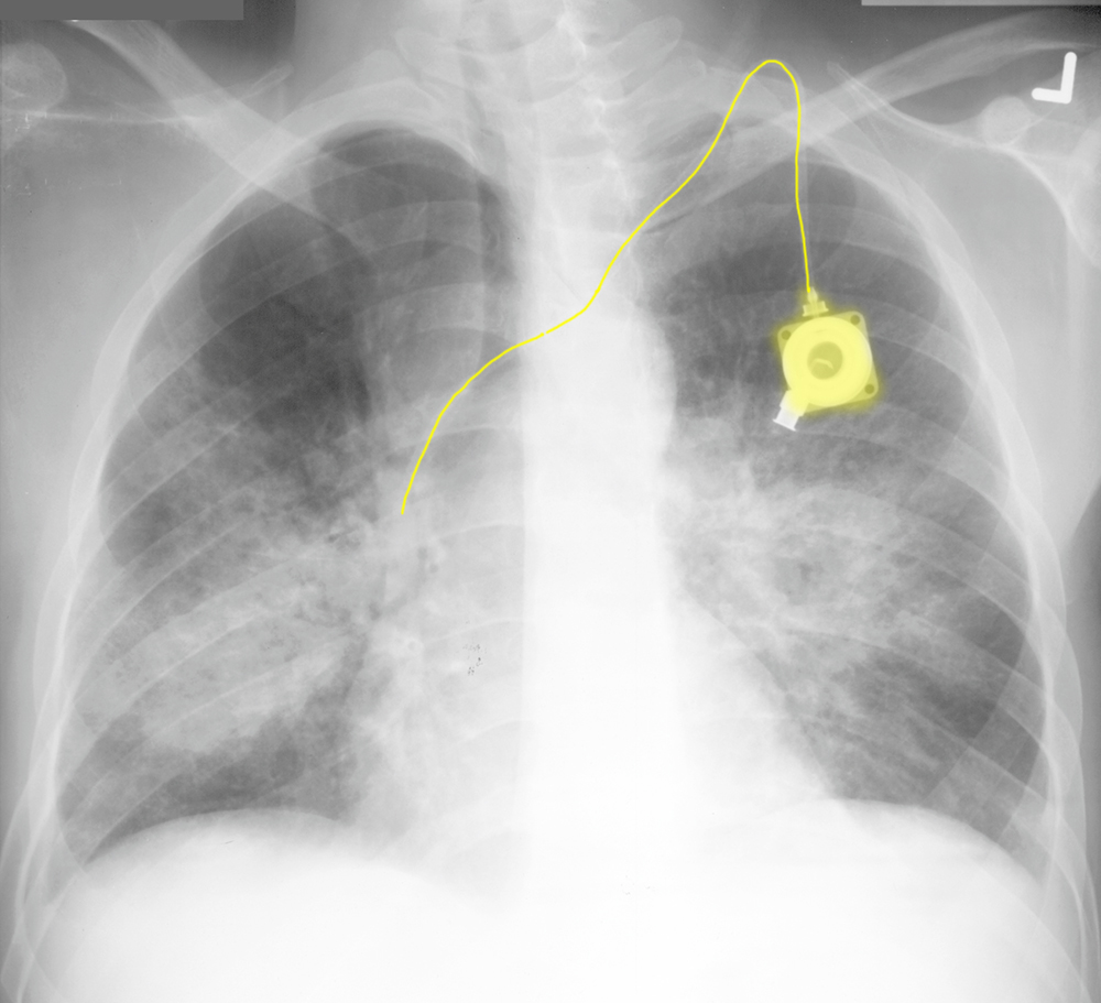
Hint 4: The yellow indicates the presence of a permanent pacer, and sternal wires from prior cardiac surgery, in this case a valve replacement. The combination of all of these findings (enlarged heart, pleural effusion, interstitial edema, and prior cardiac issues) suggest congestive heart failure.
Comparison Case 1:
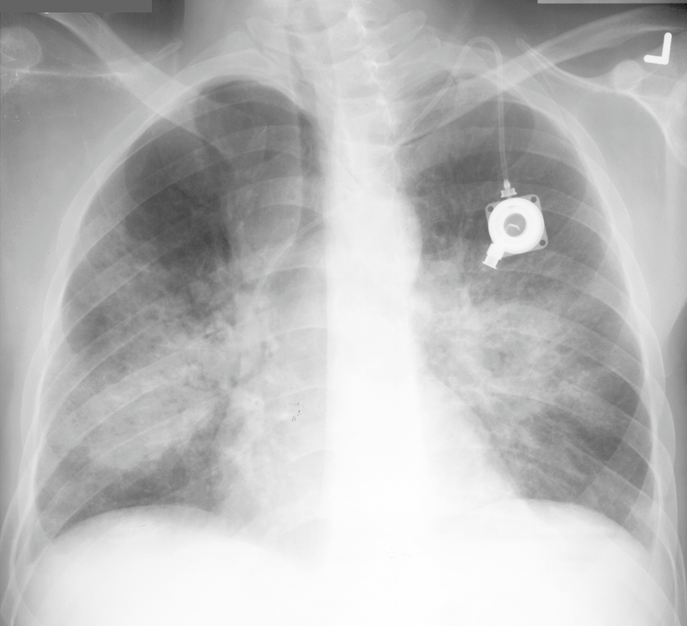
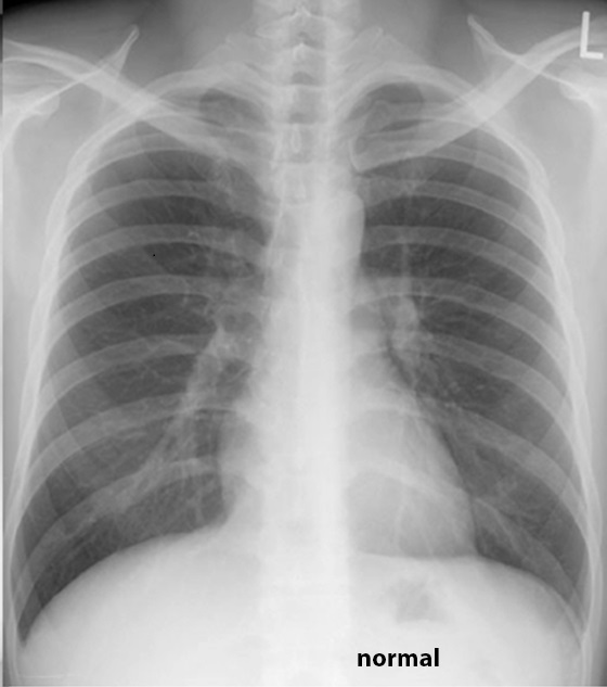
Hint 1: The heart is not enlarged (the line from right heart margin to midline is longer than the line from left heart margin to ribs).
Hint 2: There is diffuse bilateral haziness in the lungs, consistent with an alveolar pattern, which can be seen in pulmonary edema, but is NOT specific.
Hint 3: There are tiny bilateral pleural effusions.
Hint 4: There is a portacath in place via the left subclavian. The combination of these four findings (heart not enlarged, bilateral alveolar lung opacities, small effusions, and port) suggest something OTHER than congestive heart failure. In this case, the patient had pulmonary lymphoma.
