

Case 2
This is a 34 year old woman with vague abdominal pain for several months and infertility.
Question 1:
a) What are technical parameters of the study?
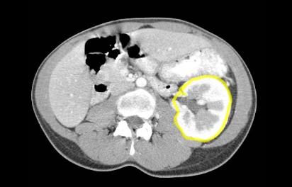
This is an axial CT scan displayed in soft tissue windows, with oral and intravenous contrast.
b) What do you think of her urinary system?
She seems to have only one kidney, on the left.
c) Do you think this is likely to be a congenital or acquired abnormality?
This single left kidney is much larger than a normal kidney would be, indicating that this is likely congenital, rather than due to removal of the right kidney. When a kidney is removed in an adult, the other kidney does not typically enlarge to this degree, although many patients can regain renal function after nephrectomy over time due to compensatory mechanisms in the nephrons of the remaining kidney.
Case 2
These are selected images from the prior study on our young patient with one kidney.
Question 2:
a) What does the appearance of the left kidney on this image indicate about the timing of the scan after IV contrast administration?
The cortex appears much more dense than the medulla. This is normal if the scan is performed relatively early after contrast administration. The first place the filtered contrast will appear is in the cortex (where the glomeruli are). A short time later, it will start to make its way into the medulla, and then shortly thereafter into the collecting system.
b) What is outlined in blue on the lower CT slide (link below)?
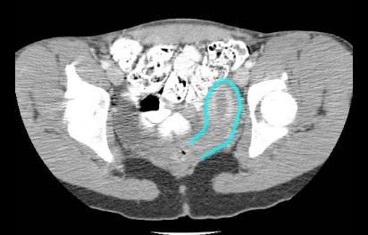
The blue outline indicates the uterus, which is not normal in appearance. Instead of being midline and pear-shaped, it is on the left and tubular in shape.
c) Why does it make sense that someone with a congenital single kidney might have a uterine anomaly as well?
The kidneys and uterus share common embryologic precursors, so an anomaly of one can be mirrored by an anomaly of the other. In this patient, the right kidney and the right portion of the uterus failed to form, resulting in a single (large) kidney on the left and a malformed uterus, called a 'unicornuate' uterus (only one horn).
Case 2
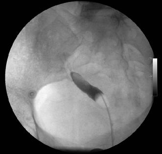
This is a study from a different young female patient with similar symptoms of infertility.
Question 3:
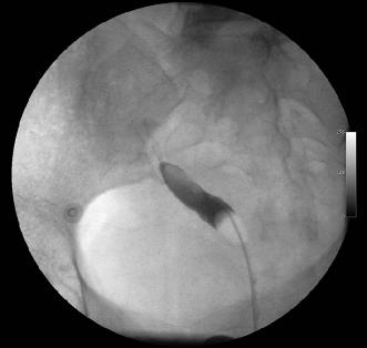
a) What is this study?
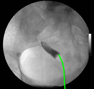
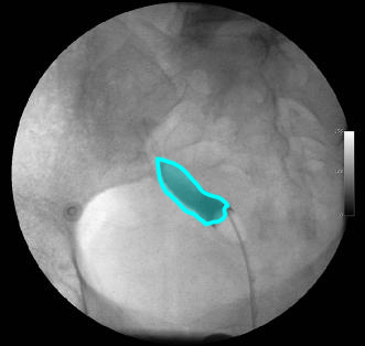
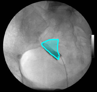
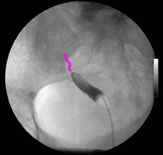
This is a hysterosalpingogram. A link to a normal comparison in a different patient is shown below. This uterine cavity does not appear triangular, like a normal uterus. Instead, it is tubular and extending only to the patient's right side. This is the appearance of a unicornuate uterus, like our original patient had, but on the opposite side in this case.
b) What other type of study might be indicated in this patient?
Given the presence of a unicornuate right uterus, it would probably be prudent to look at the right and left kidneys for other anomalies. The easiest way to do this would be with a renal ultrasound. In this patient, the kidneys were normal.
Case 2
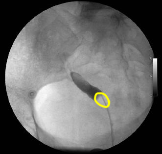
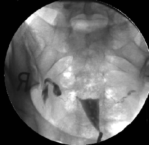
This is our patient's study again.
Question 4:
Why might this anomaly lead to infertility?
If the uterus is only half there, it is likely too small to support a normal pregnancy. Also, on this single image, there is very little contrast in the uterine tube and no evidence of spill into the peritoneal cavity, so the uterine tube may not be patent, which would lead to infertility.
