

Case 1
This study is from a 23 year old man with seizures and headaches for many years.
Question 1:
a) What type of study is this?
This is an axial head CT scan without IV contrast. The image is displayed with brain windows, but since these are the only windows routinely used (other than bone windows) for viewing the brain, this is not generally specified.
b) What general terms would you use to describe the main abnormality?
There are irregular, large chunky calcifications in the brain bilaterally, most marked in the left temporal and right parietal regions.
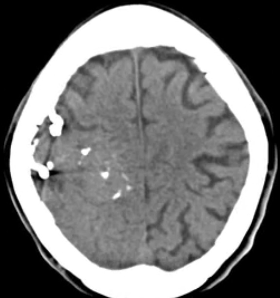
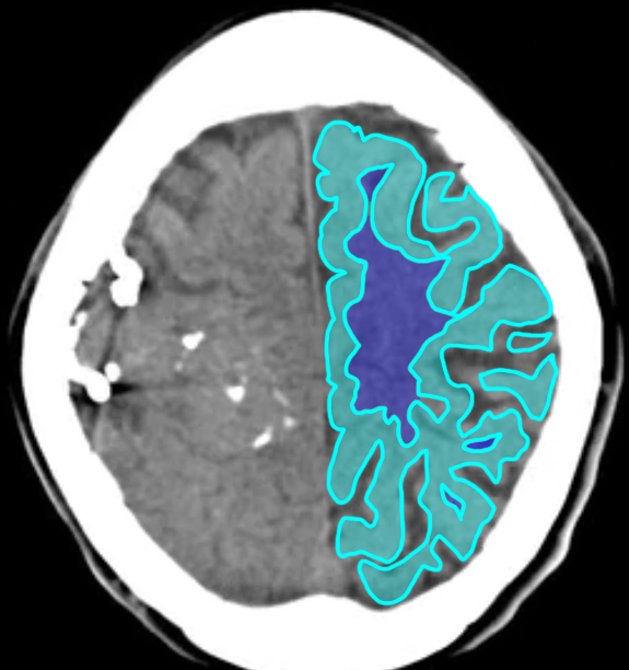
c) Which part of the brain appears DENSER on these images-white matter or grey matter?
Grey matter is slightly denser (lighter in color) than white matter (which appears darker). This is best seen on images high over the convexity of the brain, as indicated in the labeled image below.
Case 1
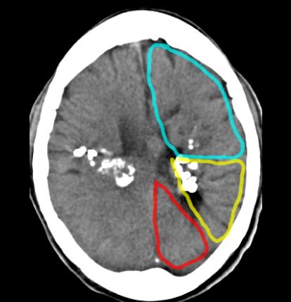
This is a selected image from the prior head CT scan. Try to identify the lobes of the brain that are on this slice. They are outlined below.
Question 2:
a) What are the three lobes indicated on the outlined image?
The frontal lobe is outlined in blue, the parietal lobe in yellow and the occipital lobe in red.
b) What structure forms the boundary between the frontal and parietal lobes?
The central sulcus forms the boundary between the frontal and parital lobes of the brain.
Case 1
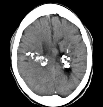
This is another selected slice from this patient's head CT, with labels below.
Question 3:
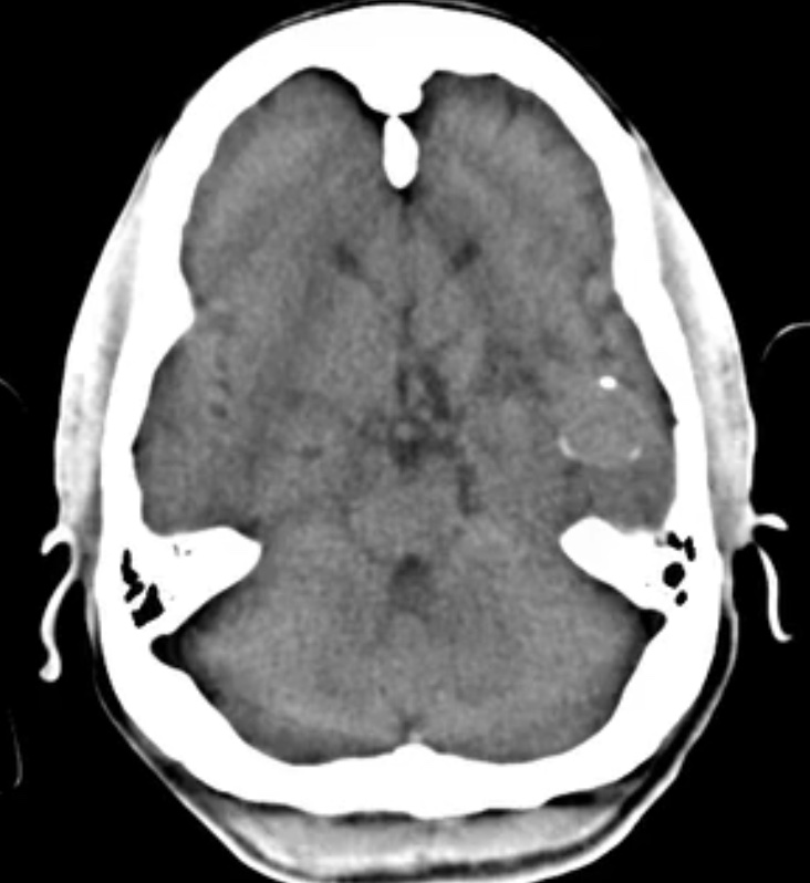
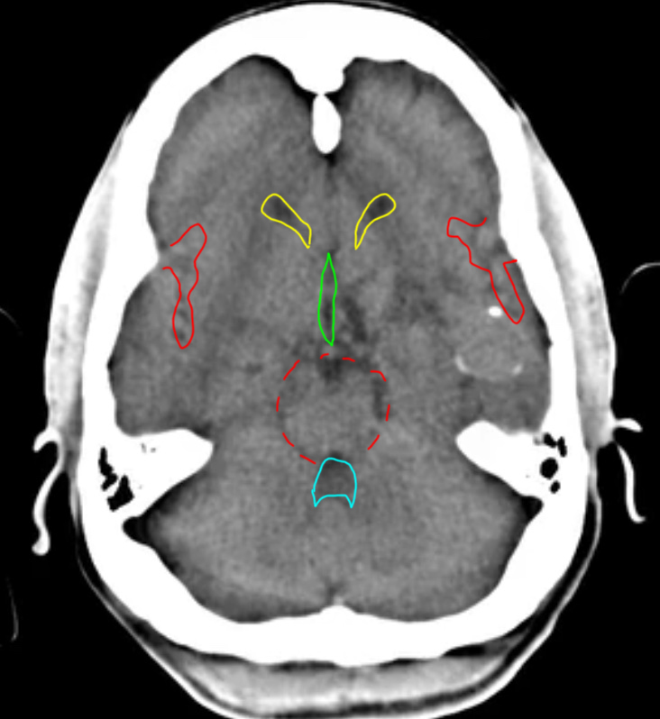
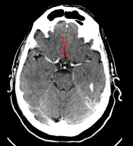
a) How can you tell that this study is done without IV contrast?
There are no enhancing cerebral vessels evident on this image. Bright vessels would generally be seen in the regions indicated below by the red dotted lines, the Sylvian fissures and region of the circle of Willis.
b) What are the other structures labeled below?
The lateral ventricles are outlined in yellow. The 3rd ventricle is outlined in green. The 4th ventricle is outlined in blue. Bright vessels may be seen in these regions as well after IV contrast administration.
Case 1
These sets of images are from a different patient and are normal.
Question 4:
a) What is the difference between the first set of images and 'series 2', indicated below?
The initial set of axial head CT images have no IV contrast. Series 2 has IV contrast. For most routine head CT scans, scanning begins about 5 minutes after contrast injection, to allow the contrast to equilibrate throughout the tissues and vessels. In special circumstances (to assess perfusion or to perform CT angiography), scanning may be started sooner. One of the things contrast helps with in brain imaging is showing breakdown of the blood-brain barrier. Contrast will be seen in areas where this barrier is disrupted and it can take some time for this to accumulate.
b) CT is preferred over MR for what types of neurologic problems?
CT is faster and more readily available than MR, so it is often a first screening study, particularly in trauma (where it can also show bony abnormalities better, such as fractures). It is preferred for detection of hemorrhage and calcifications, which may be more difficult to detect with MR. So it is generally the first study done in suspected stroke to check for evidence of hemorrhage, even though MR can be more sensitive for early determination of extent of stroke injury.
Case 1
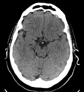
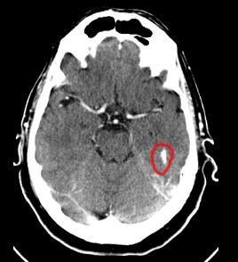
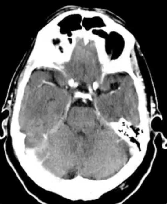
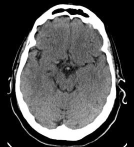
These are selected images at a similar levels from the two previous axial head CT series.
Question 5:
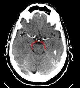
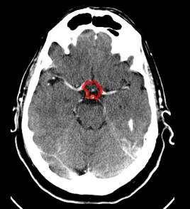
a) What is circled in red on the contrast enhanced image below?
This is tricky because that particular slice is slightly lower than the one from the non-contrast series. We are just at the top of the petrous temporal bone, and this high attenuation area represents a slice through the very uppermost part of that bone. This is easier to figure out when you look at the slice immediately BELOW the circled slice (use link to see the slice below).
b) What does hemorrhage look like on a head CT?
Because the brain parenchyma overall is relatively low in density, acute hemorrhage appears brighter (whiter) than surrounding brain. So it is important to make sure whether white objects that are seen are blood vessels (normal), part of the skull bones (normal), calcifications (some normal and some abnormal), or hemorrhage (abnormal).
c) How should a head CT be done to optimize visibility of hemorrhage?
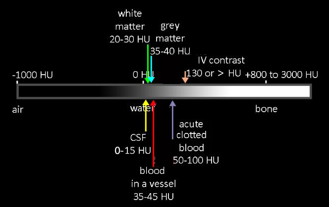
Optimal visualization of any hemorrhage will be on a non-contrast study. In this way, you will not mistake vessels for hemorrhage. You will still have to deal with bony structures and calcifications.
Case 1
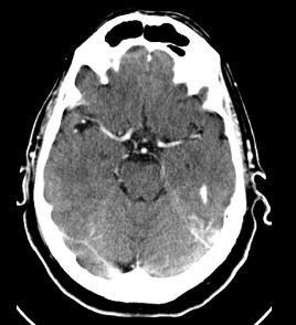
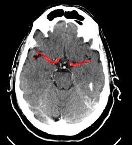
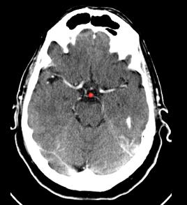
This is the same normal axial head CT image with IV contrast present. Try to identify the branches listed below before clicking on the labels.
Question 6:
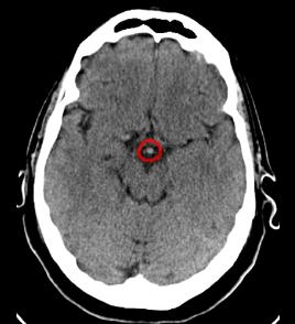
In a normal patient, how should the density of flowing blood in a cerebral vessel compare to brain parenchyma?
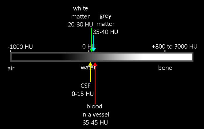
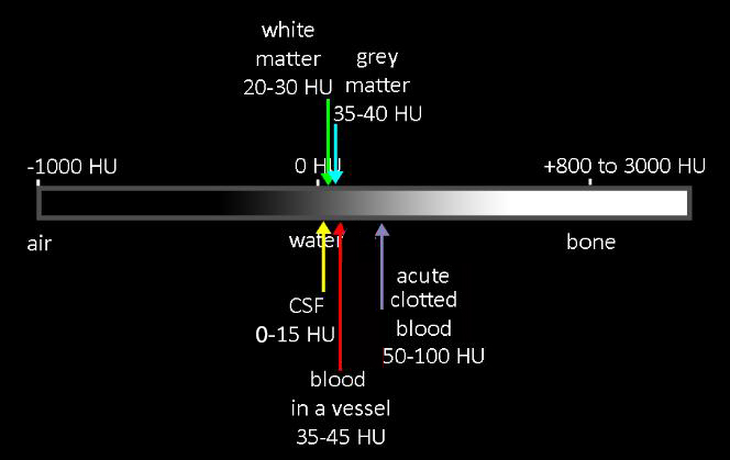
Flowing blood in a vessel should be similar in density to grey matter, and will be slightly denser than white matter.
Case 1
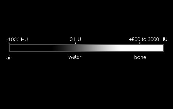
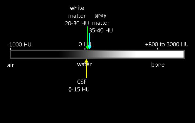
This diagram shows the position of brain tissues and hemorrhage on the density spectrum of the body.
Question 7:
What might lead to higher density than expected in blood vessels on a non-contrast CT scan?
Normal blood is only very slightly more dense than grey matter (but the difference between unopacified blood and white matter is a bit more apparent). The measured density of blood on CT depends on hematocrit, so any process that increases the hematocrit can make unopacified blood look more dense than usual. Things that can increase hematocrit include dehydration and the rare condition polycythemia vera (patients who produce abnormal amounts of red blood cells). Hematocrit is the proportion of red blood cells to serum and is normally about 45-52% for men and 37-48% for women.
Case 1
This is another study on our original patient, with seizures and headaches for many years, and irregular calcifications bilaterally on head CT.
Question 8:
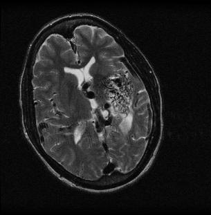
a) What type of study is this?
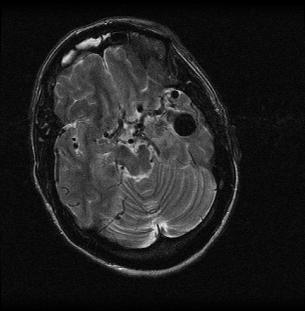
This is a brain MR in the axial plane with T2 weighting (fluid appears bright). Vessels appear dark, so this study was not done with bright blood technique (a different set of MR parameters that can make flowing blood appear white).
b) Is it as easy to see the calcifications on this study as on the CT scan?
No. MRI is not as sensitive as CT for detection of calcifications, since they will be low signal on most pulse sequences and may be missed, if small. Special sequences can be used to show calcifications better, but routine T1 and T2 weighted sequences may not show them well.
c) How does the appearance of the brain abnormalities look different on the MR images than the CT images in terms of shape?
The MR shows a tangled network of fine twisting tubular structures in the regions where the dense calcifications were shown on the original CT. This suggests the presence of a tangle of abnormal vascular channels in the area, that were not appreciated on the CT images. Notice that the vessels of the circle of Willis also appear dark. This congenital malformation is called an AVM (arteriovenous malformation). If small, they can be resected or treated by IR with intravascular coiling. This one is so large that treatment would be difficult without risk of damage to adjacent brain.
Case 1
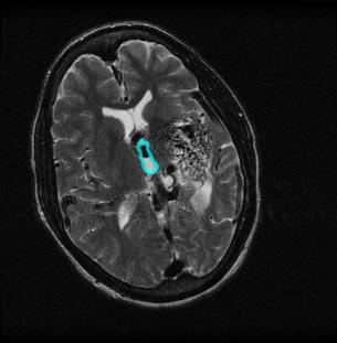
This is a selected image from the T2 weighted brain MR previously shown.
Question 9:
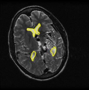
What are the three labeled structures below?
Structure 1 is the lateral ventricles, outlined in yellow. Structure 2 is the 3rd ventricle, outlined in blue. Structure 3 is the network of abnormal tangled vessels present bilaterally. This is an example of a congenital vascular anomaly that accounts for the patients symptoms of headaches and seizures.
Case 1
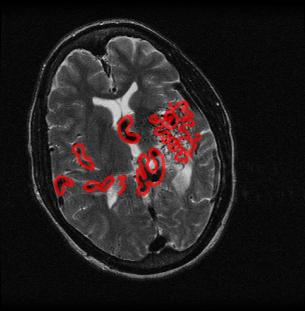
This is the same MR image, with some other vessels marked for you to identify.
Question 10:
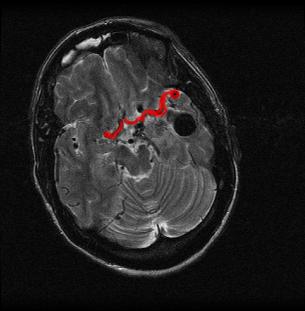
a) What are vessels 1?
These are the middle cerebral arteries.
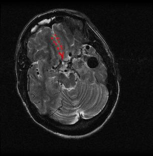
b) What are vessels 2?
These are the anterior cerebral arteries.
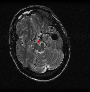
c) What are vessels 3?
This is the basilar artery.
d) What do you think of the frontal sinus?
There is high signal material in the frontal sinus, consistent with sinusitis (fluid in the sinus cavity, which should be filled with air.
