

Case 4
This 31 year old female who recently gave birth noticed that she could not smell her baby's diapers.
Question 1:
a) What is this study?
This is an axial T2-weighted MR sequence without fat suppression. Water is bright and can be clearly seen in the globes and the CSF spaces.
b) What cranial nerve could give rise to a tumor in this location?
There is a large mass located between the orbits, consistent with origin from cranial nerve I (the olfactory nerve). This could account for problems with the sense of smell, as this patient was experiencing.
Case 4
These are more images from the same patient.
Question 2:
a) What are these images?
These are coronal T1-weighted images (CSF is dark) with IV contrast (gadolinium, look for vessels lighting up in the cortical sulci and circle of Willis).
b) What is the single comparison image shown below (from the same patient)?
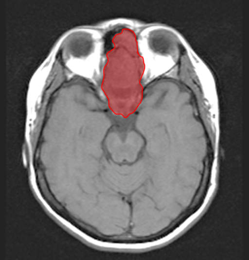
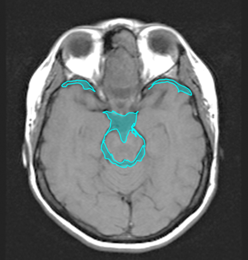
The comparison image is a T2-weighted (CSF is labeled in blue below in the region of the brainstem, temporal lobes, and suprasellar regions and is LOW signal) MRI in the axial plane. The tumor, between the orbits and extending into the left frontal sinus, is labeled below in red.
c) Does the tumor show enhancement with contrast on the Gadolinium-enhanced series (the coronal series)?
Yes, the tumor lights up brightly indicating that it is very vascular. Comparing the region of the tumor to the T1 axial image without contrast shows that the tumor is slightly lower signal than brain prior to administration of gadolinium, but is very bright on the coronal series after contrast.
Case 4
These are more images on the same patient.
Question 3:
a) What are these images?
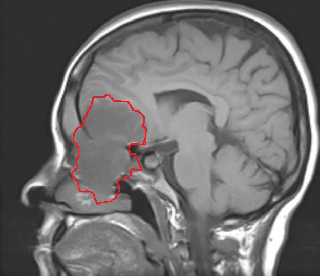
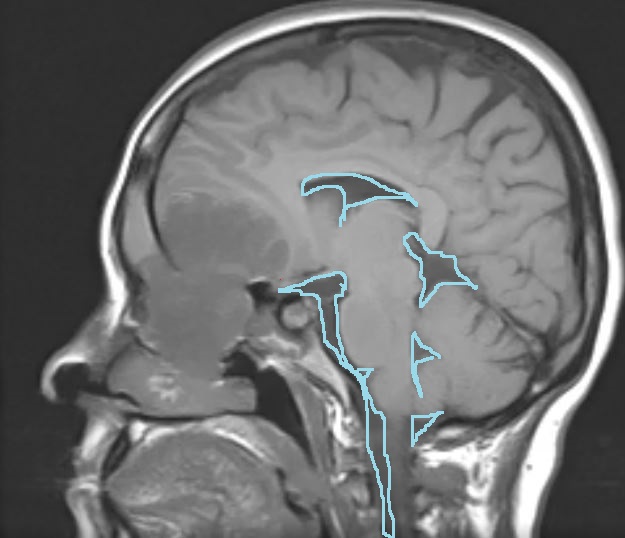
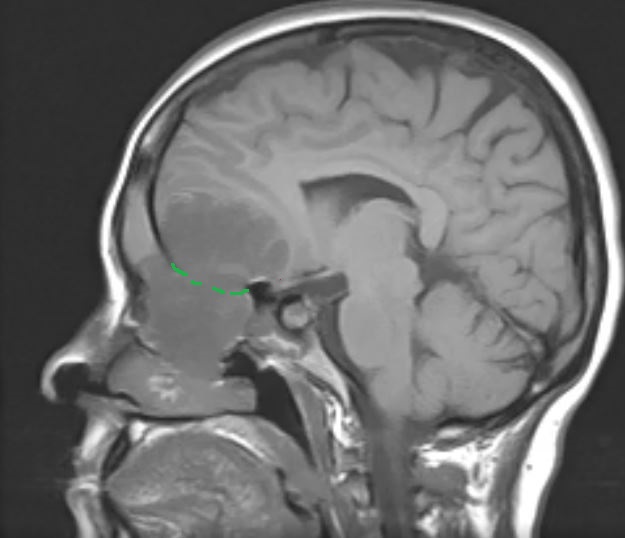
These are sagittal T1-weighted images without IV contrast or fat saturation. The tumor appears dark in comparison to the previous contrast-enhanced sequence (see labels below). This imaging plane shows the tumor extending down and up from the level of the cribriform plate, where the first cranial nerve is located. You know this is either a T1-weighted image or a FLAIR image because CSF signal is low (see below).
b) What is the appearance of grey vs white matter on these T1-weighted images? Is this relationship the same as for a CT scan?
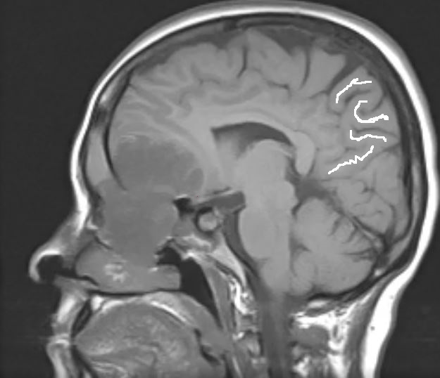
The grey matter is darker in terms of the shade of grey than the white matter (see labels below), best seen in the gyri near the brain surface on this image. This is the opposite of their appearance on CT, where the grey matter is a lighter grey than the white matter. This confirms that this is a T1-weighted image and NOT the other type of brain MR where the CSF has low signal--FLAIR imaging.
Case 4
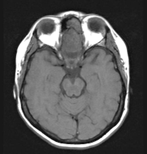
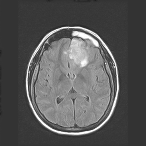
There are many other specialized MR sequences that are used in imaging the brain and spinal cord. This image is an example of FLAIR imaging (Fluid Attenuated Inversion Recovery).
Question 4:
a) What is the appearance of CSF on this FLAIR image from our patient?
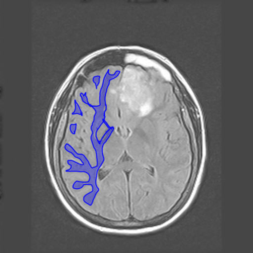
The CSF in the ventricles on this image is dark. This might make you call this a T1-weighted image. But FLAIR allows the radiologist to selectively turn down the signal from fluid in the CSF space without and provide better visualization of subtle abnormalities in the periventricular regions.
b) How can you tell a FLAIR sequence from at T1-weighted sequence?
On T1-weighted MRI, the grey matter is darker than the white matter. On T2-weighted sequences (including FLAIR), the grey matter is lighter than the white matter. Check the labels below to show the dark appearance of white matter on these images.
c) What are the other sequences shown below used for?
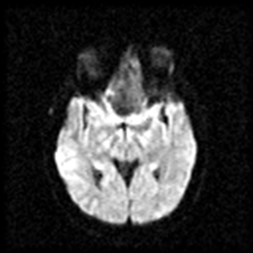
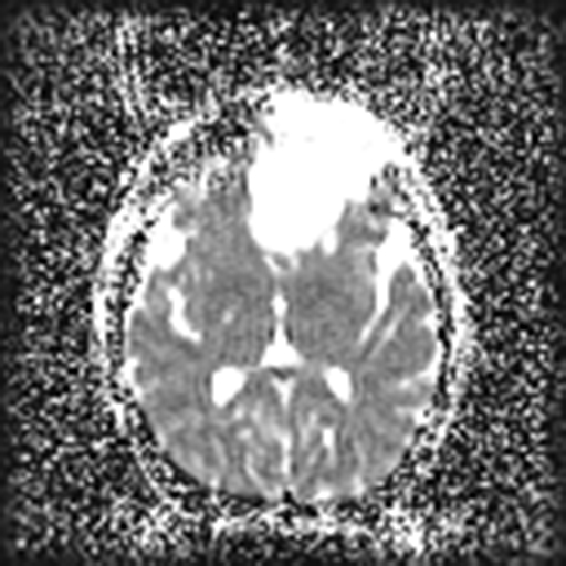
The links below show other specialized sequences that image different aspects of fluid in the body: diffusion-weighted imaging (DWI) and apparent diffusion coefficient (ADC) imaging. Both are related to the state of water in various body compartments and how freely it can move within the local microarchitecture. Both play important roles in early detection of stroke and in imaging of tumors. You can see that there is abnormal signal within this patient's tumor on both sets of images--the tumor is DARK on DWI and BRIGHT on ADC.
Case 4
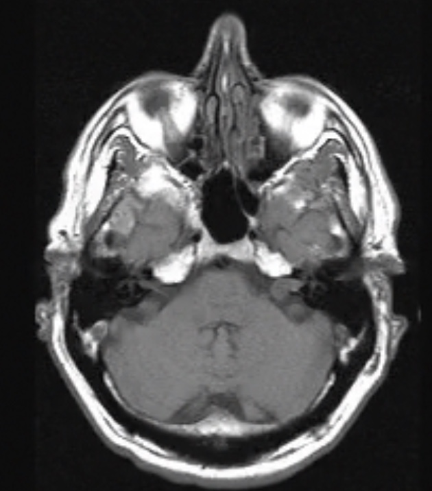
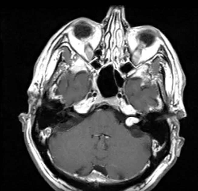
This is a different patient with left sensorineural hearing loss, tinnitus and vertigo.
Question 5:
a) Problems with what cranial nerve could give rise to this set of symptoms?
These symptoms seem related to problems with the 8th cranial nerve (vestibulocochlear).
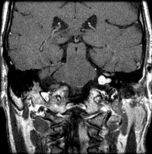
b) What are these images?
These are MRI images (cortical bone is dark) with T1-weighting (CSF in ventricles is dark) and gadolinium enhancement (vessels are lighting up). Check the selected image below to see more clearly where the patient's abnormality is located.
Case 4
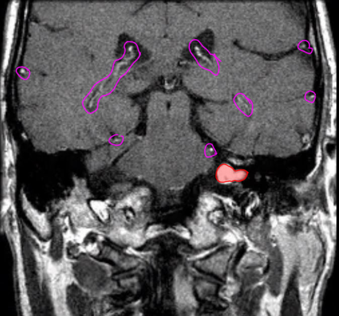
These are selected images of the same patient. The tumor is outlined in red on the labeled images below.
Question 6:
a) How can you tell that gadolinium was given for these two selected images?
Look for enhancing vessels in the same places you would see on CT--along brain surface in sulci and centrally in the region of the circle of Willis (purple circles on labeled images).
b) What is outlined in blue on image 1 with labels?
Blue is outlining the CSF space, including the extension into the normal right internal auditory canal, containing the normal right 8th cranial nerve and the 7th cranial nerve (facial nerve).

Case 4
These are more MRI images on the same patient.
Question 7:
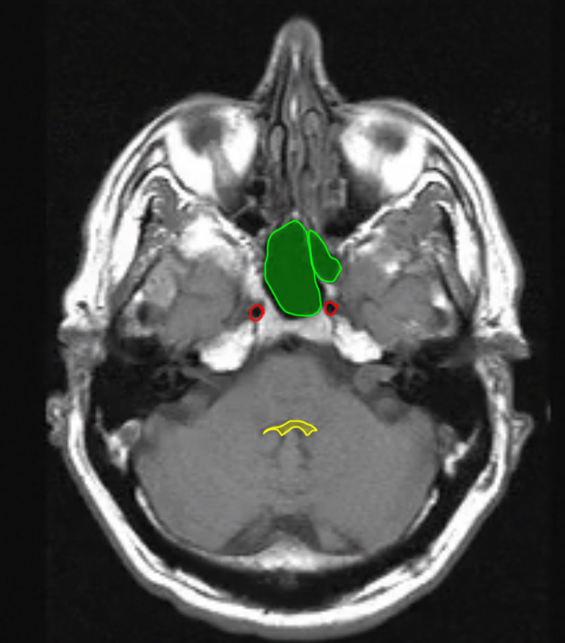
a) What are the structures outlined below?
red-carotid arteries green-sphenoid sinus yellow-4th ventricle
b) Does the patient's tumor show enhancement?
Yes, this left vestibular schwannoma is very vascular and enhances brightly comparing the pre and post-gadolinium T1-weighted images. Analysis of the enhancement pattern of various CNS tumors is very complex, and not all tumors enhance to this degree.
c) What condition would be suggested if the patient were found to have a similar tumor on the right as well as the left?
Presence of bilateral vestibular schwannomas (sometimes also called acoustic neuromas) would strongly suggest the diagnosis of neurofibromatosis, a congenital condition associated with many tumors in many parts of the body.
