

Case 3
This is a 60 year old man with new onset of right-sided weakness, numbness, slurred speech and confusion.
Question 1:
a) What is this study?
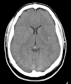
This is an axial head CT without intravenous contrast, the typical initial study done in a case such as this where stroke is a major diagnostic consideration.
b) Do you see any abnormality?
On the left side in the temporoparietal region there is an area of high attenuation, worrisome for hemorrhage. It is for this reason that noncontrast CT is performed, since the hemorrhage might be missed if it was obscured by nearby enhancing vessels.
Case 3
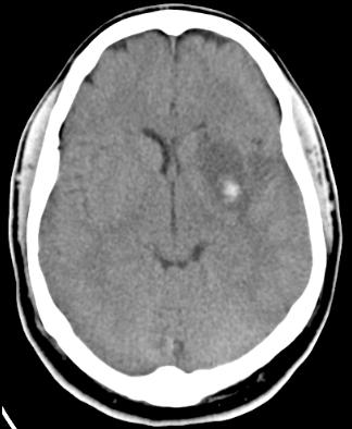
This is a followup study on this patient, whose symptoms progressed.
Question 2:
a) How does this study compare to the initial exam?
On the followup CT, the area of hemorrhage is larger and there is a larger area of low attenuation surrounding it.
b) What density is edema on a head CT?
Edema (water) is lower attenuation than brain parenchyma, similar to the density of the CSF. So it will look dark compared to brain.
c) What do you think of the ventricles on this study?
There is now asymmetry of the anterior horns of the lateral ventricles, which was not apparent on the initial series. This indicates increased intracranial pressure as this hemorrhagic stroke is progressing.
Case 3
This is a selected image from the original CT scan. Try to identify the hemorrhage and adjacent edema before clicking on the 'abnormality' label below.
Question 3:
Why is it important to identify that this stroke is hemorrhagic?
The initial treatment of stroke usually involves anticoagulation to try to break up the clot that is blocking brain blood flow. However, if the stroke is hemorrhagic, anticoagulation is contraindicated. Intravascular interventions may be indicated to stop the bleeding and remove any remaining clot.
Case 3
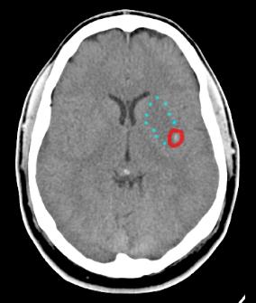
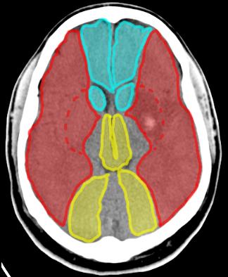
The link below shows the vascular territories of the anterior, middle and posterior cerebral arteries on a selected CT image from our patient with a hemorrhagic stroke on the left.
Question 4:
a) What are the three vascular territories shown below?
Blue is the anterior cerebral territory. Red is the middle cerebral territory. Yellow is the posterior cerebral territory. The middle cerebral gives rise to dee pbranche (lenticulostriates) that supply the basal ganglia, where this hemorrhage appears to be.
b) What vascular territory is giving rise to the hemorrhage in this patient?
The hemorrhage is in the territory of the middle cerebral artery. This is important to determine in case an intravascular intervention is planned.
c) What other kind of imaging might be helpful in a patient with a negative head CT but symptoms suggestive of stroke?
CTA (CT arteriography) is most often done next, if the patient has symptoms and history that make vascular intervention to remove clot appropriate. If clot is seen on CTA, the patient can be taken directly to the angiography suite to have the clot removed and potentially reverse the neurologic deficits. If the patient is not a candidate for this procedure, MRI can be done to better define the extent of ischemia and help in treatment planning.
Case 3
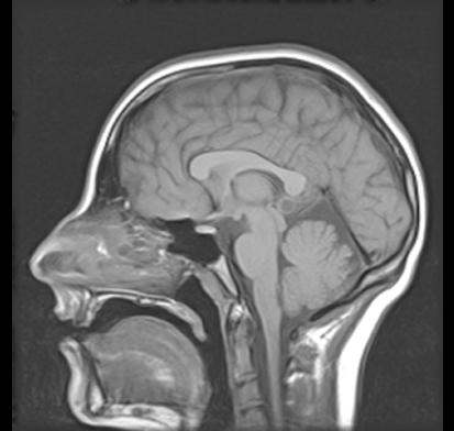
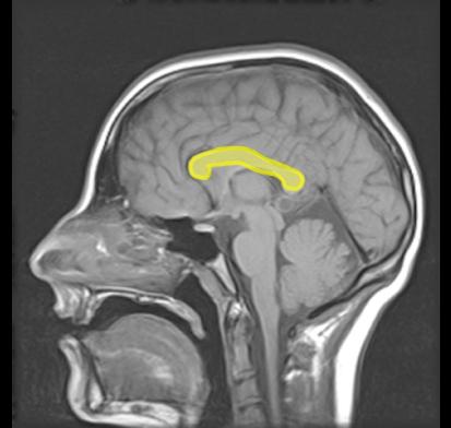
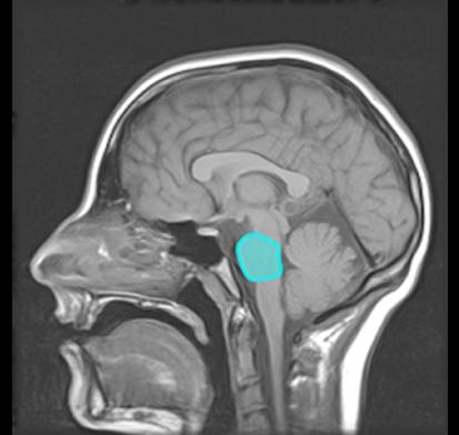
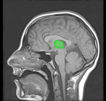
This is an imaging study on a 75 year old woman with confusion and right-sided weakness. Her head CT was normal. Try to identify the listed structures before clicking on the labels below.
Question 5:
a) What is this study?
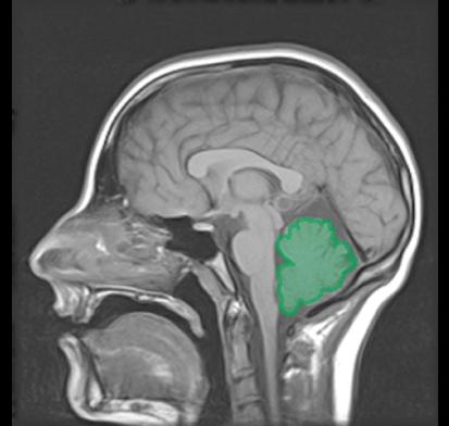
This is a sagittal T1-weighted MR image in the midline, without fat suppression.
b) What advantage doe MR have over CT for examination of brain structures?
Individual brain structures are much more clearly depicted on MR than on CT. While the brain tissues are all fairly similar in density, their structure makes them look different on MR, based on their chemical composition.
