

Case 4
Case 4 (inspiration), Part 1
Question 1:
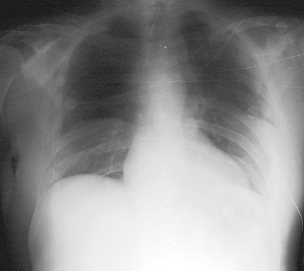
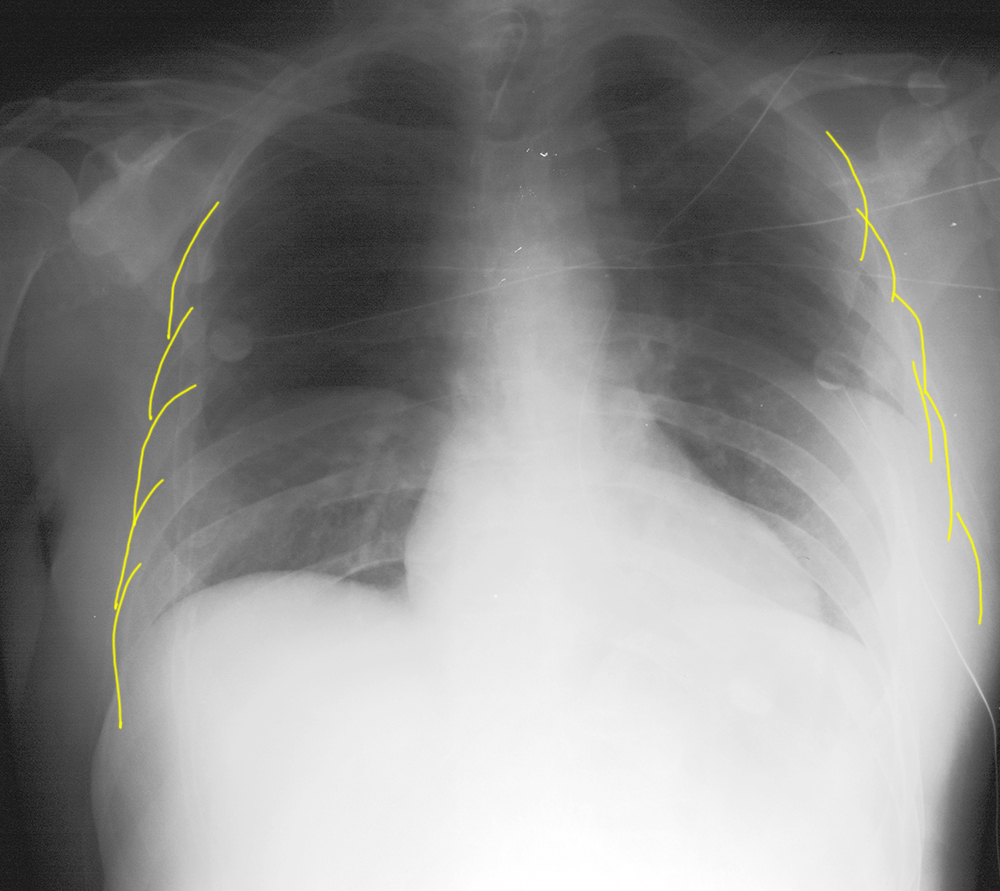
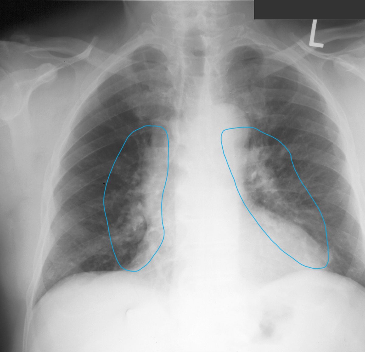
a) What is wrong on this image?
×
Answer:
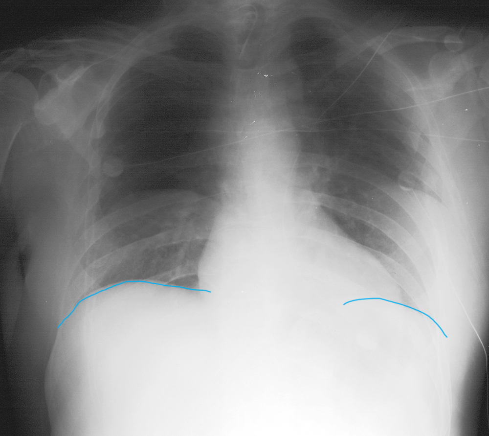
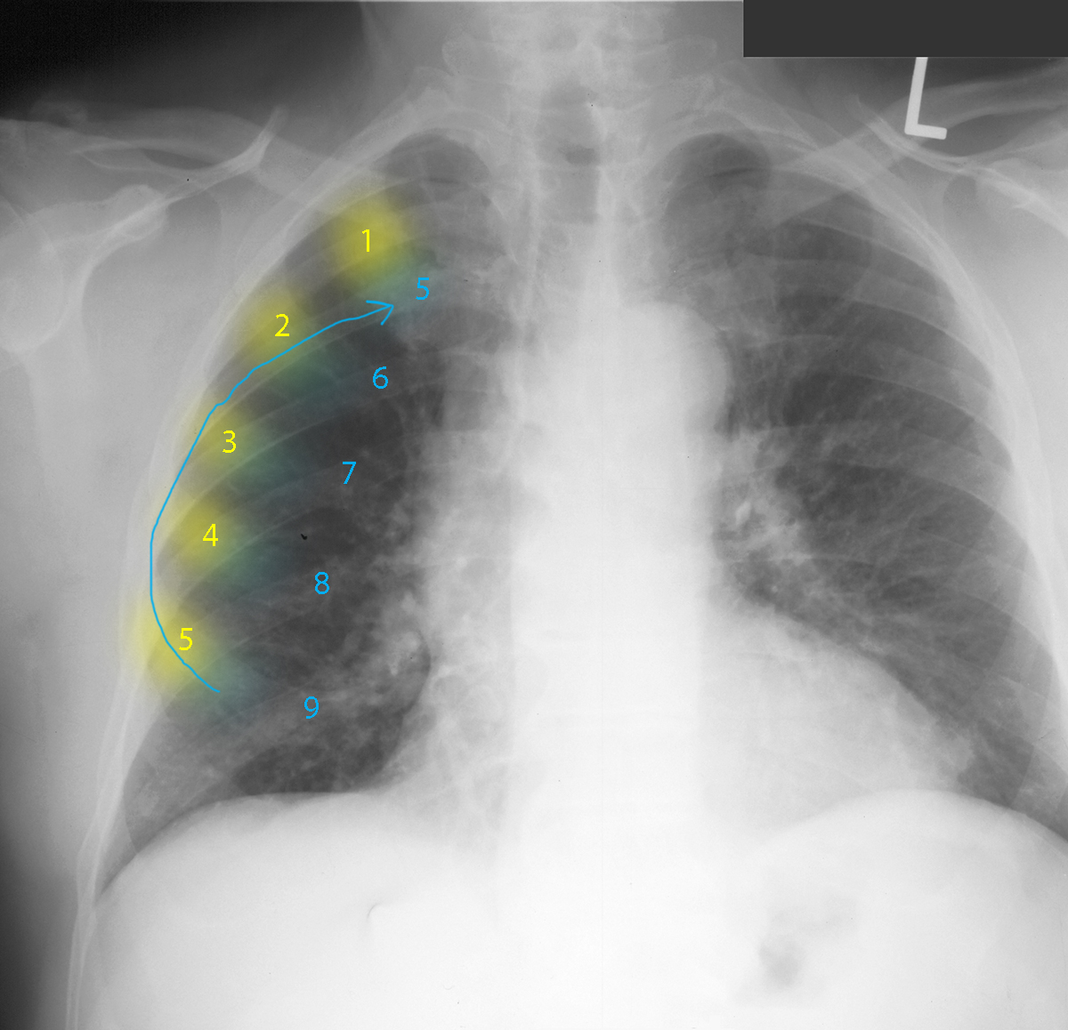
This is an example of the effects of a low level of inspiration. Only 9 posterior ribs are visible above the diaphragms.
This is an example of the effects of a low level of inspiration. Only 9 posterior ribs are visible above the diaphragms.
b) Why does this matter?
×
Answer:
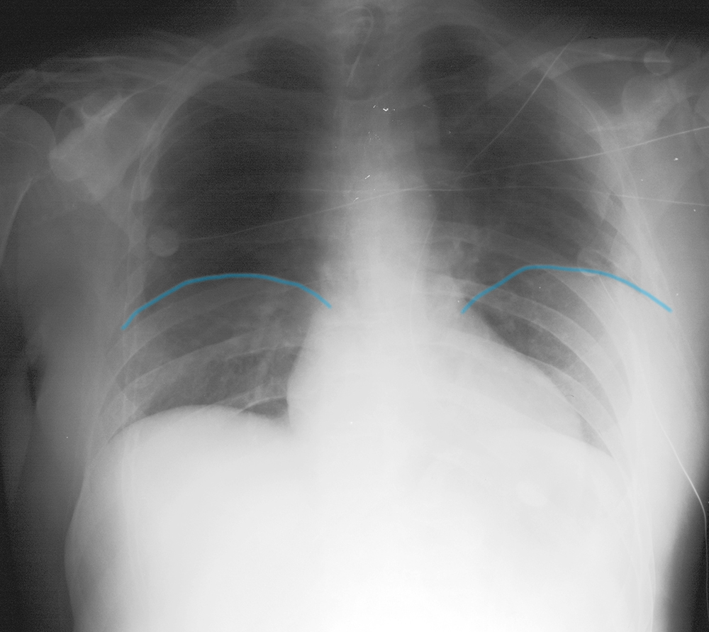
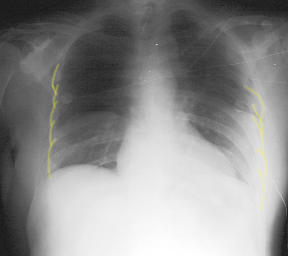
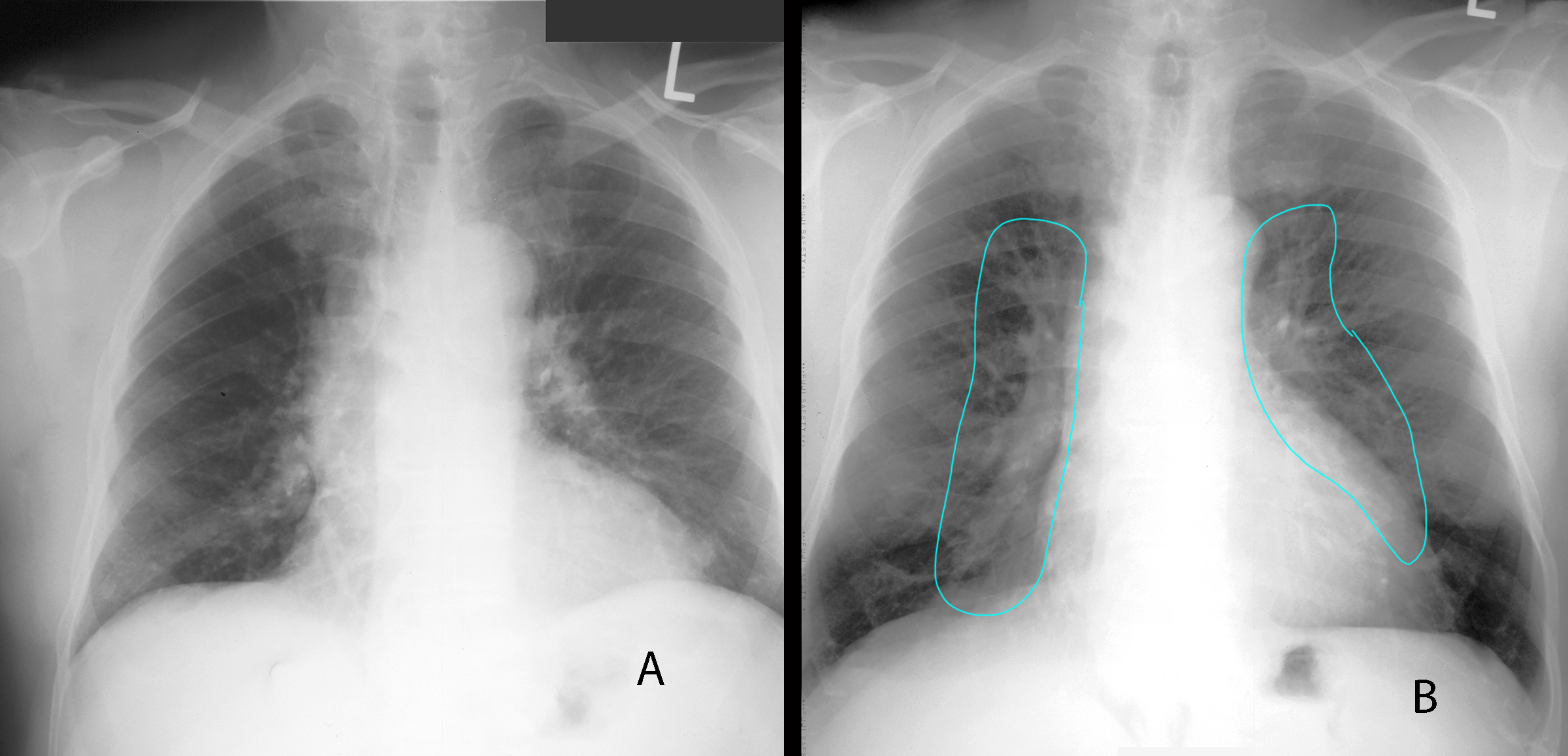
As you can see from the labels below, the line segments overlying the heart are longer than those overlying the lungs, suggesting cardiomegaly. But this can be seen with high diaphragm position, which spreads the heart out more and makes measurement inaccurate. Also, the hilar regions look very hazy, which could represent edema. This can also be an artifact of crowding of pulmonary vessels on a low inspiration image.
As you can see from the labels below, the line segments overlying the heart are longer than those overlying the lungs, suggesting cardiomegaly. But this can be seen with high diaphragm position, which spreads the heart out more and makes measurement inaccurate. Also, the hilar regions look very hazy, which could represent edema. This can also be an artifact of crowding of pulmonary vessels on a low inspiration image.
Case 4
Case 4, Part 2
Question 2:
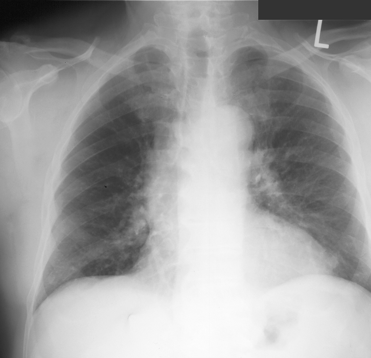
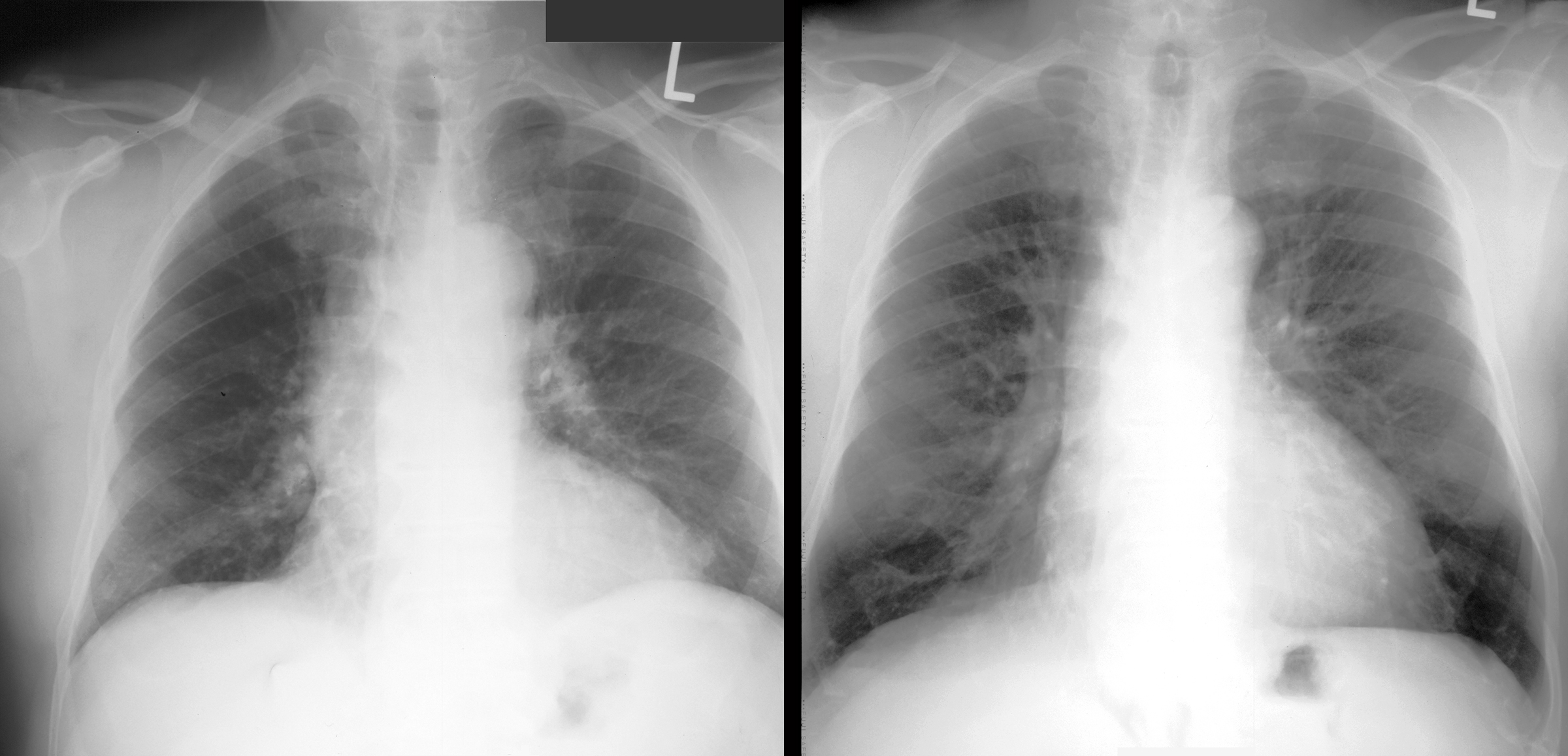
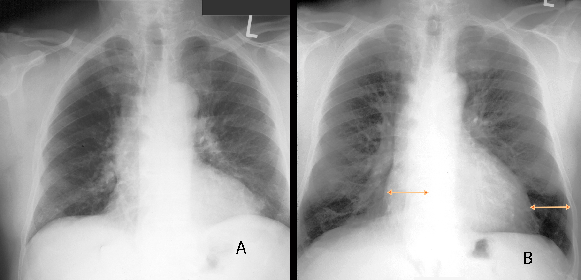
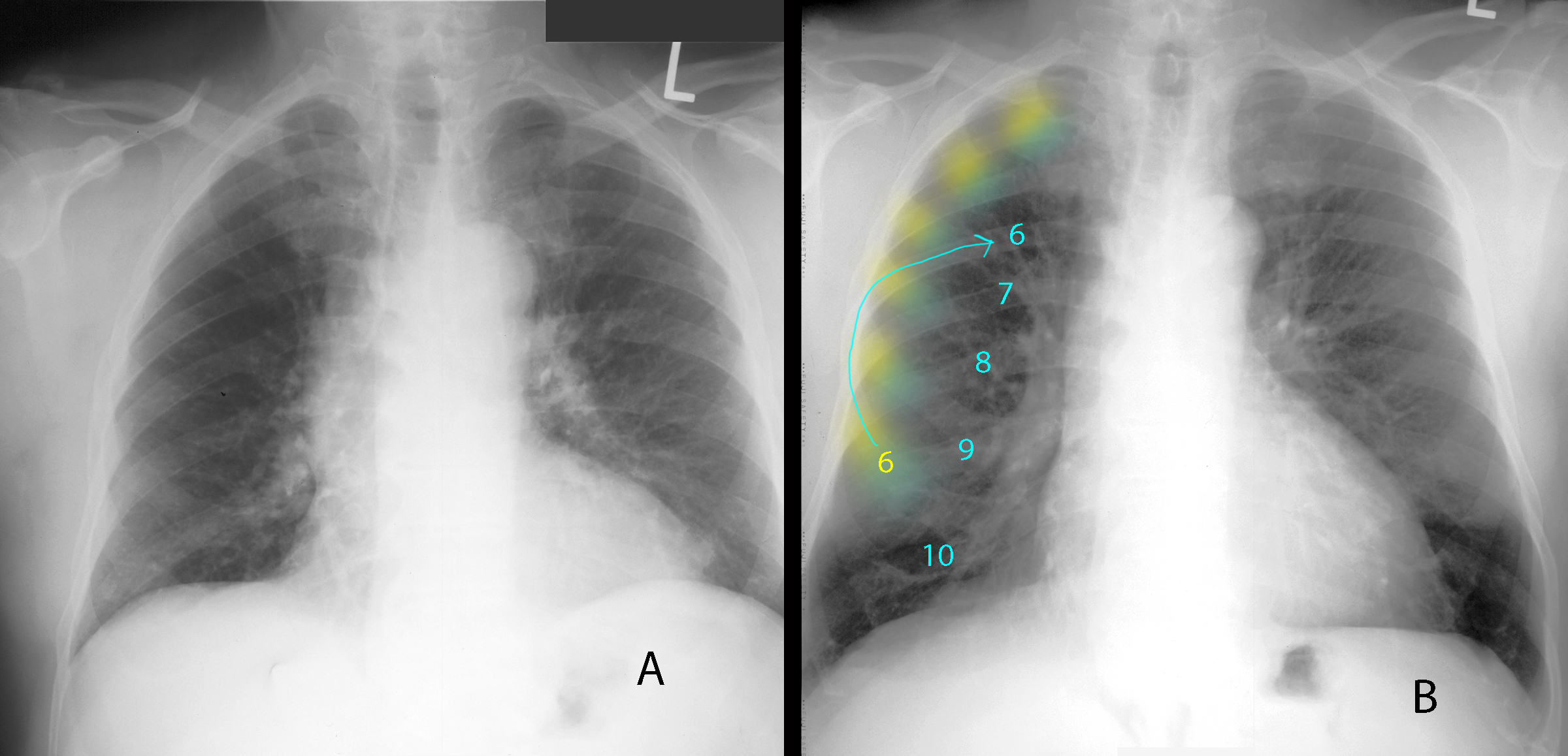
For the previous case, the chest radiograph was immediately repeated as the low inspiration was evident, shown on the right. How does the repeat image compare to the original?
×
Answer:
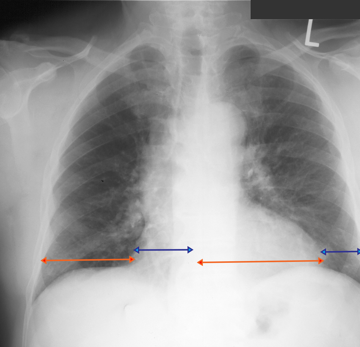
On image B, there are 10 posterior ribs above the diaphragm. The heart now measures normal in diameter and the hila are no longer hazy. Adequate inspiration is very important for assessing the heart and the pulmonary vasculature.
On image B, there are 10 posterior ribs above the diaphragm. The heart now measures normal in diameter and the hila are no longer hazy. Adequate inspiration is very important for assessing the heart and the pulmonary vasculature.
Case 4
Case 4, Part 3
Question 3:
What is wrong with this image?
×
Answer:
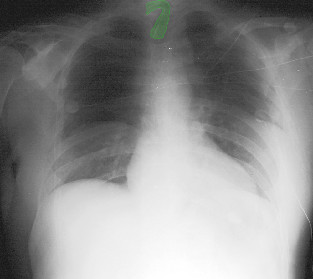
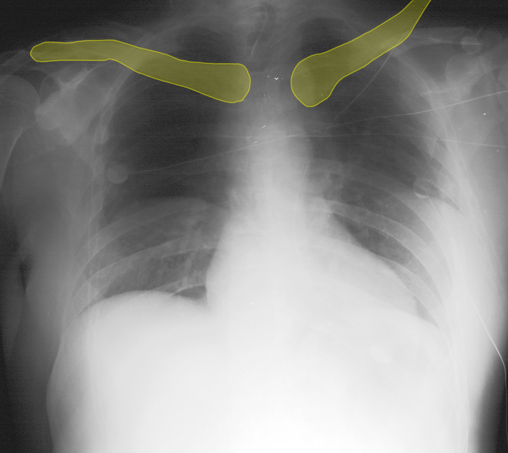
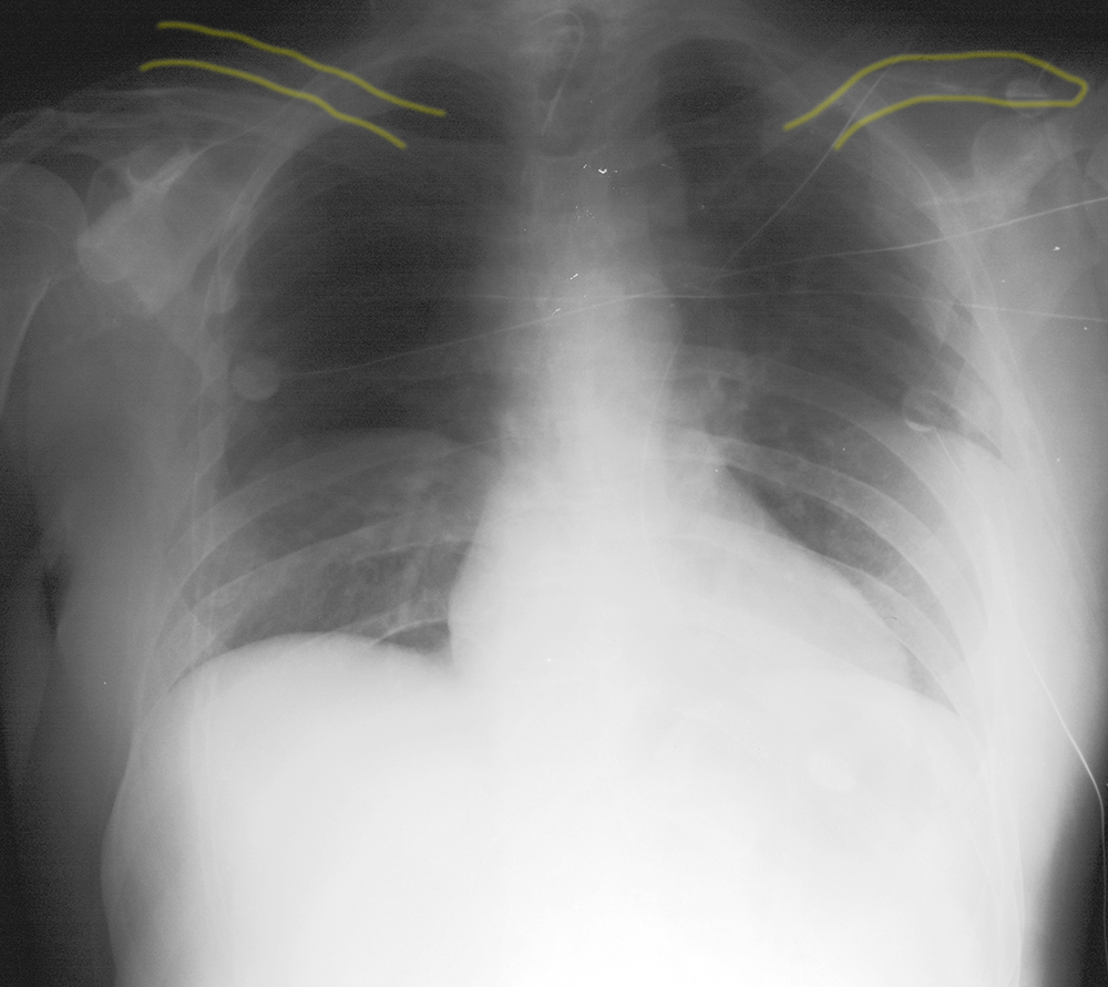
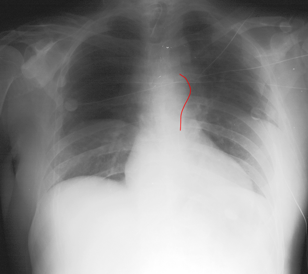
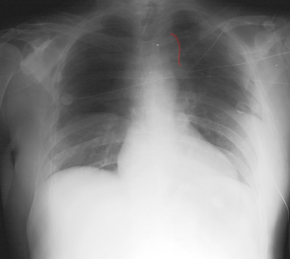
There are many mysteries indicated below. At first glance, the image may look overall too dark and a bit blurry. But with more careful analysis, you can see two sets of clavicles, two aortas, and two sets of diaphragms, as well as two sets of lateral ribs. This is a double-exposure. When the image is acquired on the digital receptor, it must be erased before another exposure is obtained. If this step is not completed, you can get two superimposed images, as in this case. This is not a common problem, but does occasionally happen, again more often for portable radiography.
There are many mysteries indicated below. At first glance, the image may look overall too dark and a bit blurry. But with more careful analysis, you can see two sets of clavicles, two aortas, and two sets of diaphragms, as well as two sets of lateral ribs. This is a double-exposure. When the image is acquired on the digital receptor, it must be erased before another exposure is obtained. If this step is not completed, you can get two superimposed images, as in this case. This is not a common problem, but does occasionally happen, again more often for portable radiography.
