

Case 3
Case 3 (rotation), Part 1
Question 1:
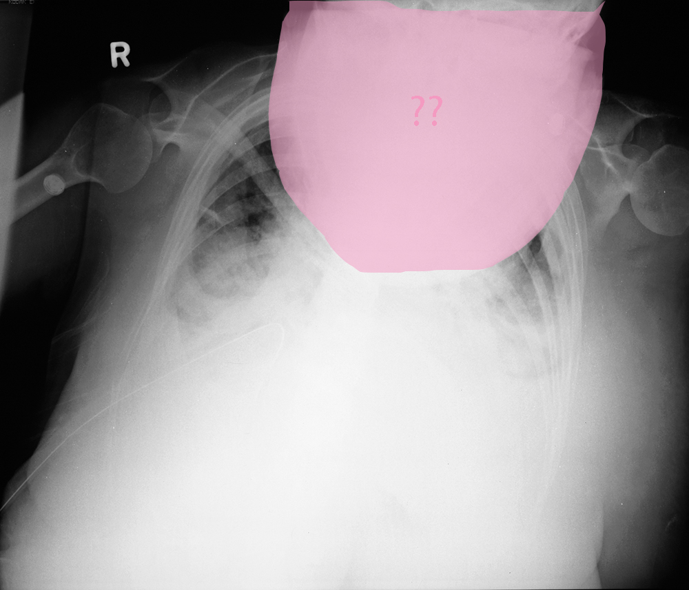
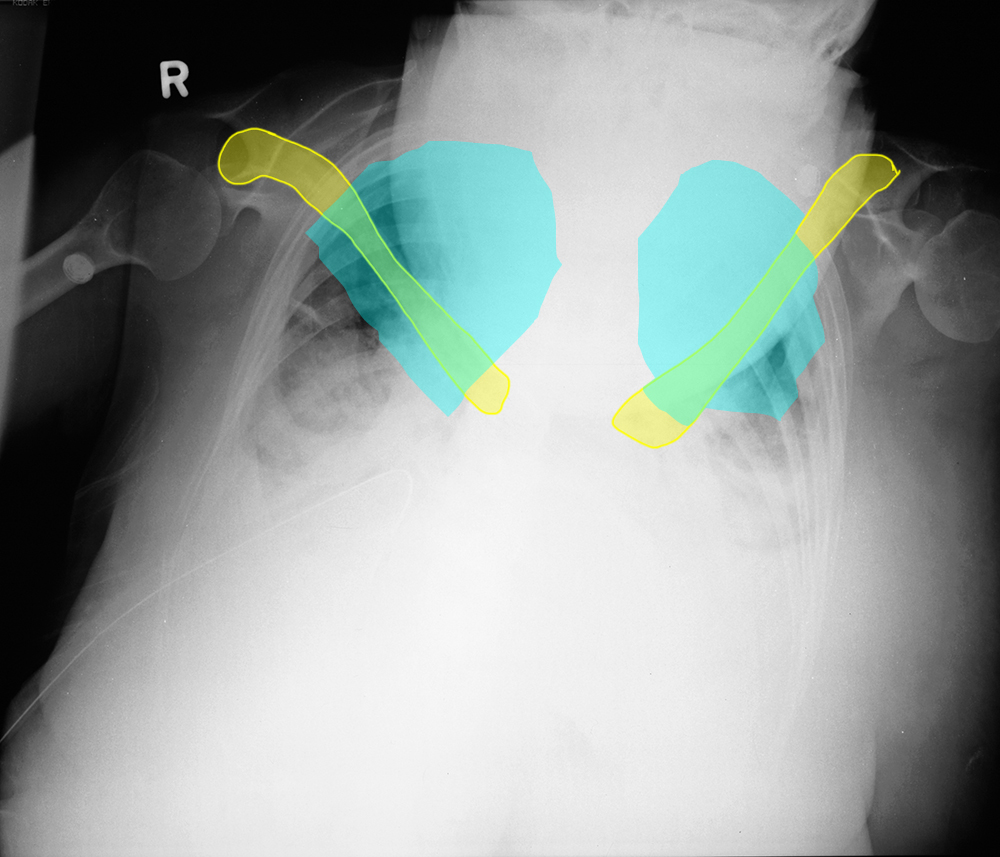
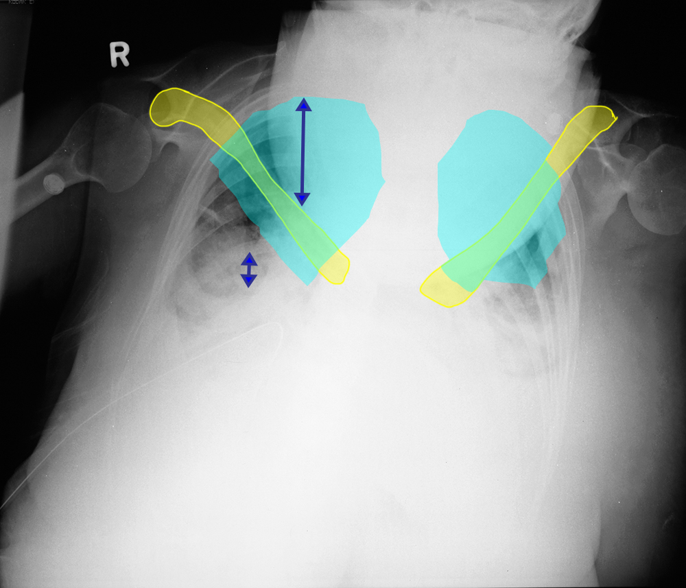
a) What is wrong with this image?
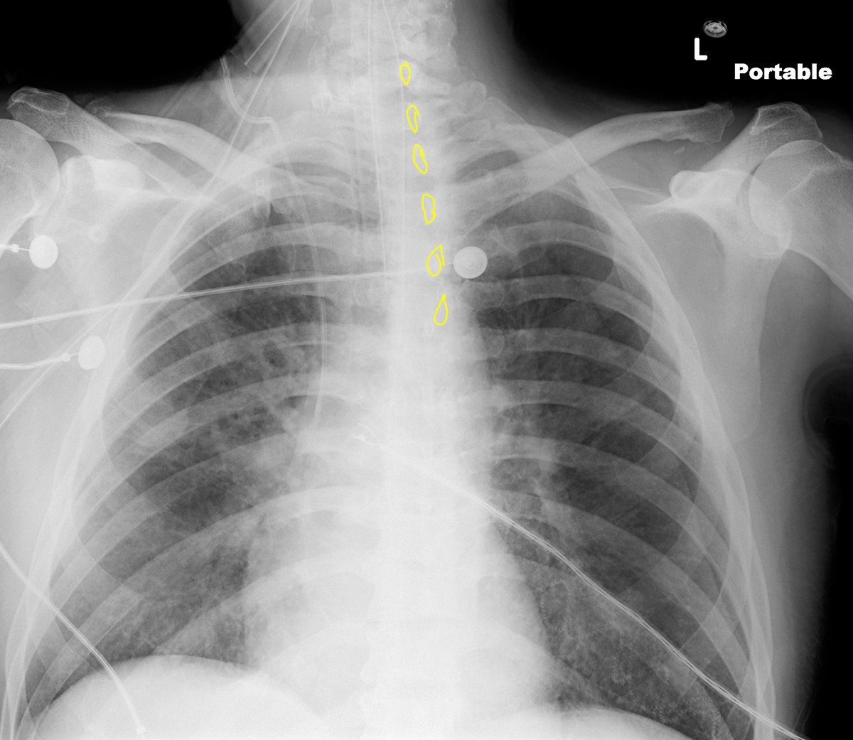
The mystery marked below is the patient's head. They are in a very kyphotic position, so that their head and neck are overlying the upper part of their chest. If you look at the clavicle position and the estimation of where the lung apices are (partly obscured by the head), you can see that the clavicles are very low in position.
b) Why does this matter?
Clearly, the head is obscuring the upper lungs and mediastinum. But even with less marked lordotic position, the contours of the mediastinum may be altered, making it harder to appreciate subtle abnormalities. Sometimes, lordotic positioning can also make the diaphragms look abnormal and may simulate lower lobe atelectasis or pneumonia.
Case 3
Case 3, Part 2
Question 2:
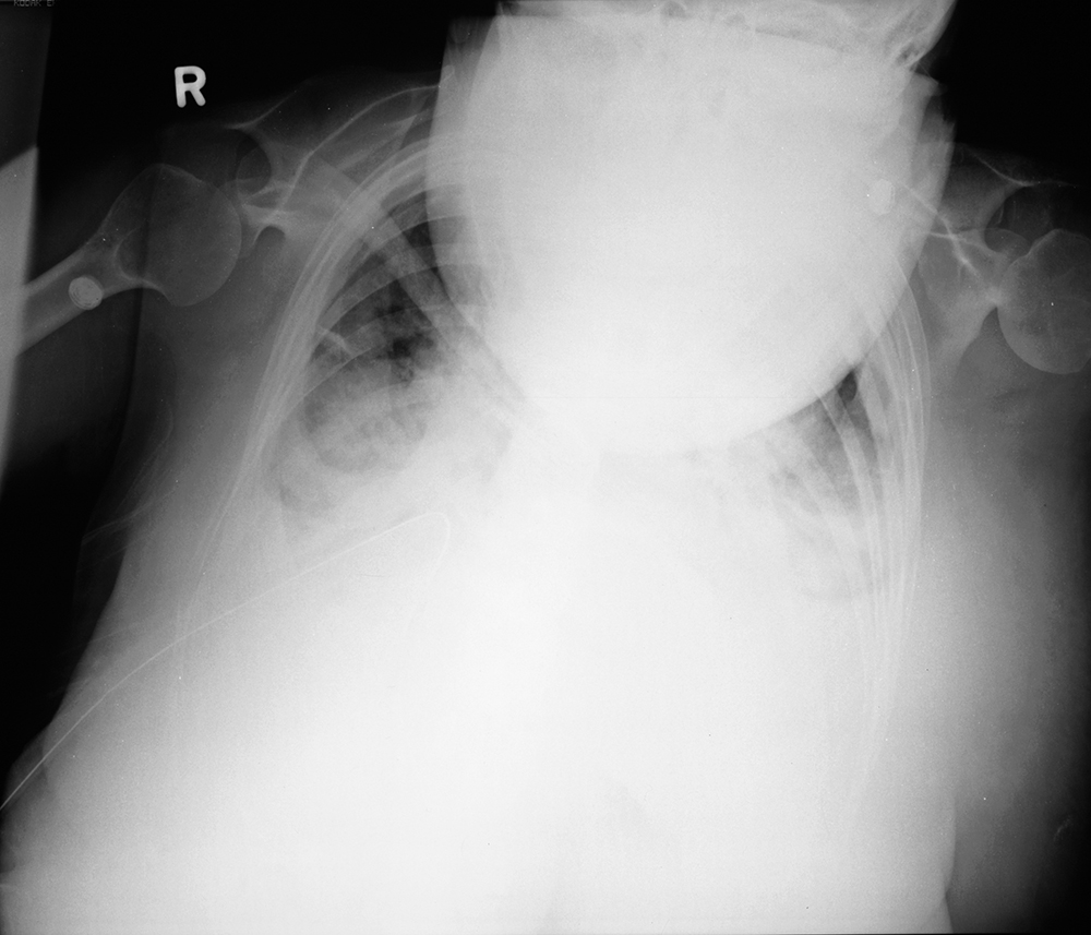
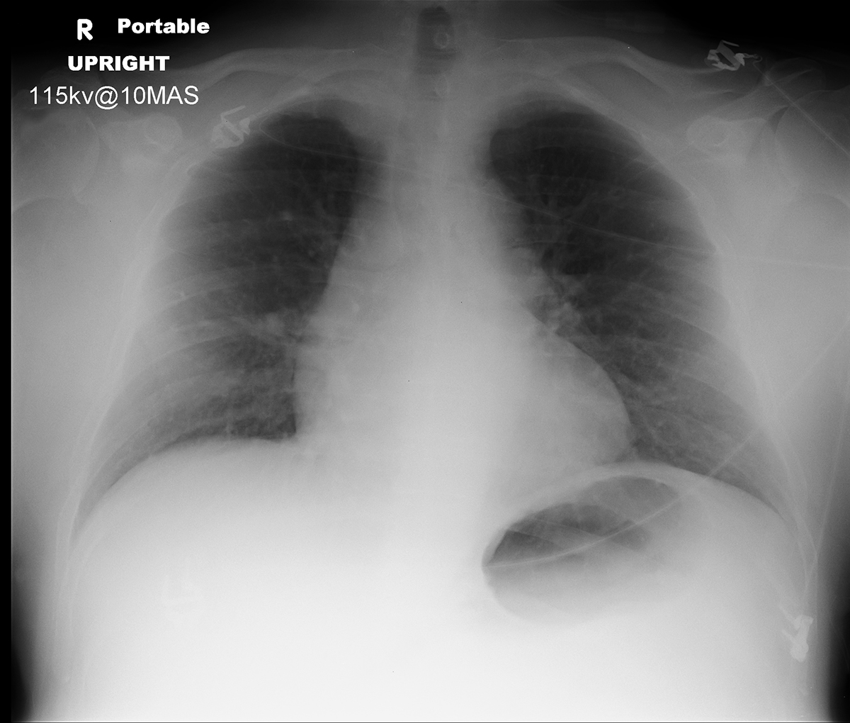
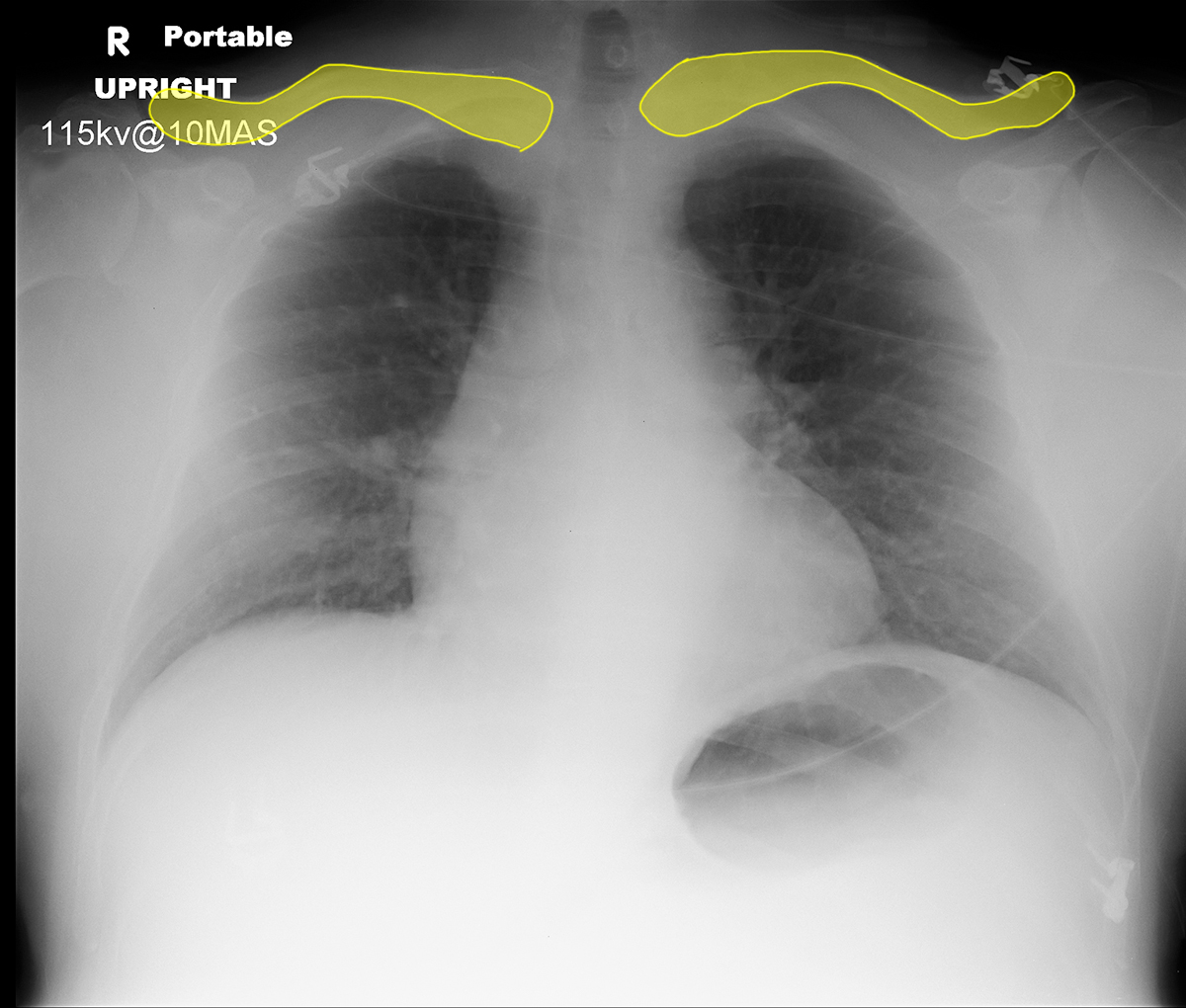
a) What is wrong with this image?
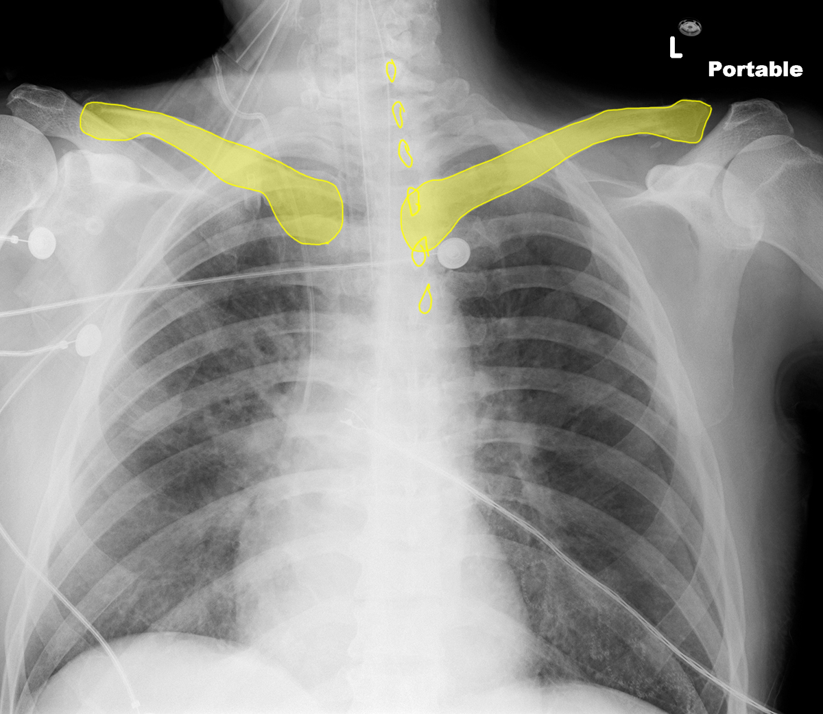
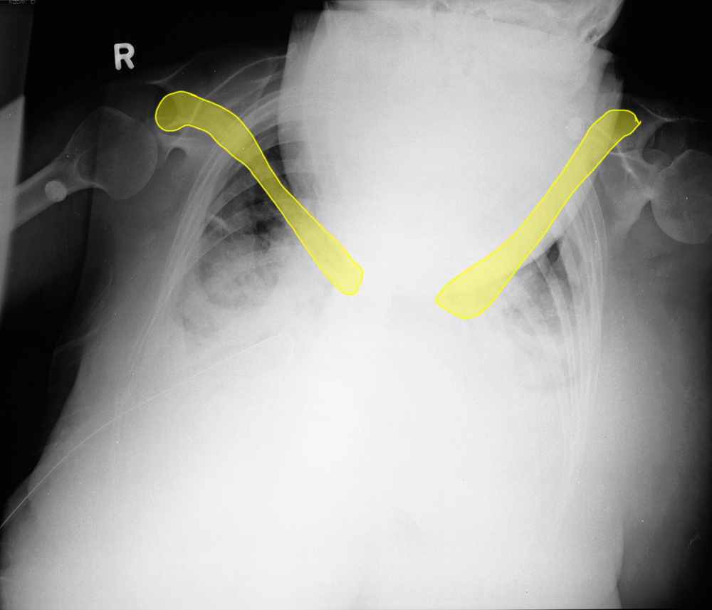
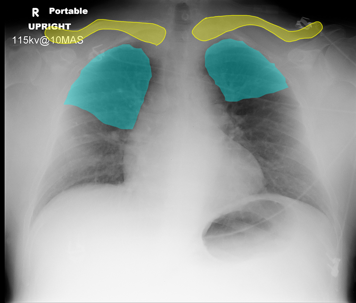
If you look at the labels below, you will see that there is evidence of lordotic positioning, with no lung visible above the clavicles.
b) Why does this matter?
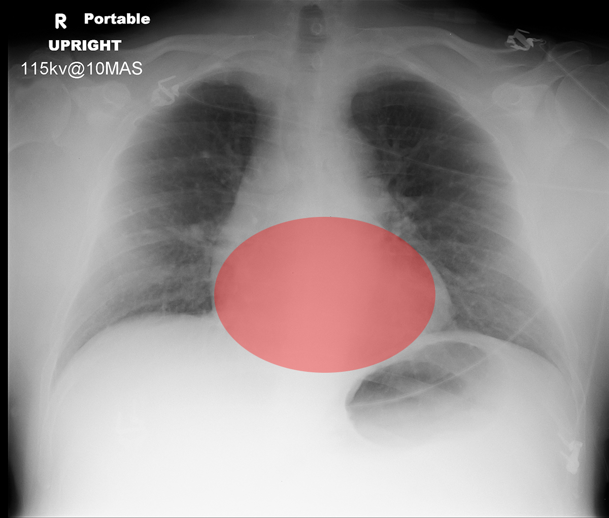
As for kyphotic positioning, lordosis can alter the appearance of mediastinal contours and the diaphragm. In this case, the heart has an unusual oval contour, as if you are looking at it from below instead of from anteriorly.
Case 3
Case 3, Part 3
Question 3:
a) What type of image is this and what technical problem is present?
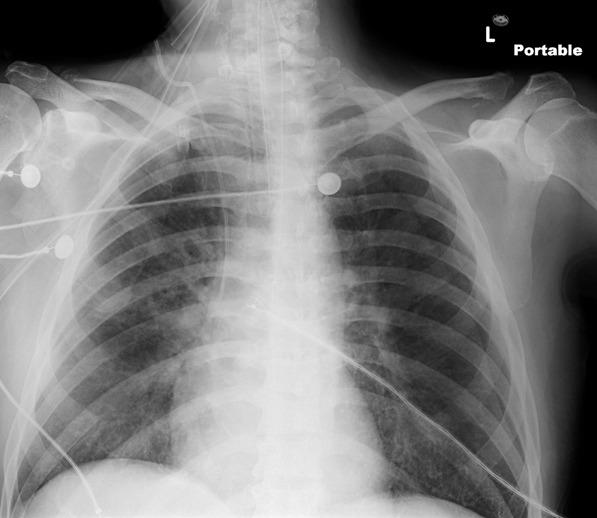
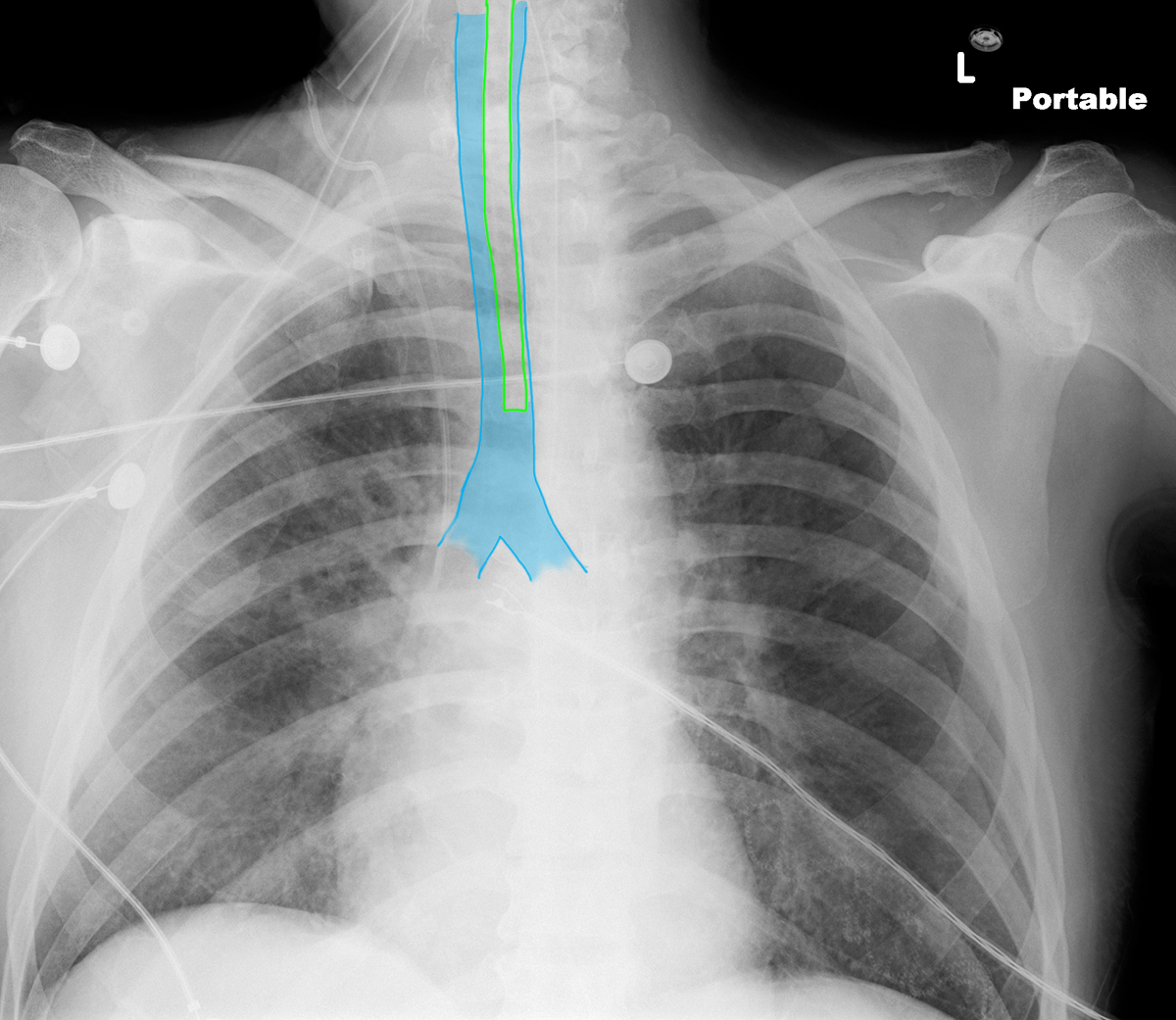
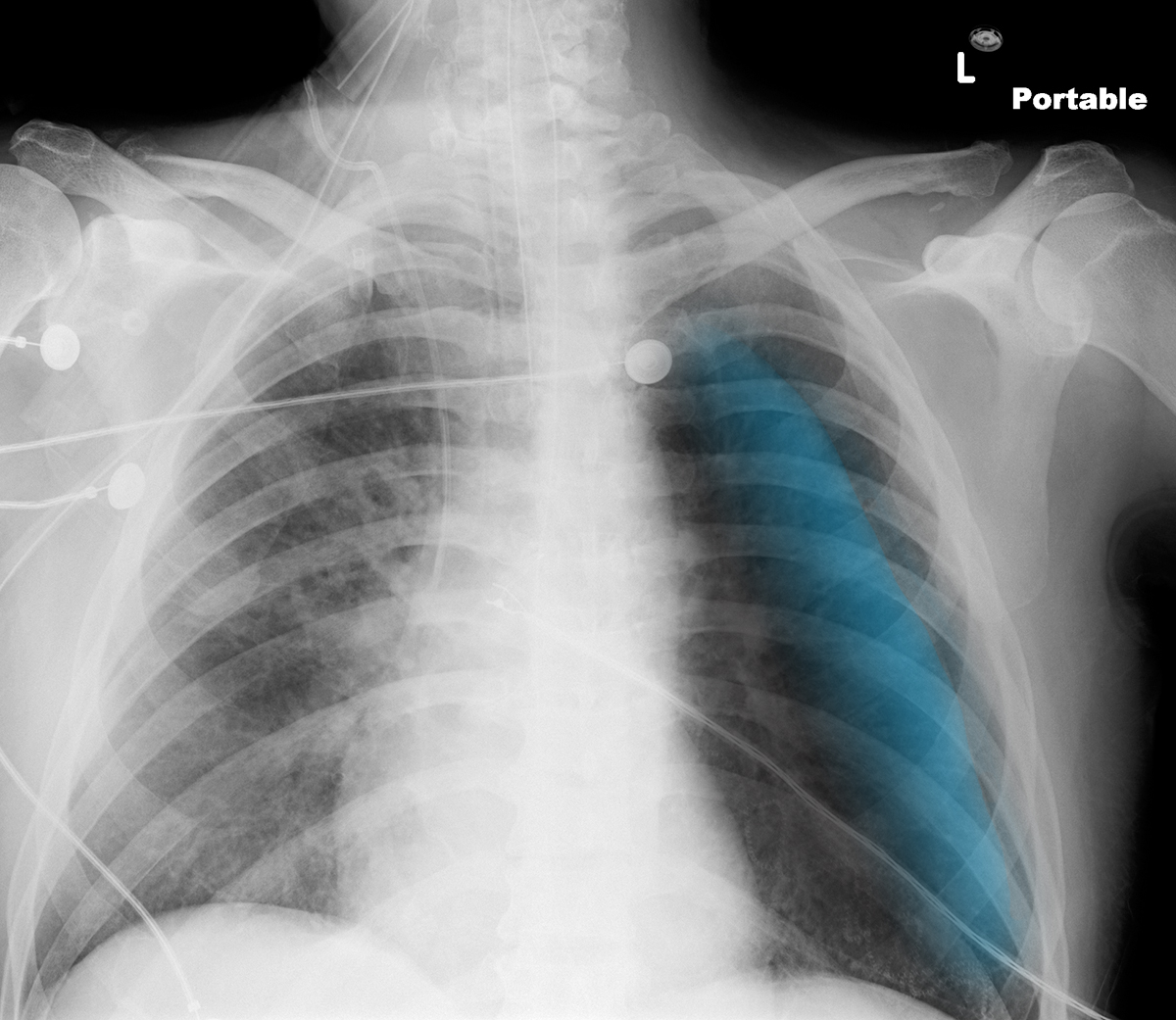
This is a portable chest radiograph, with many support lines present. Portable radiographs are prone to many technical problems, as it is often difficult to position patients who may be unable to cooperate with placement of imaging apparatus. This case shows rotation in the axial plane. The anterior part of the patient is rotated to the left side of the image.
b) Why does this matter?
Rotation in the axial plane can displace structures, like the trachea as in this example. Rotation in the axial plane can also alter the contours of the mediastinum.
c) What is abnormal in the left hemithorax?
There is a gently curved interface extending from the upper to the lower left hemithorax, indicated by the label below. This shape and position could represent a pneumothorax, but there is no white line seen at the boundary between collapsed lung and pleural space, as would be evident in a pneumothorax. This is a common artifact, a skin fold. Bunched up skin will produce a denser area with this contour and can simulate a pneumothorax. For diagnosis of pneumothorax, always look for the white pleural line. If you are uncertain, repeating the image will often solve the problem, a skin folds move around or may be done on the next image.
