

Case 2
Case 2 (exposure and contrast), Part 1
Question 1:
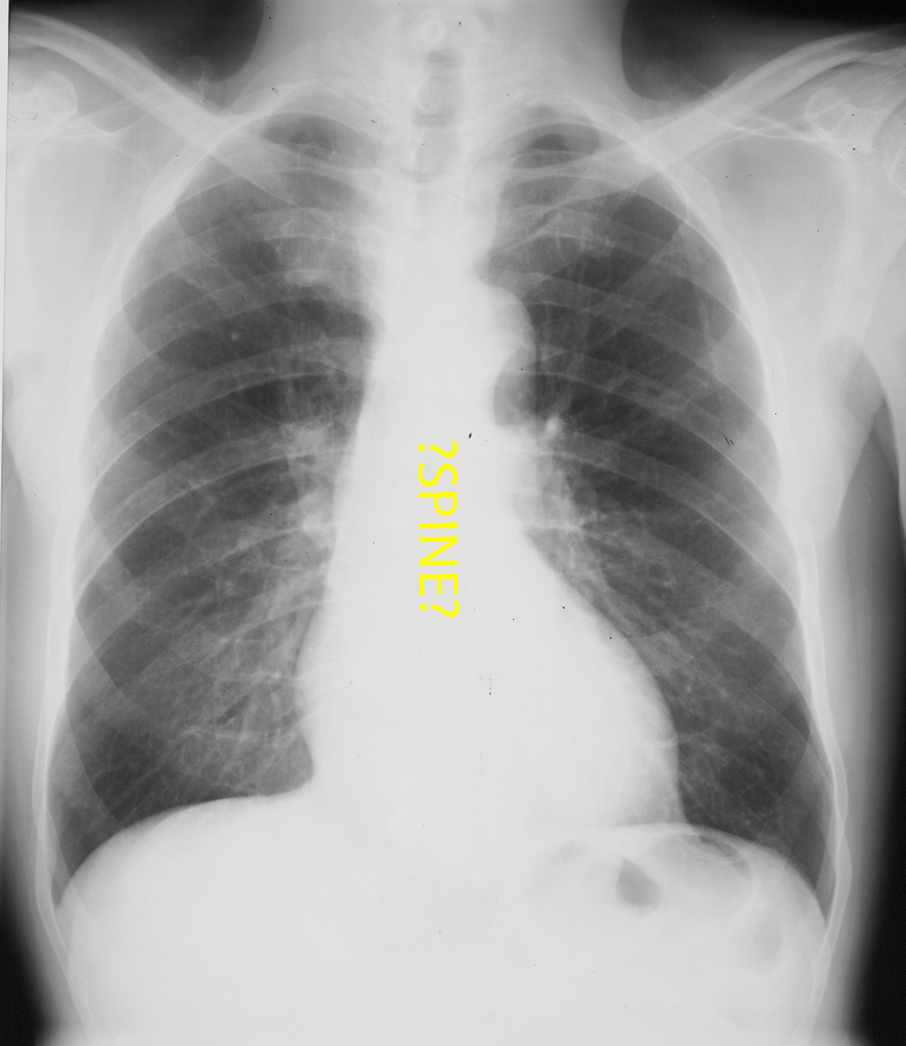
a) What is wrong with this image?
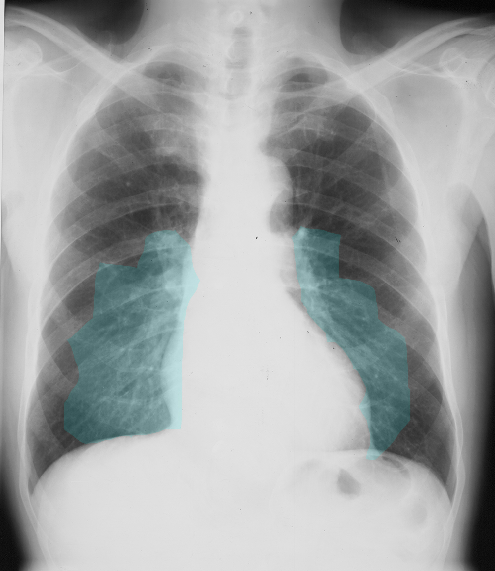
This is an example of an image where not enough photons have reached the receptor. This can be called 'under-exposure' or 'under-penetration', since the x-rays penetrate the patient's body to produce an image. This image is overall too light. If you cannot see any of the spine through the mediastinum, that is under-penetration. In the film era, this was more of a problem than with digital images, as the technologist can correct for slightly dark or light images after they are acquired by adjusting the brightness of the image.
b) Why does this matter?
If the image is overall too light (under-penetrated), this can make the lungs look abnormally light as well, and may simulate subtle interstitial or alveolar lung disease.
Case 2
Case 2, Part 2
Question 2:
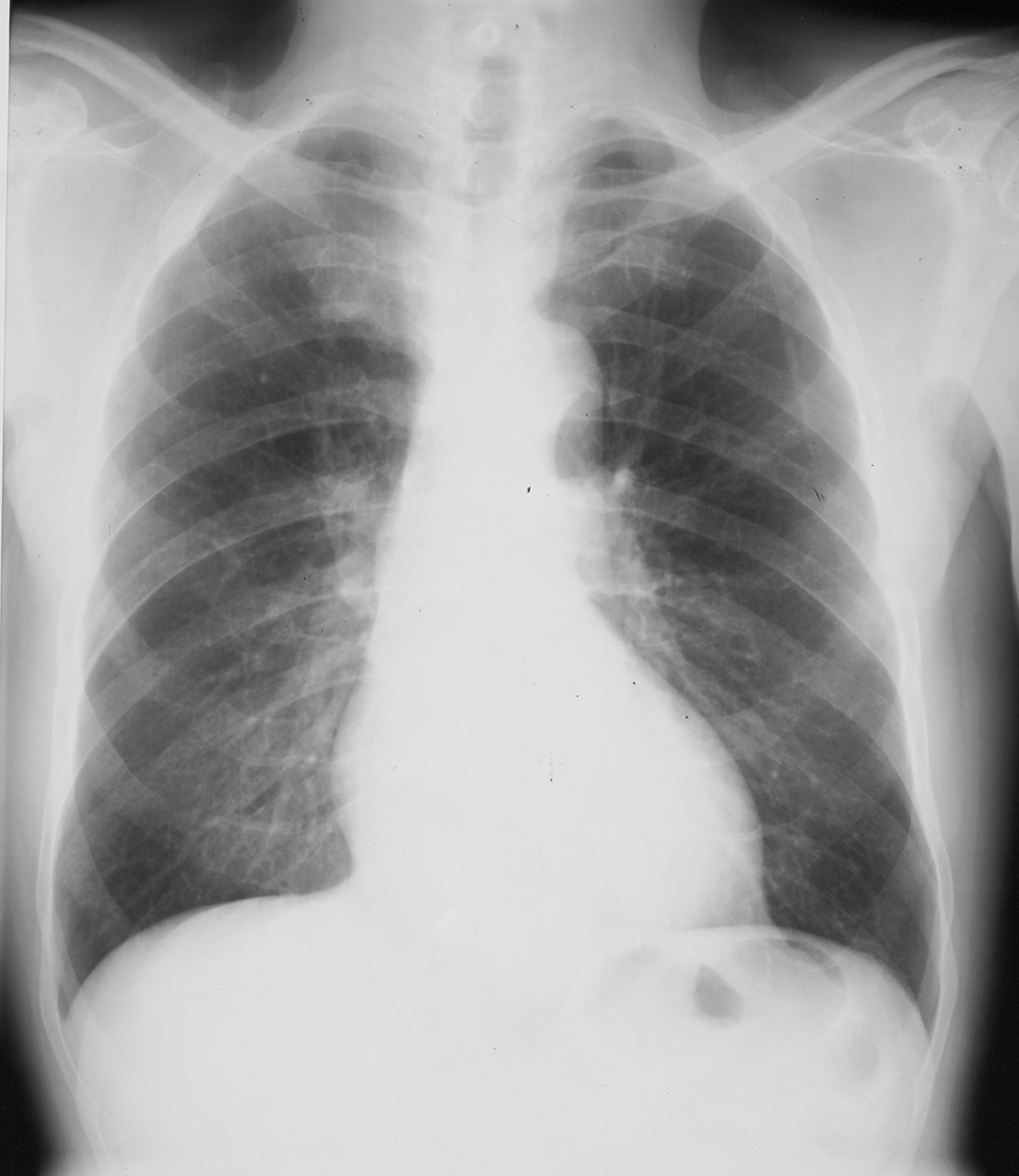
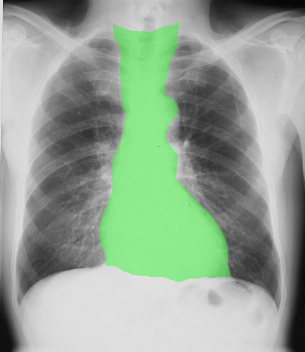
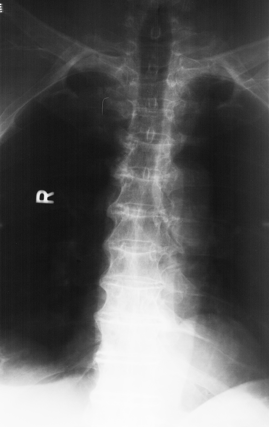
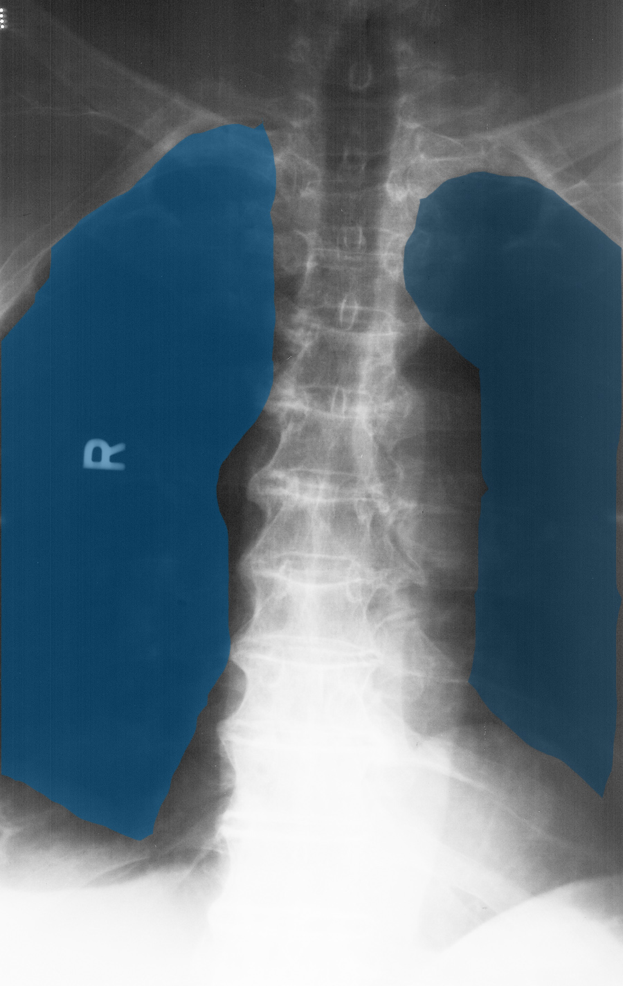
a) What is wrong with this image?
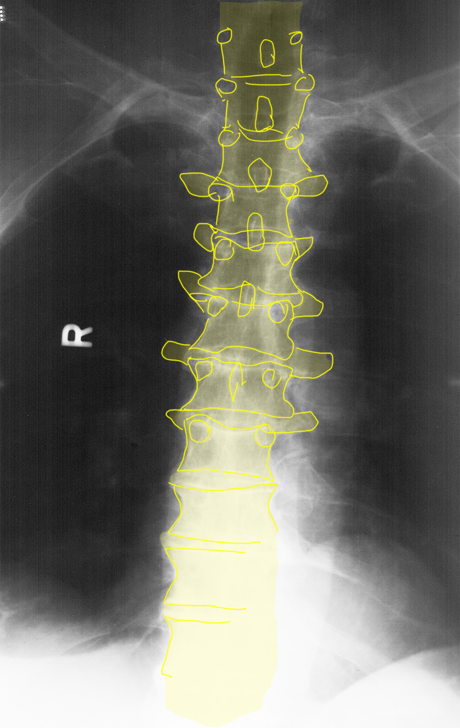
In this case, you see the spine TOO well! Every part of the thoracic spine is very clear, but the lungs look very black. This image is over-penetrated (over-exposed). In fact, it was actually part of a thoracic spine series, not a chest radiograph, but an over-exposed chest radiograph would have a similar appearance. Again, over and under exposed images are less commonly seen with digital imaging, but most often are a problem with portable chest radiography where the exposure is more difficult to optimize.
b) Why does this matter?
When the lungs look very black like this, it may be mistaken for emphysema or even pneumothorax.
Case 2
Case 2, Part 3
Question 3:
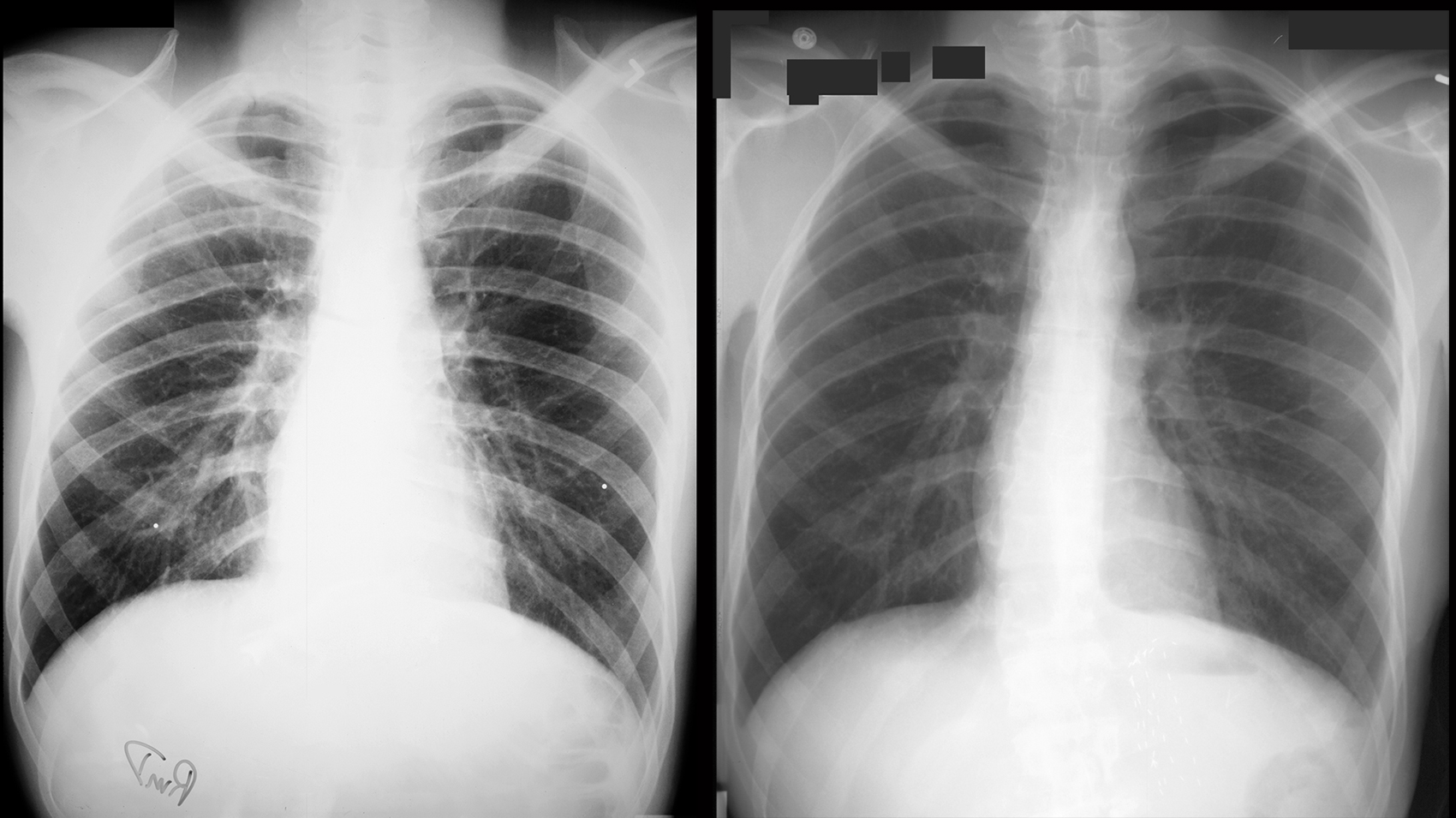
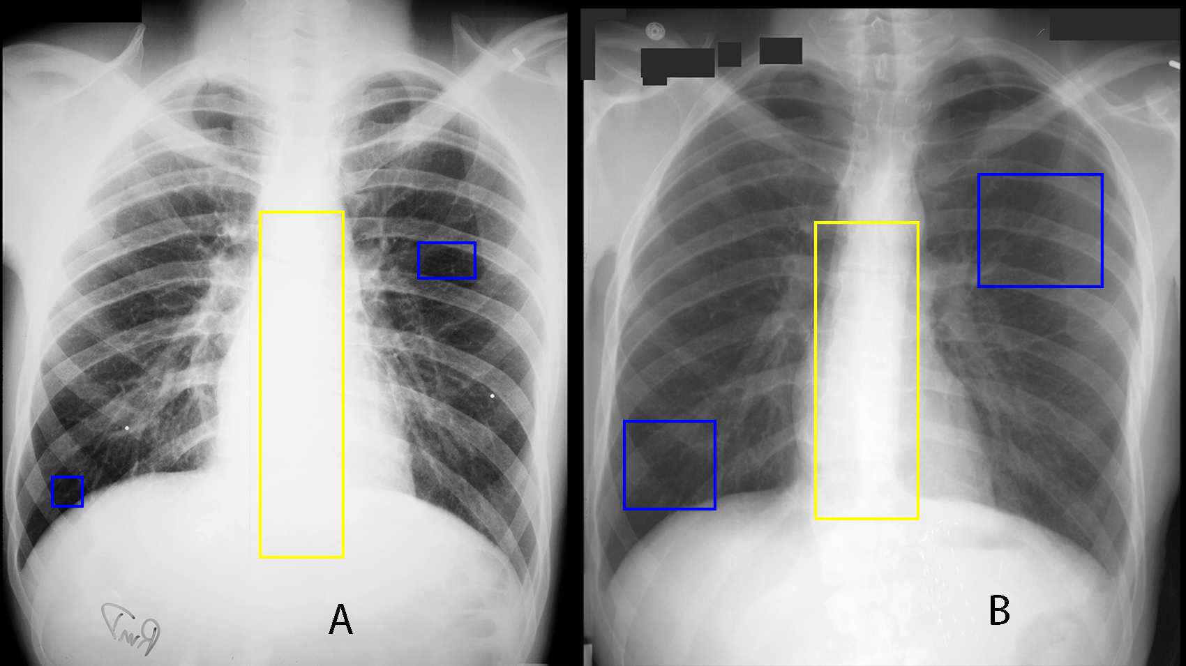
These are two images of the same patient, done one day apart. What is wrong with the image on the left?
The left image is both TOO WHITE (in the mediastinum, with no spine seen) and TOO DARK (lungs look very black). This image has very high contrast. For a chest radiograph, because many of the findings in the lungs and mediastinum depend on detection of very subtle density differences, we prefer a very LOW contrast image, as is demonstrated on the image on the right. The left image was done with the wrong technique--with settings suitable for bone or contrast radiography. where HIGH contrast is desirable.
