

Imaging Anatomy Abdomen Case 2
54 year old female with epigastric pain
Question 1:
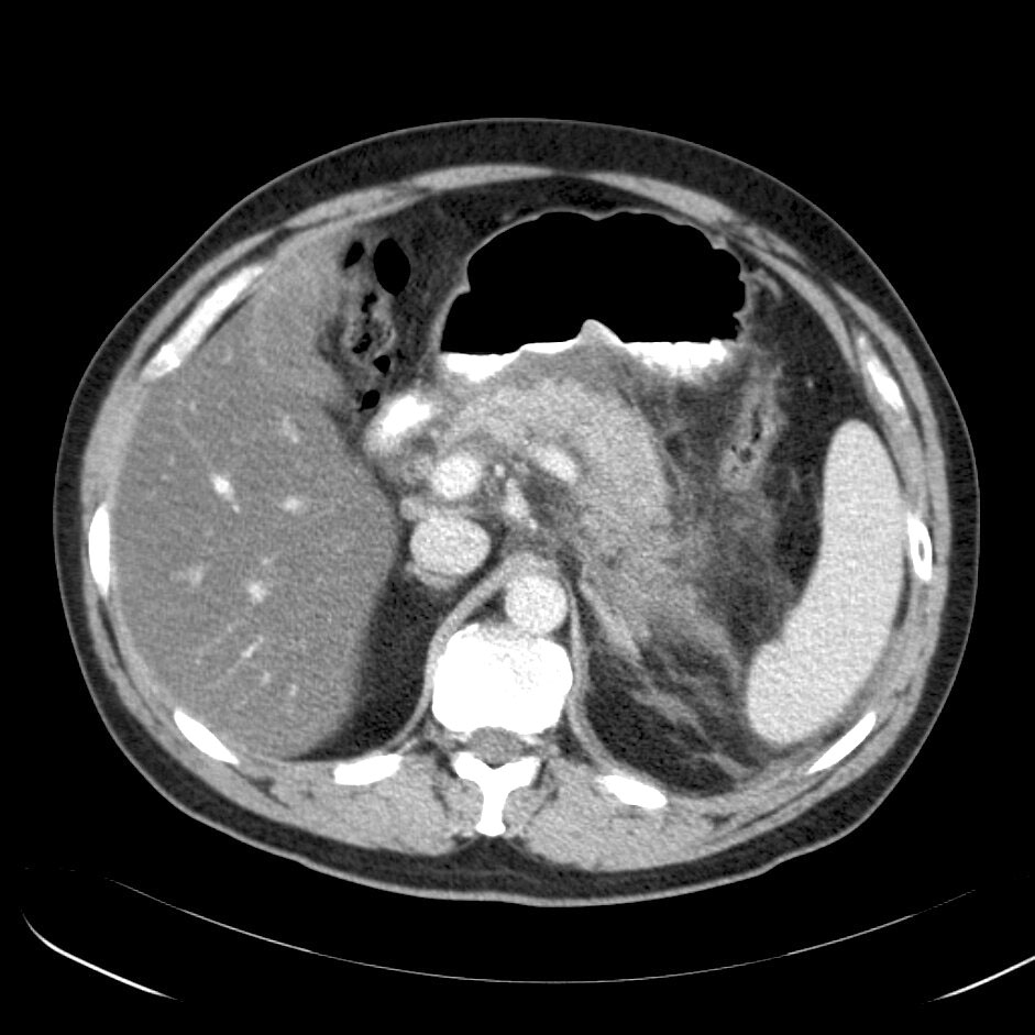
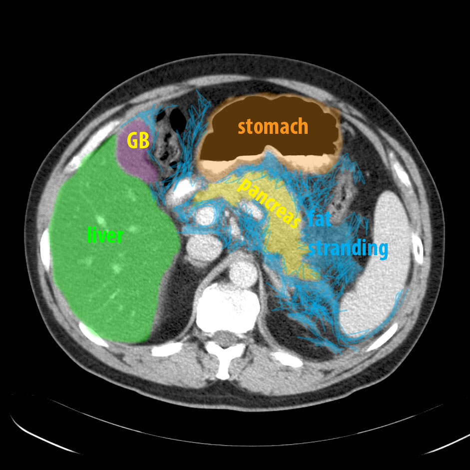
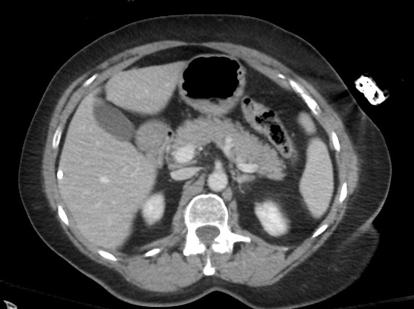
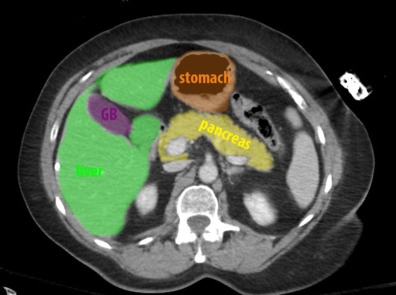
Review the axial CT image series and the select CT image below. Which abdominal organ is surrounded by abnormal appearing fat?
The fat surrounding the pancreas is abnormal in density. It is closer to water density than normal fat, making the margins of the pancreas indistinct. This is a case of pancreatitis, or inflammation of the pancreas.
steps for cross-sections:
1. what is it? 2. what imaging plane?
3. for CT, contrast/window? 4. where are we?
Imaging Anatomy Abdomen Case 2
This is another patient with abdominal paIn, with a normal comparison below from a different patient
Question 2:
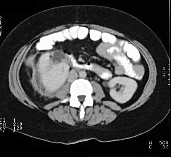
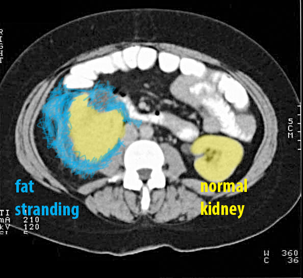
What organ is abnormal? Focus on the appearance of the intra-abdominal fat
In this case, the fat surrounding the kidney is abnormally high in density. This change in the appearance of fat is most often due to edema and is called 'fat stranding'. This often is a result of infection, although tumor infiltration or hemorrhage can have a similar appearance. Inflammation of the kidney is called 'pyelonephritis'. If you look at the patient's left kidney, you can see how sharp the margin of the organ is adjacent to normal dark fat in the surrounding connective tissue, enclosed by Gerota's fascia. On the right, the compartment of Gerota's fascia is filled with fluid and appears very indistinct, obscuring the margin of the kidney.
steps for cross-sections:
1. what is it? 2. what imaging plane?
3. for CT, contrast/window? 4. where are we?
Imaging Anatomy Abdomen Case 2
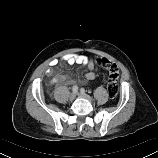
This is another patient with abdominal pain.
Question 3:
a) What organ is abnormal in this case?
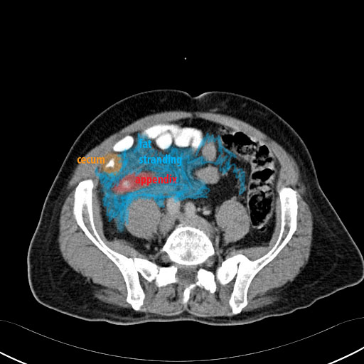
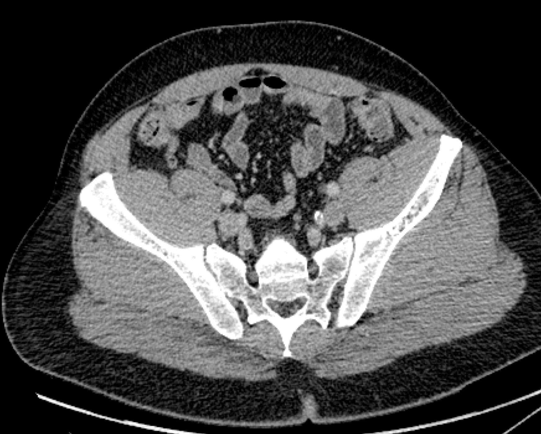
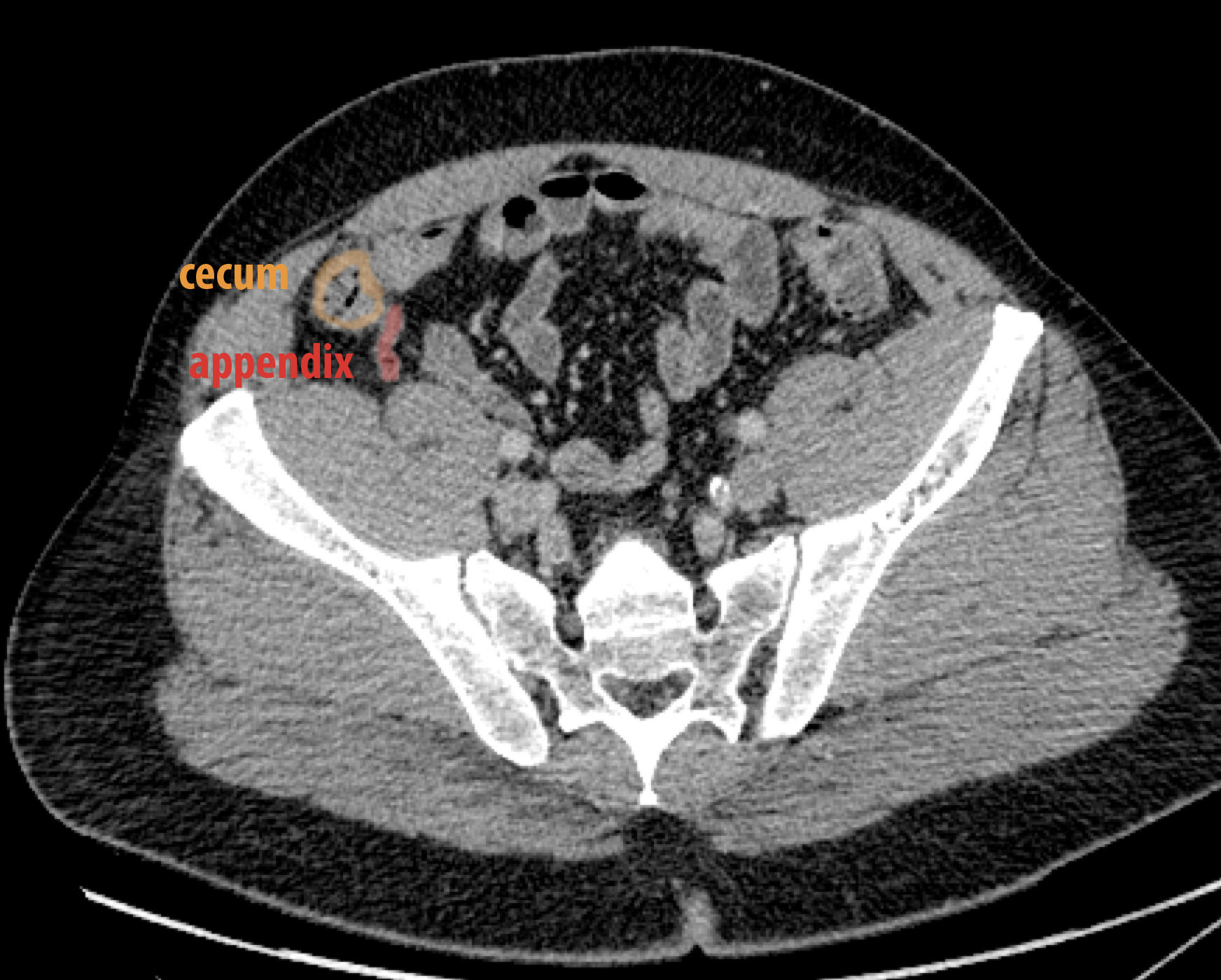
There is extensive fat stranding in the right lower quadrant of the abdomen, in the region of the cecum and appendix. This is a patient with appendicitis, or inflammation of the appendix. The cecum is also abnormal with a thickened wall due to edema from the infection in the region. On the normal comparison, the appendix is quite small with sharply defined margins from surrounding dark normal fat, and the wall of the cecum is thin and sharply defined.
b) What are five things that can cause fat stranding?
The most common thing is infection/inflammation, as in these three examples (pancreatitis, pyelonephritis and appendicitis). Other things to consider if the clinical situation warrants are tumor infiltration, hemorrhage, edema, or scarring/fibrosis.
steps for cross-sections:
1. what is it? 2. what imaging plane?
3. for CT, contrast/window? 4. where are we?
This is the last page of the mini-consolidation Abdomen Imaging Anatomy cases. To return to the main page for Foundations 2 Imaging Anatomy, click HERE. To return to the first page of the Abdomen cases for mini-consolidation, click HERE.
