

Case 2
These cases review imaging findings for focal opacities in the lung on radiography and CT. You are shown three asymptomatic patients.
Question 1:
Patient one is shown first. Click below to see the other cases. For each, how would you describe the finding? Consider size, shape, and density.
×
Answer:
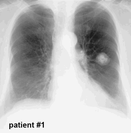
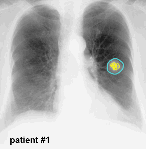
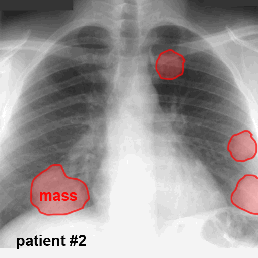
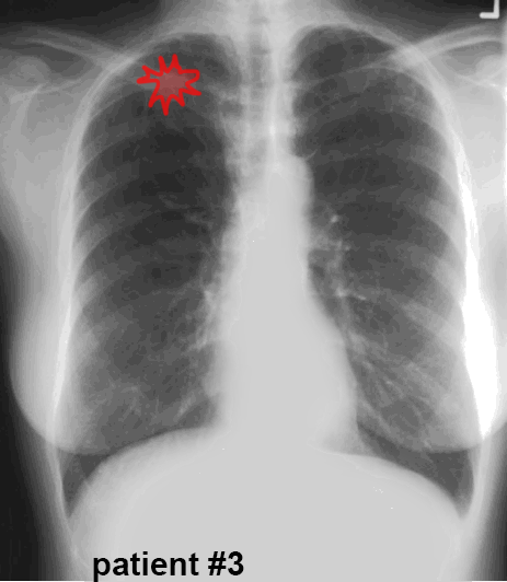
For Patient 1, there is a rounded mass in the left mid-lung that contains a relatively large area of coarse calcification. The mass has smooth margins. For masses that are round, smooth and under 3 cm in diameter, you can use the term 'nodule'. The presence of calcium suggests a benign etiology. For patient two, there are multiple opacities, not all of them round and one at the right base that is definitely too large to call it a nodule. These should be called masses, based on irregular shape and size over 3 cm. For patient three, there is a subtle mass overlying the clavicle on the right. It is fairly small but is NOT round. It has a spiculated margin, with spiky projections in every direction. This must also be called a mass because it is not round, even though it is likely less than 3 cm in diameter.
For Patient 1, there is a rounded mass in the left mid-lung that contains a relatively large area of coarse calcification. The mass has smooth margins. For masses that are round, smooth and under 3 cm in diameter, you can use the term 'nodule'. The presence of calcium suggests a benign etiology. For patient two, there are multiple opacities, not all of them round and one at the right base that is definitely too large to call it a nodule. These should be called masses, based on irregular shape and size over 3 cm. For patient three, there is a subtle mass overlying the clavicle on the right. It is fairly small but is NOT round. It has a spiculated margin, with spiky projections in every direction. This must also be called a mass because it is not round, even though it is likely less than 3 cm in diameter.


Case 2
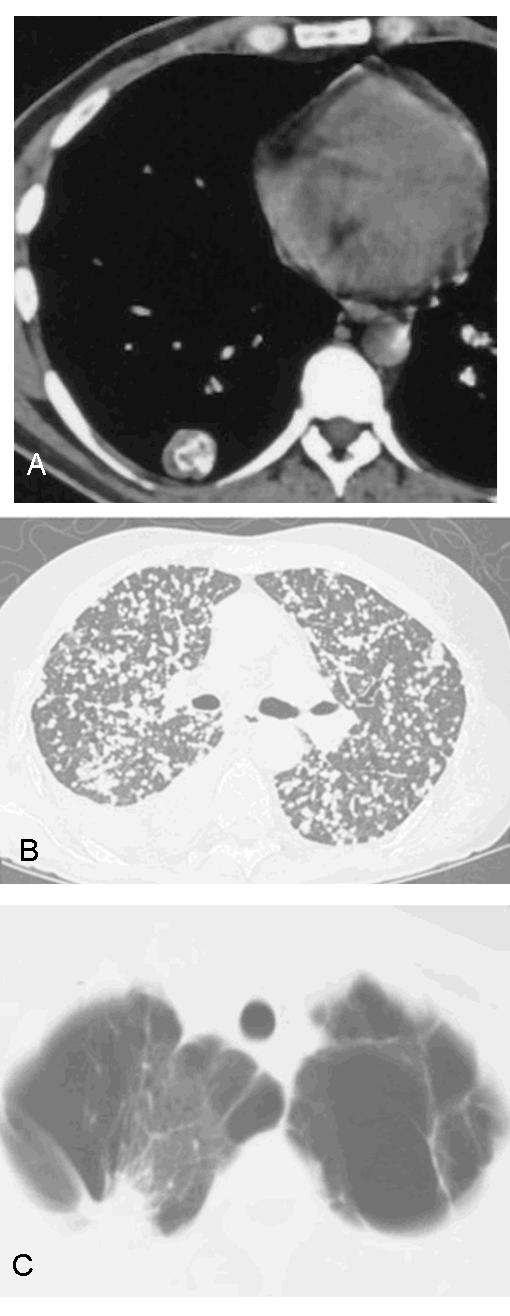
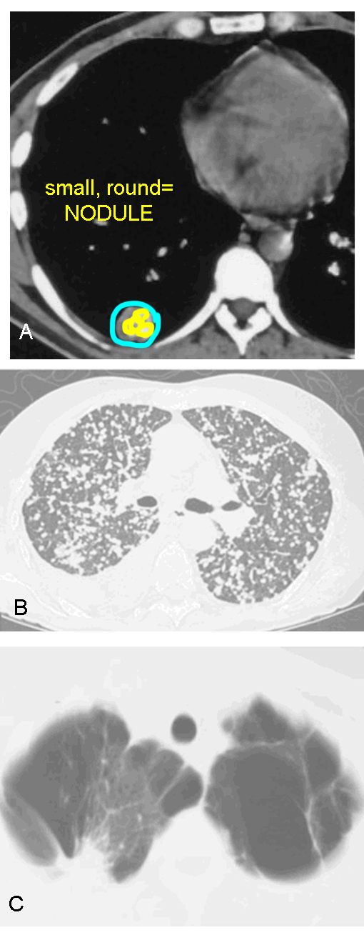
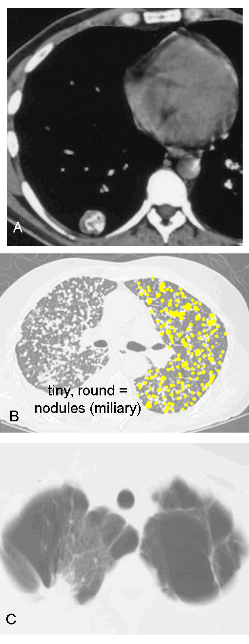
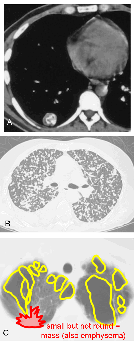
These CT images are from various patients with focal opacities in their lungs.
Question 2:
For each case, what would you call the opacities or opacities? To decide, consider size and shape, and note any other interesting findings.
×
Answer:
Case A is the same as Case 1 from the previous page--a small rounded opacity with internal calcification, which we would call a nodule, and be confident that it is benign based on the calcification. Case B shows innumerable tiny rounded opacities that can also be called nodules, and based on their tiny size and distribution, these can be called 'miliary' nodules, suggesting TB. Case C shows a small but very irregular shaped opacity, which must be called a mass, likely a lung cancer.
Case A is the same as Case 1 from the previous page--a small rounded opacity with internal calcification, which we would call a nodule, and be confident that it is benign based on the calcification. Case B shows innumerable tiny rounded opacities that can also be called nodules, and based on their tiny size and distribution, these can be called 'miliary' nodules, suggesting TB. Case C shows a small but very irregular shaped opacity, which must be called a mass, likely a lung cancer.
