

Case 3
These cases demonstrate another pattern of abnormality in the lungs that can lead to specific diagnoses--cystic lung disease.
Question 1:
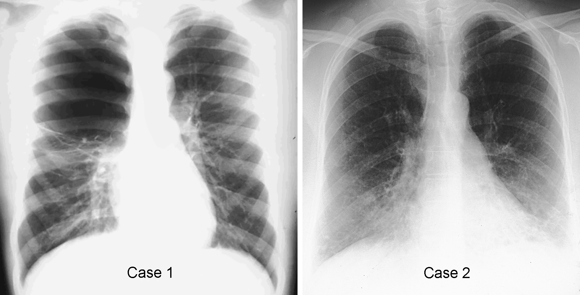
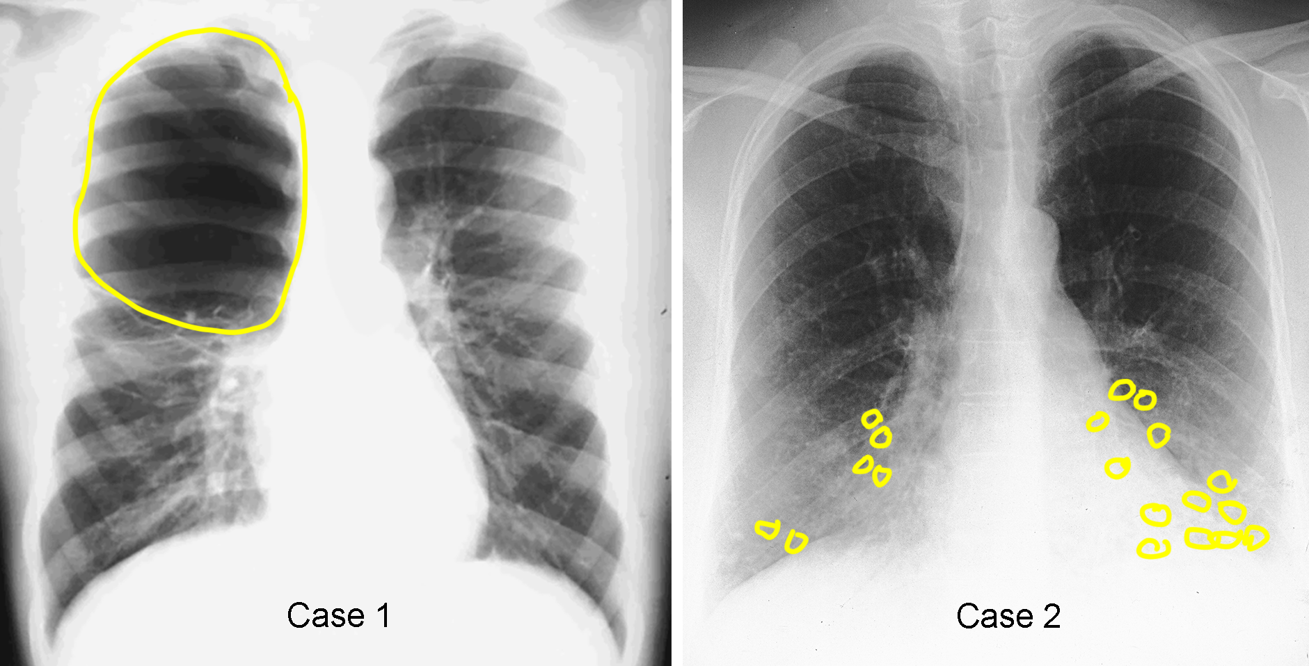
What is the definition of a lung cyst? What diagnoses might produce lung cysts? Where are the cysts located in Case 1 vs Case 2?
×
Answer:
A cyst in the lung is defined as an air-filled rounded structure, usually with a visible wall. Some areas of emphysema can be considered cysts, but often they do not have a visible wall. If bronchiectasis has a rounded appearance, it can be called 'cystic bronchiectasis'. Trauma to the lung can result in a cyst forming due to damage to lung tissue. Scarring from fibrosis can lead to formation of rows of cysts due to destruction and retraction of tissue, called 'honeycombing'. In Case 1, a large cyst fills the upper right hemithorax. Depending on symptoms, this might be congenital or acquired. In Case 2, the radiographic findings are subtle. Many types of cysts are best seen on CT scans. But there are some faint rounded lucencies in both bases, left greater than right.
A cyst in the lung is defined as an air-filled rounded structure, usually with a visible wall. Some areas of emphysema can be considered cysts, but often they do not have a visible wall. If bronchiectasis has a rounded appearance, it can be called 'cystic bronchiectasis'. Trauma to the lung can result in a cyst forming due to damage to lung tissue. Scarring from fibrosis can lead to formation of rows of cysts due to destruction and retraction of tissue, called 'honeycombing'. In Case 1, a large cyst fills the upper right hemithorax. Depending on symptoms, this might be congenital or acquired. In Case 2, the radiographic findings are subtle. Many types of cysts are best seen on CT scans. But there are some faint rounded lucencies in both bases, left greater than right.
Case 3
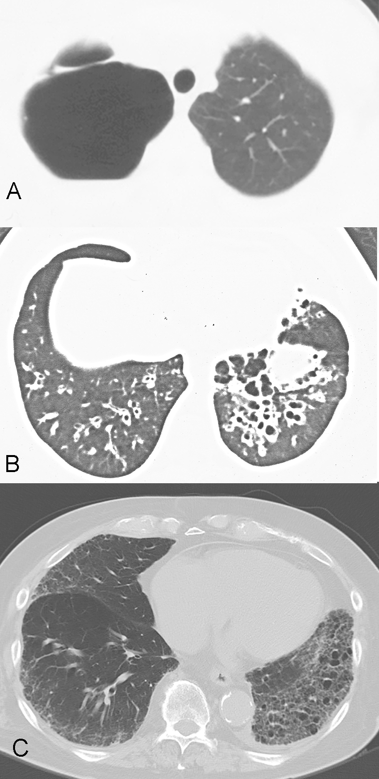
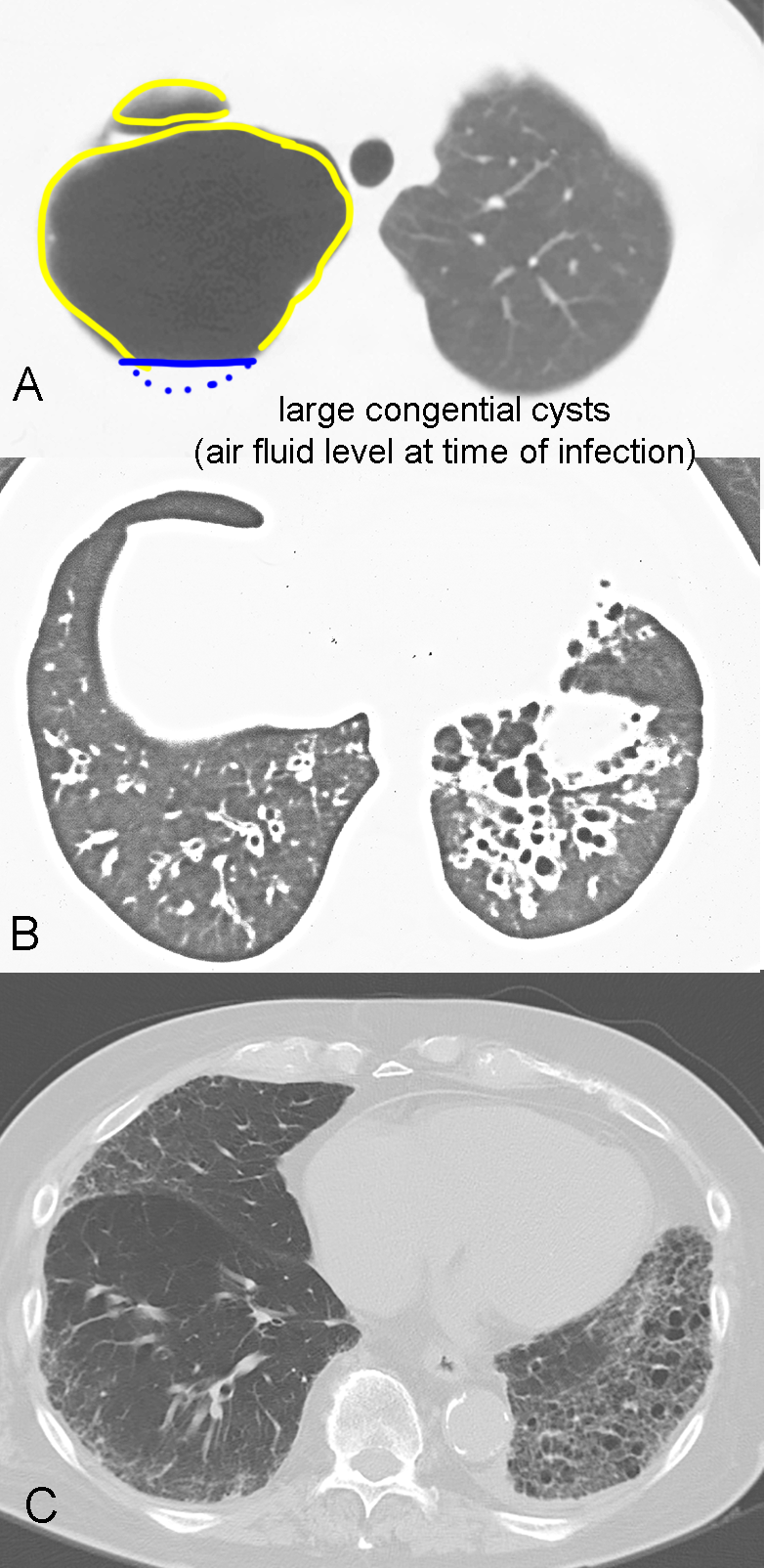
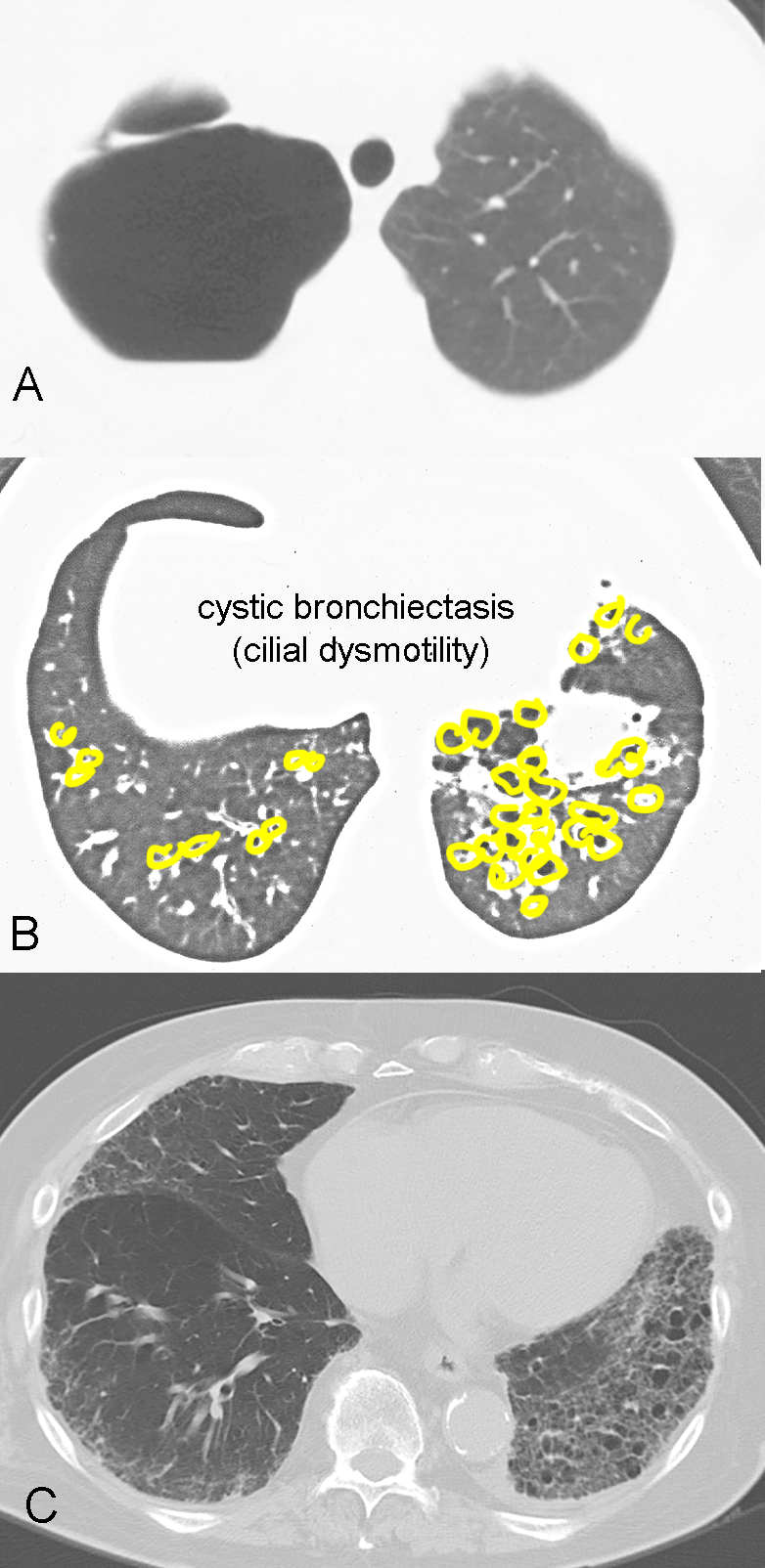
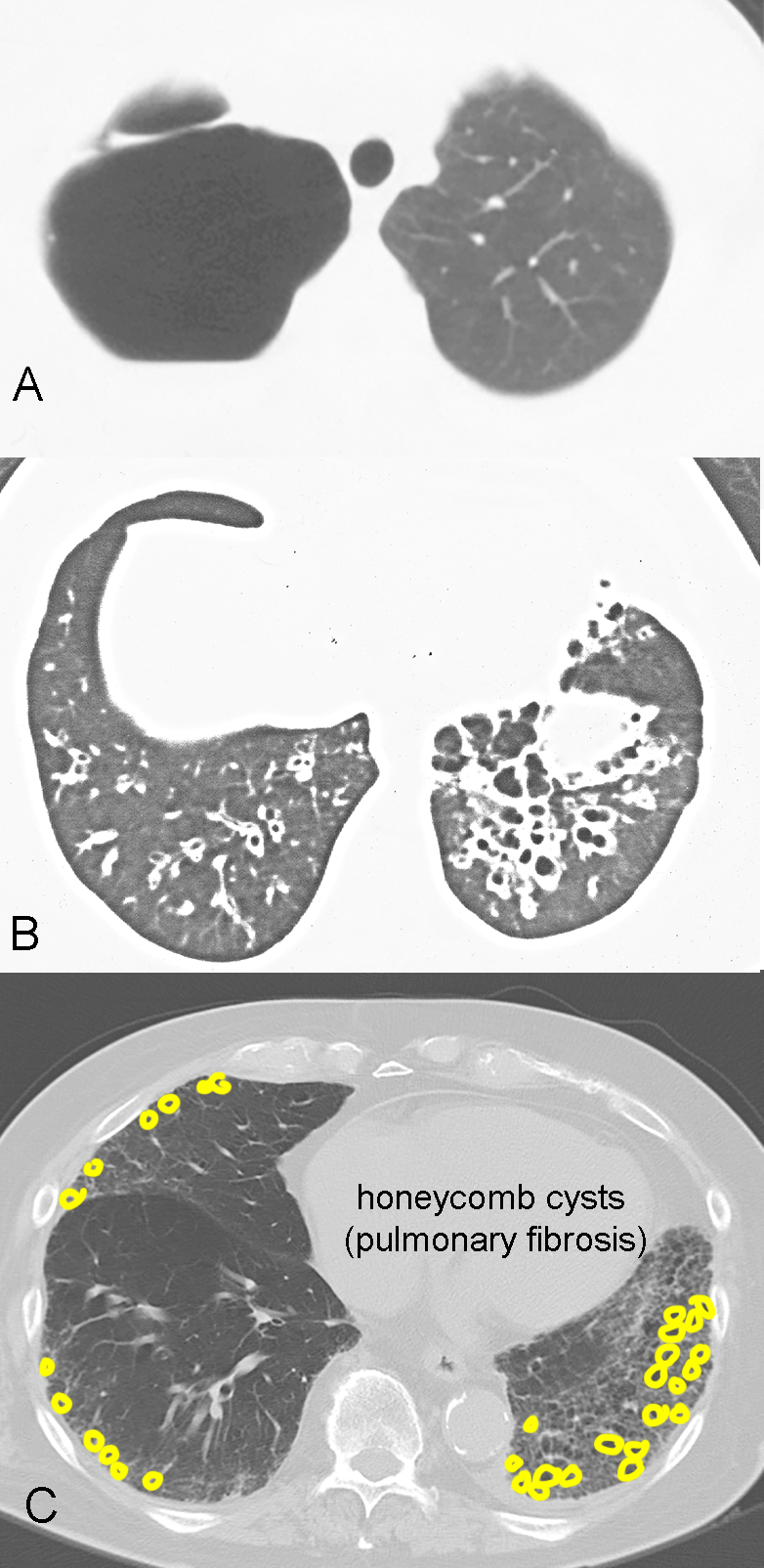
Here are CT scans from the two previous cases and one more example of cystic lung disease.
Question 2:
Which of the cystic lesions shown do you think would be visible on CXR? Which would be the hardest to see?
×
Answer:
Case A is the large right upper lung cyst, probably congenital. It was easy to see on the CXR. At the time of this CT image, the patient had developed an infection, so there is now an air fluid level within the cyst. Case B is the faintly visible cystic disease that could be seen on the CXR, and was cystic bronchiectasis. Case C would be very difficult to recognize as cysts on CXR, as the cysts are very small and have thin walls. This is honeycombing, which usually requires a CT scan to detect.
Case A is the large right upper lung cyst, probably congenital. It was easy to see on the CXR. At the time of this CT image, the patient had developed an infection, so there is now an air fluid level within the cyst. Case B is the faintly visible cystic disease that could be seen on the CXR, and was cystic bronchiectasis. Case C would be very difficult to recognize as cysts on CXR, as the cysts are very small and have thin walls. This is honeycombing, which usually requires a CT scan to detect.
