

Case 2
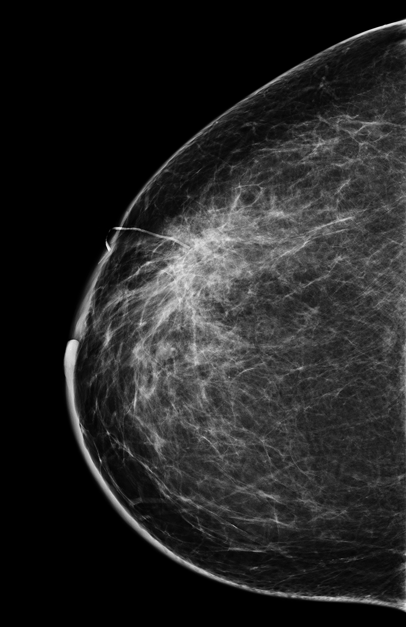
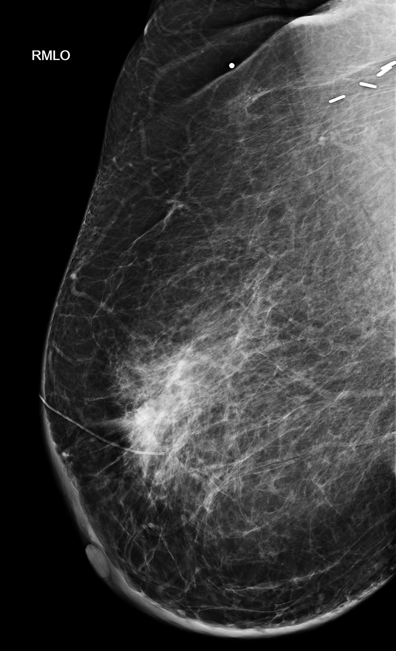
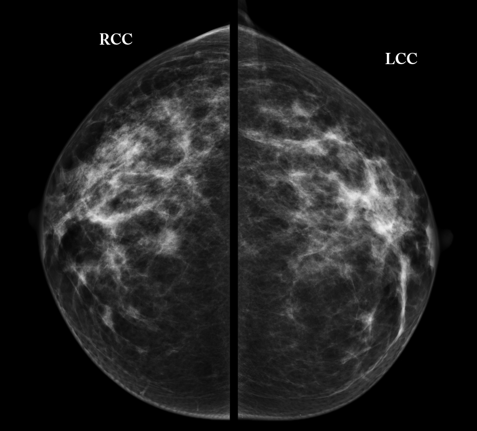
Presentation and HPI--53 year old female with lump in the right breast for several weeks. No family members with breast cancer. She missed her routine yearly mammogram last year. No other symptoms or past medical history.
Further Explanation:
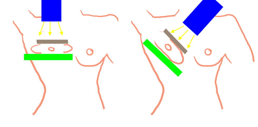
Case 2
This is a series of images done with digital tomosynthesis, a new way to get slices of the breast tissue rather than one view. This is also sometimes called '3D mammography'
Further Explanation:
Case 2
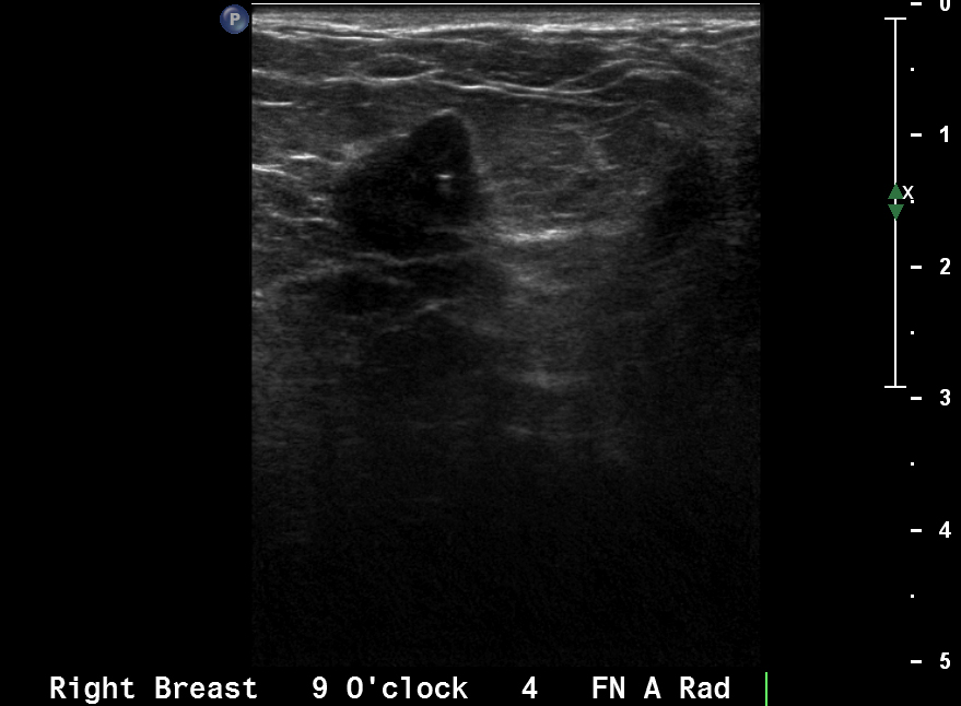
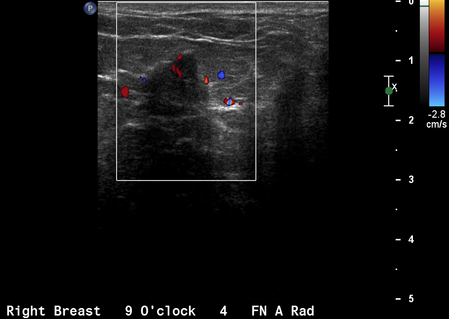
This is another type of study of the breast that is usually done either to evaluate a lump (felt by patient or doctor) or to further evaluate an abnormality seen on a mammogram.
Further Explanation:
Case 2
Outcome--the patient had a biopsy showing breast cancer, and went on to a surgical resection (lumpectomy) with radiation treatment, and is doing well.
Further Explanation:
