

Case 4
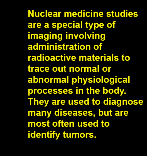
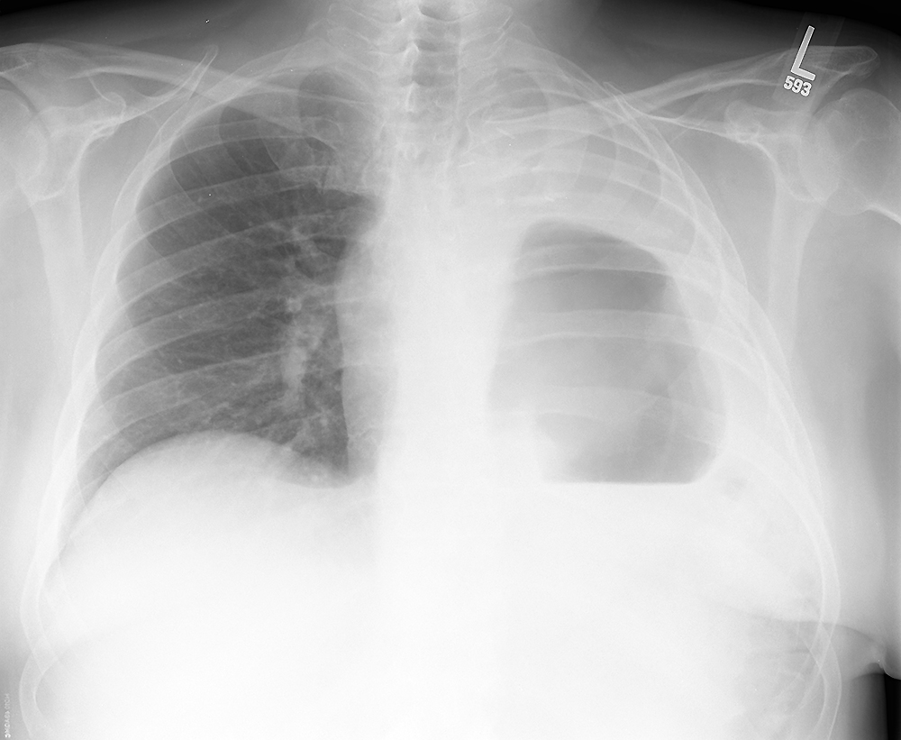
Presentation and HPI--65 year old male with a long history of cigarette smoking who now has left chest pain and shortness of breath. He is coughing up blood for the past month. His past medical history includes high blood pressure and emphysema.
Further Explanation:
Case 4
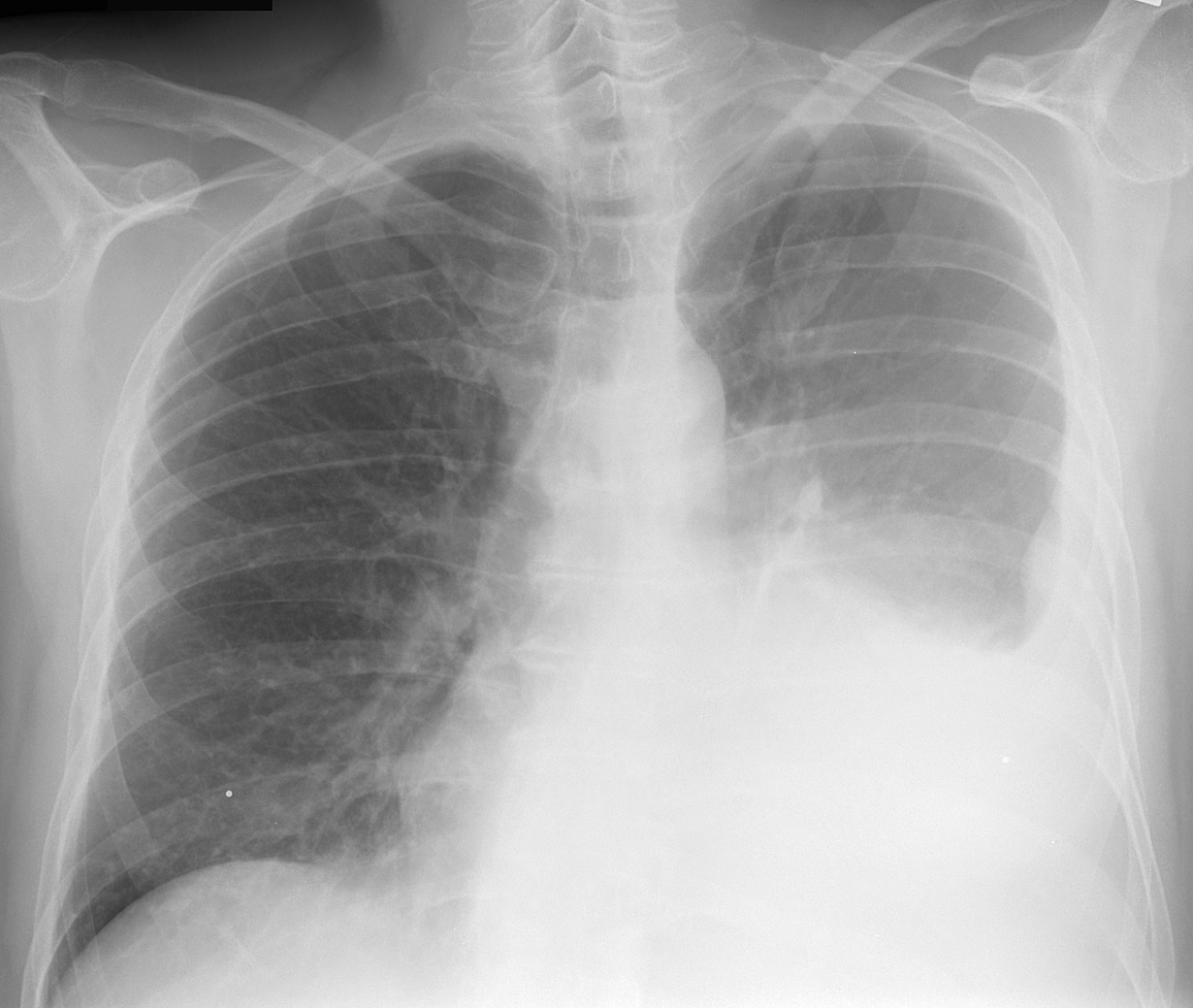
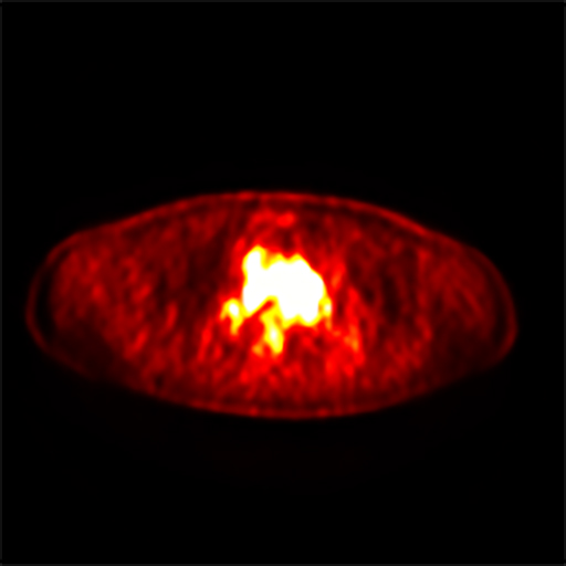
This is an example of a nuclear medicine study--another type of radiologic study but in this case, a radioactive tracer is introduced into the body, and a radiation detector is brought close to the patient to determine where the tracer has gone. In this case, the tracer was injected into a vein in the patient's arm, and was designed to home in on actively growing tumor cells. This particular type of nuclear medicine study is called a PET scan (positron emission tomography).
Further Explanation:
Case 4
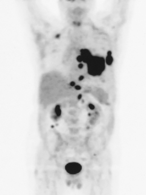
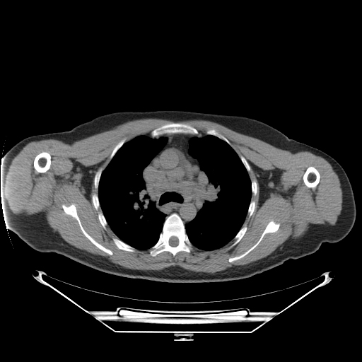
This is a different patient who also has shortness of breath and cough, but is not coughing up blood. Because a PET image alone is very low resolution, and does not always show exactly what organ is taking up the tracer, it is often helpful to do both a CT and a PET scan at the same time. The CT shows anatomy better and the PET shows pathology. In this case, the CT showed enlarged lymph nodes in the center of the chest (called the mediastinum) which are also taking up the PET tracer, but this was another cause of increased cell growth that is NOT cancer. This patient has an inflammatory condition called sarcoidosis.
Further Explanation:
Case 4
Outcome-The tumor was too extensive to remove with surgery, so the patient was treated with radiation and chemotherapy with good response. For cure of lung cancer, surgery is usually needed, but newer chemotherapy treatments can produce lasting remissions in some patients.
Further Explanation:
