

Case 1
This case concerns the first checklist item for one organizational scheme for reading chest radiographs: A, or airway.
Question 1:
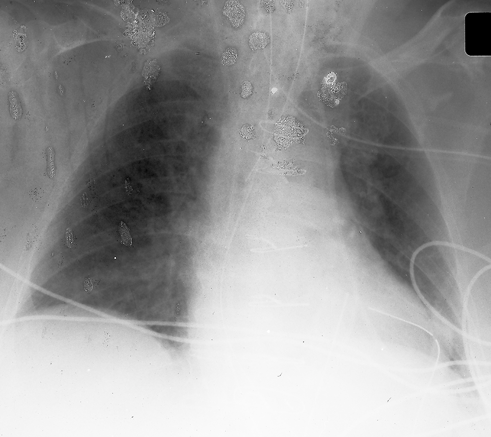
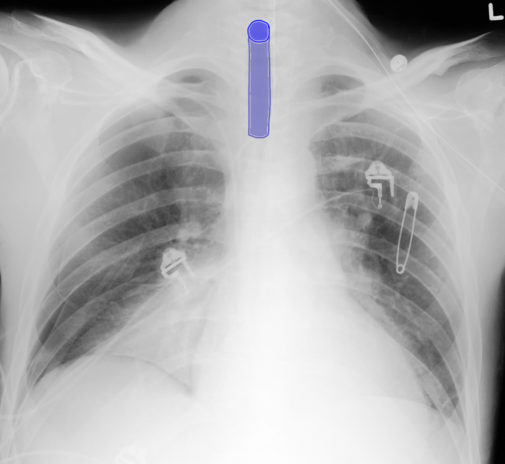
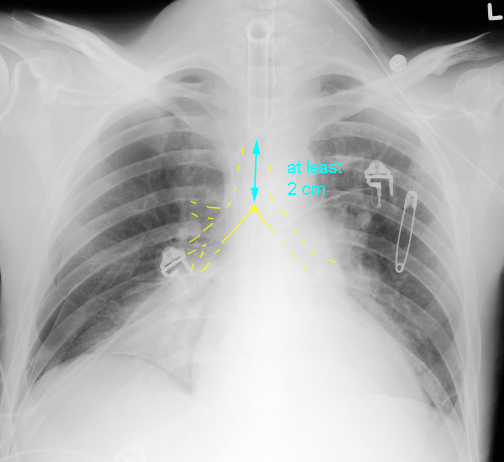
What do you think of this patient's airway? Is the support line in appropriate position?
×
Answer:
There is an artificial tube overlying the upper trachea, and based on its shape at the top, it seems to be coming toward you, suggesting that it is a tracheostomy tube, which exits toward the front in the neck. If you look at the links below, you can see that the tip of a tube in the trachea should be at least 2 cm above the carina. The distance from one posterior rib to the next posterior rib in most adults with reasonable inspiration is nearly 2 cm.
There is an artificial tube overlying the upper trachea, and based on its shape at the top, it seems to be coming toward you, suggesting that it is a tracheostomy tube, which exits toward the front in the neck. If you look at the links below, you can see that the tip of a tube in the trachea should be at least 2 cm above the carina. The distance from one posterior rib to the next posterior rib in most adults with reasonable inspiration is nearly 2 cm.

Case 1
This patient had an endotracheal tube placed and this portable radiograph was done to check the position of the tube.
Question 2:
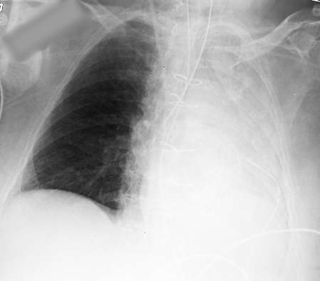
What do you think of the quality of this image? Are you confident that you can see the endotracheal tube well? Click the link below to see a repeat study on this patient, who was becoming harder to ventilate.
×
Answer:
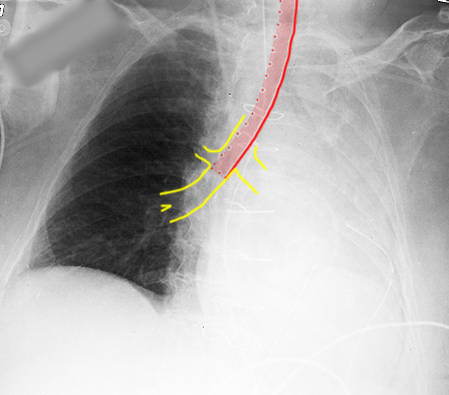
This image is of very poor quality, with lots of overlying artifact and probable motion. This is often a problem with portable chest radiographs. It is very hard to see the endotracheal tube well, and to distinguish it from other lines and tubes in the area. On the second image shown below, you can see that the left lung is now opaque. The ETT is in the right mainstem bronchus.
This image is of very poor quality, with lots of overlying artifact and probable motion. This is often a problem with portable chest radiographs. It is very hard to see the endotracheal tube well, and to distinguish it from other lines and tubes in the area. On the second image shown below, you can see that the left lung is now opaque. The ETT is in the right mainstem bronchus.
