

Case 2
The next item on our pCXR checklist is B, for bones. It is often hard to see bones on portable radiographs, but we will also also look for things often seen near the ribs, such as chest tubes or pleural processes.
Question 1:
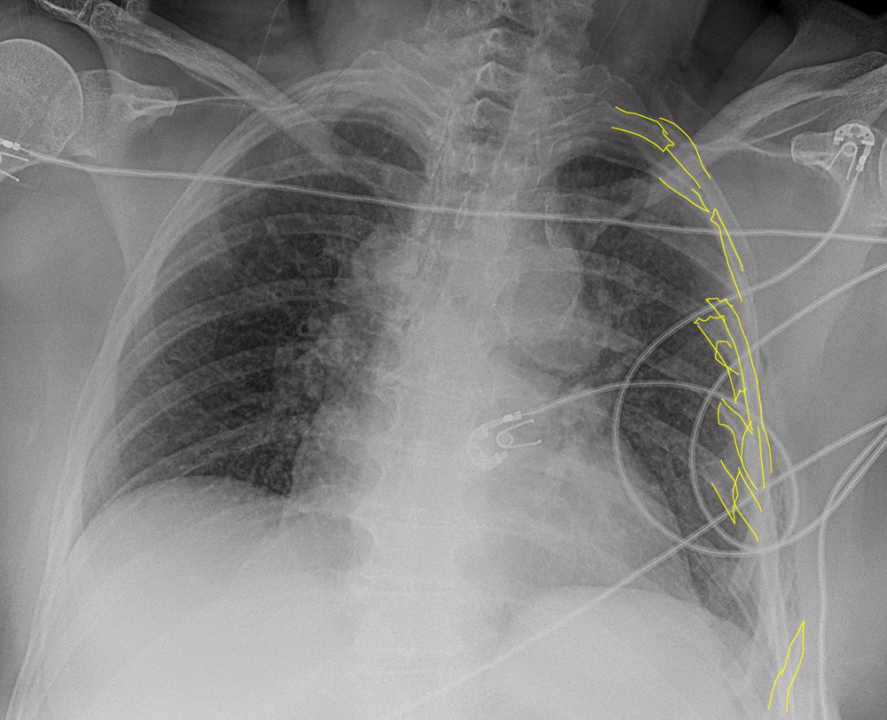
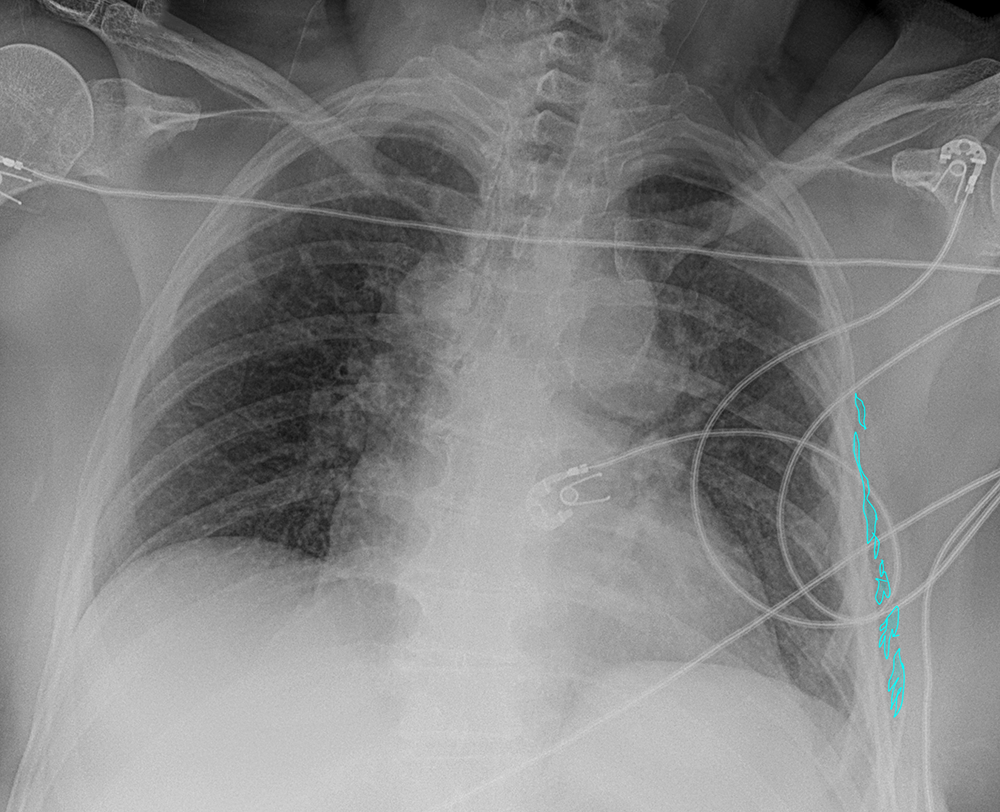
This patient has been having a lot of shortness of breath and has left chest pain after a motor vehicle collision. What do you think of their ribs? What was done to correct this (look at followup CXR below)
As mentioned above, ribs can be hard to see on portable radiographs. But the ribs on the left look very abnormal, and it appears that there are many overlapping ribs consistent with fractures. If enough ribs are damaged, it can interfere with breathing, and this condition is called a 'flail chest'. As the patient tries to breath in, the chest collapses instead of expanding. There is another clue that there may be a rib problem if you look closely just outside the left ribs. There are tiny pockets of air in the subcutaneous tissues, that has probably leaked out there from the lung, which has been damaged by the sharp rib fragments. It is unusual to insert hardware for isolated rib fractures (which usually heal with minimal support on their own). But in the case of a flail chest, a number of ribs must be stabilized to allow the patient's respiratory mechanics to work properly. A series of metal plates and screws (and skin clips) can be seen on the followup image after internal fixation of the fractured ribs.

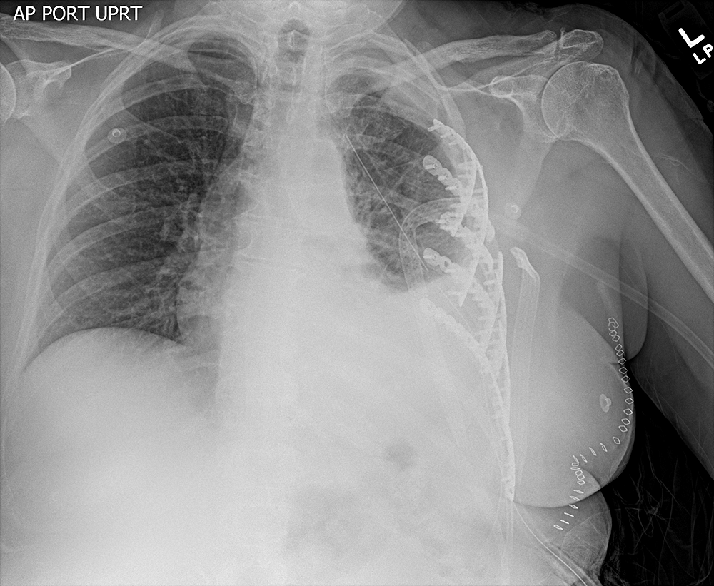
Case 2
The B item is a good time to look at the interface of lung with ribs laterally, as this is often where a pneumothorax will be visible.
Question 2:
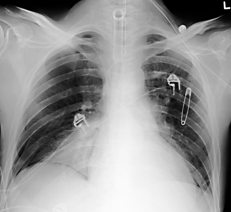
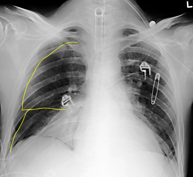
What do you think of this patient's outer lung on the right? This is the same case we looked at earlier with a tracheostomy tube in place.
There is a fine white line that runs parallel to the lung-rib interface. This is the typical appearance of a pneumothorax with the patient upright, or at least semi-upright (as is often the case for a portable CXR). This patient has a number of other abnormalities, including abnormal opacity in the right medial lung base, but the pneumothorax is the most emergent finding on this image, and should be immediately treated, usually with placement of a chest tube.
