

Case 3
The next item on our checklist is C, for cardiomediastinal region. For pCXR's, you are often checking various support lines that project over the heart.
Question 1:
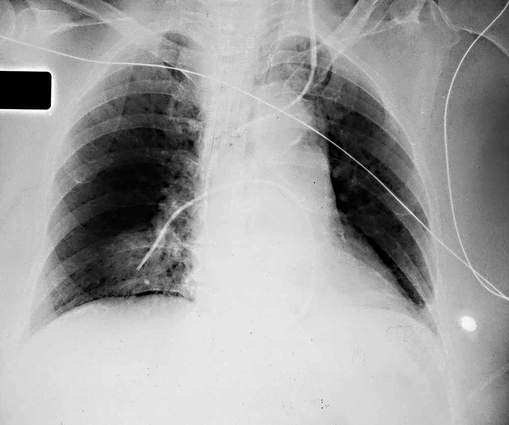
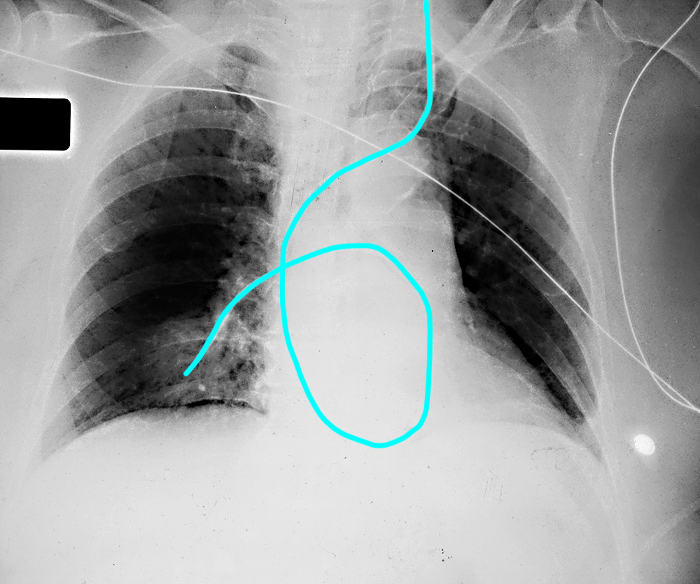
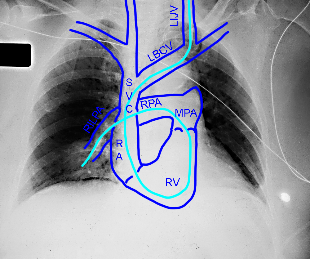
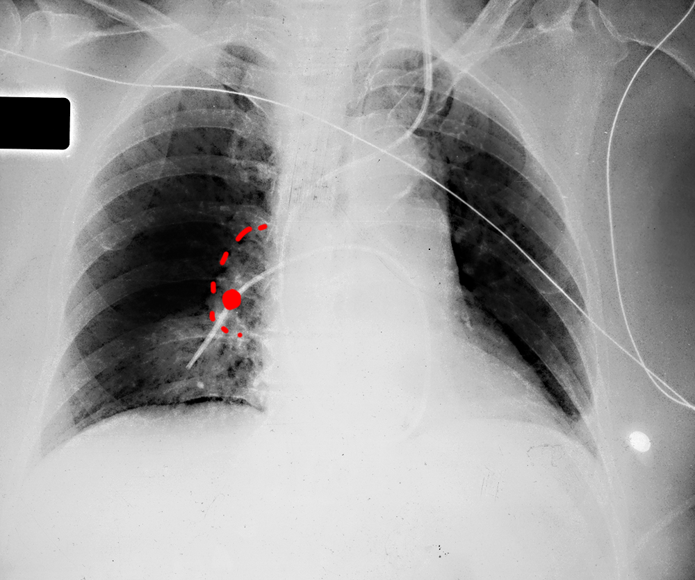
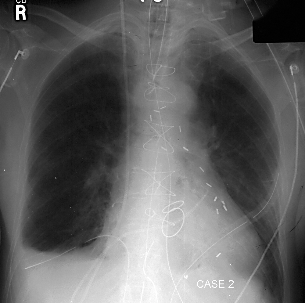
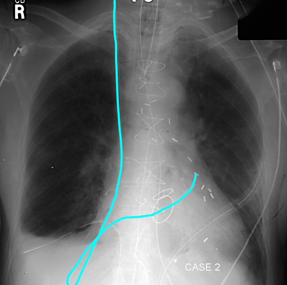
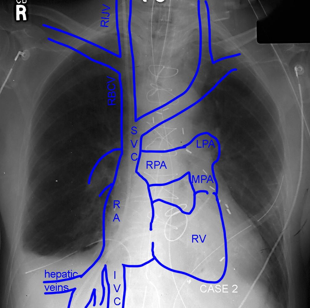
What do you think the support line is on this image? Is it is expected position? You are shown a second patient with the same type of line for you to analyze. Is it in expected position?
×
Answer:
These two patients both have pulmonary arterial lines in place, also called Swan-Ganz catheters. They are placed via the internal jugular vein most often, and flow of blood will help to move them into position in one of the interlobar (large central) pulmonary arteries, usually on the right. They are used to assess central lung vascular pressures. With such a complicated course, you can see how they might end up in the wrong place. The line in the first case is going where it should, but is just too far out into the right lung. It should be projecting over the hilum, not the lung. The second case has a more abnormal course, moving down into the IVC and hepatic veins before coming back up and through the heart to the pulmonary artery. It will not function normally in this position.
These two patients both have pulmonary arterial lines in place, also called Swan-Ganz catheters. They are placed via the internal jugular vein most often, and flow of blood will help to move them into position in one of the interlobar (large central) pulmonary arteries, usually on the right. They are used to assess central lung vascular pressures. With such a complicated course, you can see how they might end up in the wrong place. The line in the first case is going where it should, but is just too far out into the right lung. It should be projecting over the hilum, not the lung. The second case has a more abnormal course, moving down into the IVC and hepatic veins before coming back up and through the heart to the pulmonary artery. It will not function normally in this position.
Case 3
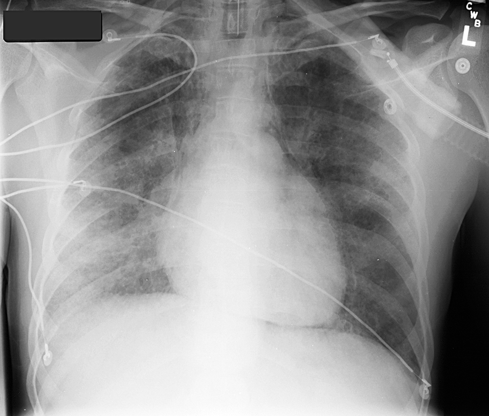
This patient has been on positive pressure ventilation for a while due to severe lung disease
Question 2:
What do you think of their mediastinum? How could positive pressure ventilation lead to this finding?
×
Answer:
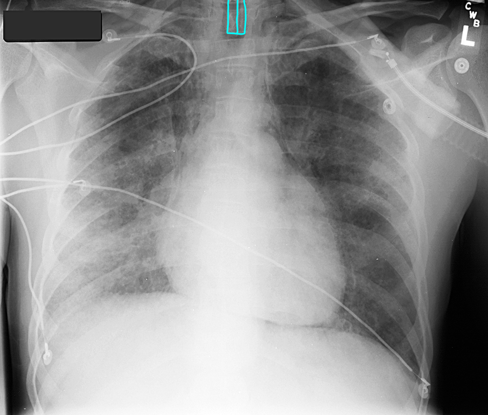
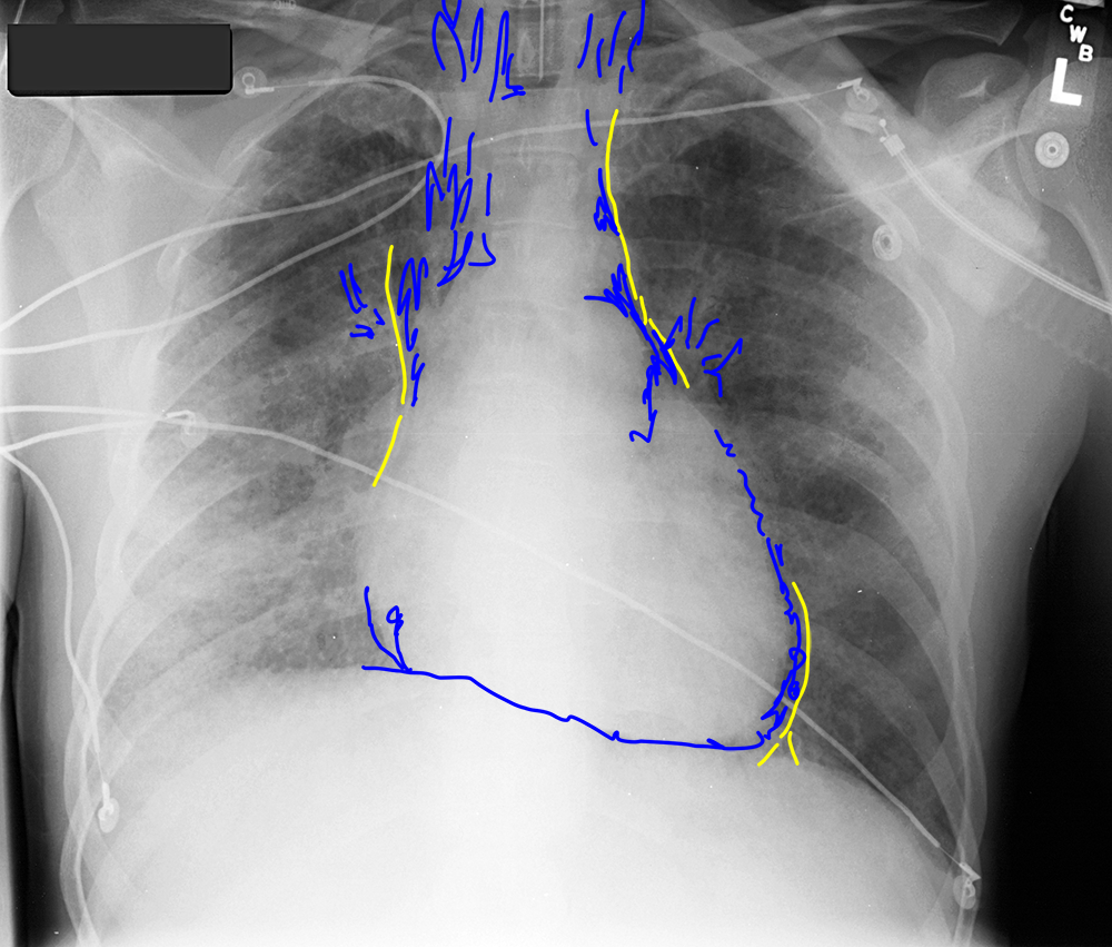
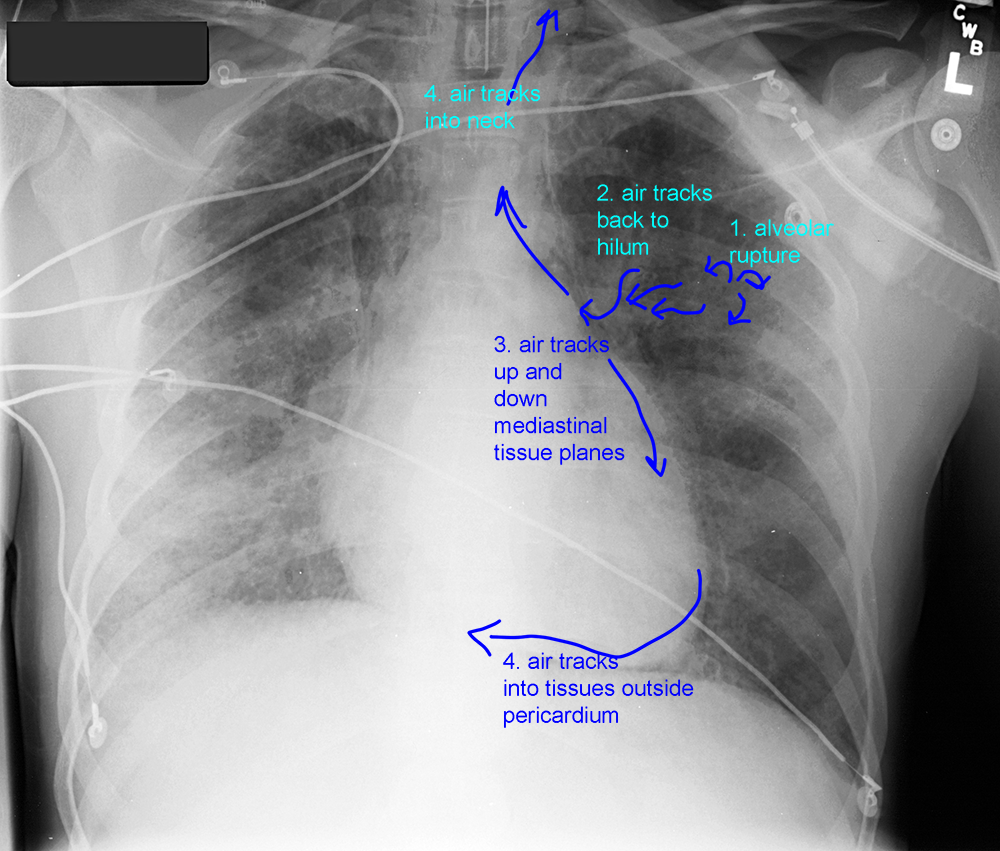
The contours of this patient's cardiomediastinum are relatively normal, but there are black streaks of air running along both sides, lifting up a thin white line in some places. This line represents the parietal pleura that is displaced laterally by air within the connective tissue planes of the mediastinum. This is called 'pneumomediastinum', and can happen due to trauma to the trachea or esophagus. In the setting of mechanical ventilation, it can also happen when pressures are high enough to rupture alveoli in the lung. This air will track back along the peribronchial connective tissues into the hilum and then into the mediastinum. It is important to recognize not because the air itself causes problems, but because it is a sign that the ventilator settings should be adjusted.
The contours of this patient's cardiomediastinum are relatively normal, but there are black streaks of air running along both sides, lifting up a thin white line in some places. This line represents the parietal pleura that is displaced laterally by air within the connective tissue planes of the mediastinum. This is called 'pneumomediastinum', and can happen due to trauma to the trachea or esophagus. In the setting of mechanical ventilation, it can also happen when pressures are high enough to rupture alveoli in the lung. This air will track back along the peribronchial connective tissues into the hilum and then into the mediastinum. It is important to recognize not because the air itself causes problems, but because it is a sign that the ventilator settings should be adjusted.
