

Case 4
The next item on our checklist is D, for diaphragm. We can also look for things like pleural processes or problems under the diaphragm like free air.
Question 1:
What position do you think this patient was in when this radiograph was taken? What do you think of the diaphragms in this patient?
×
Answer:
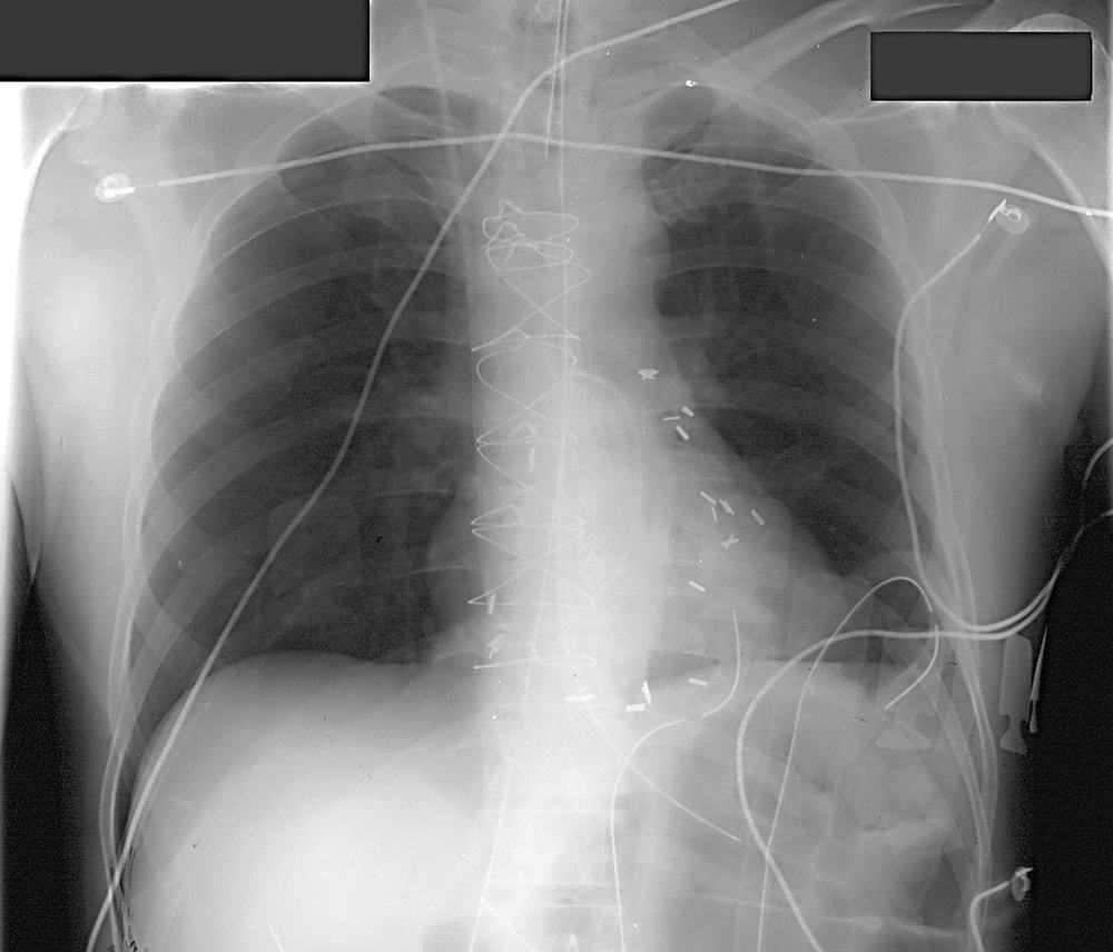
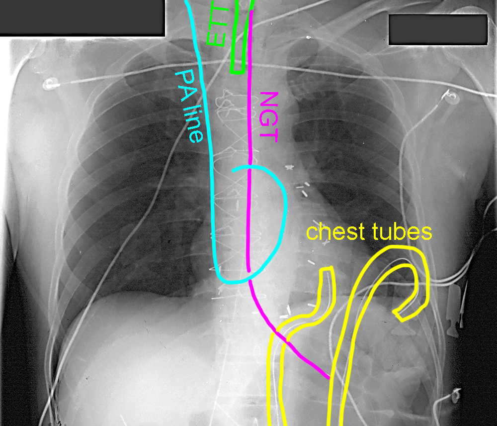
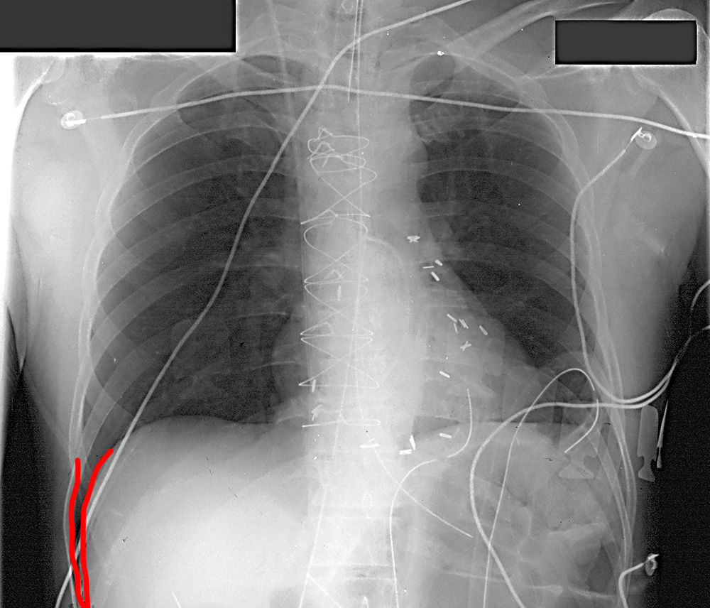
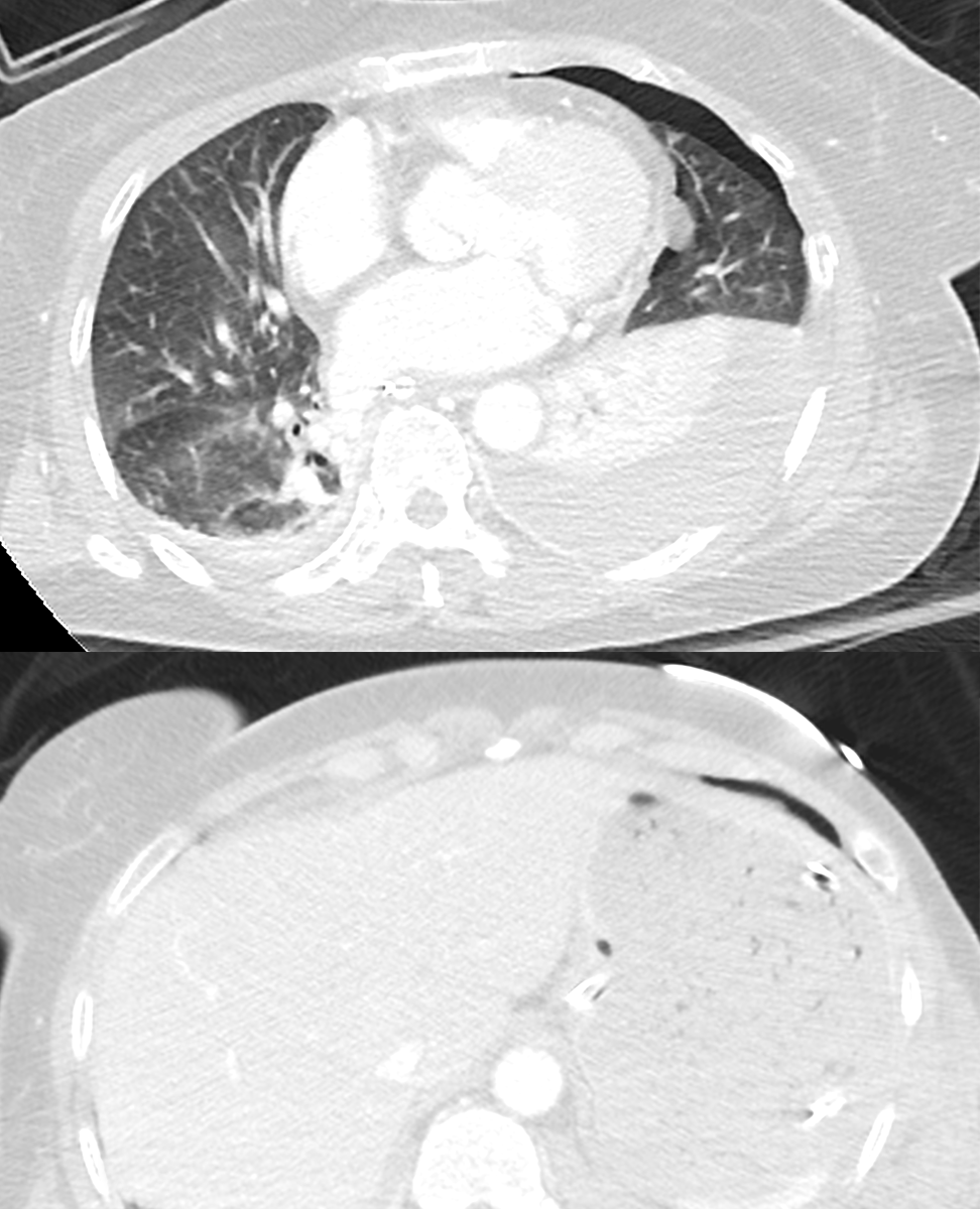
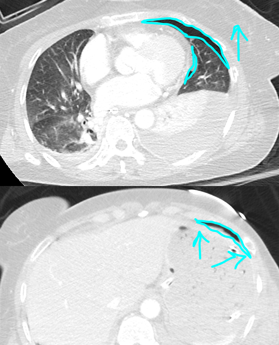
Given how many support lines are present (including a PA line with the tip a bit proximal), they were likely supine at the time of the radiograph. The costophrenic angle seems to go down much too far on the right, particularly compared to the left. Even if we assume there might be some left pleural fluid present, the right costophrenic angle looks like it extends too far inferiorly to be normal. When in the supine position (as many portable chest radiographs are taken), pneumothorax may accumulate in the region of the costophrenic angle and more anteriorly producing this appearance, called a 'deep sulcus'. It is only when the patient is upright that we look for pneumothorax in the usual place, near the apex of the lung. Check out the CT shown below from a different patient who is supine for the scan and has a left pneumothorax. The air is anterior in position in the left pleural space and extends somewhat laterally, and a bit deeper than the normal right costophrenic angle.
Given how many support lines are present (including a PA line with the tip a bit proximal), they were likely supine at the time of the radiograph. The costophrenic angle seems to go down much too far on the right, particularly compared to the left. Even if we assume there might be some left pleural fluid present, the right costophrenic angle looks like it extends too far inferiorly to be normal. When in the supine position (as many portable chest radiographs are taken), pneumothorax may accumulate in the region of the costophrenic angle and more anteriorly producing this appearance, called a 'deep sulcus'. It is only when the patient is upright that we look for pneumothorax in the usual place, near the apex of the lung. Check out the CT shown below from a different patient who is supine for the scan and has a left pneumothorax. The air is anterior in position in the left pleural space and extends somewhat laterally, and a bit deeper than the normal right costophrenic angle.
Case 4
This patient has abdominal pain as well as shortness of breath.
Question 2:
What position do you think this patient was in for this portable CXR? What is abnormal in the region of the diaphragms? What diagnoses would you consider for this appearance?
×
Answer:
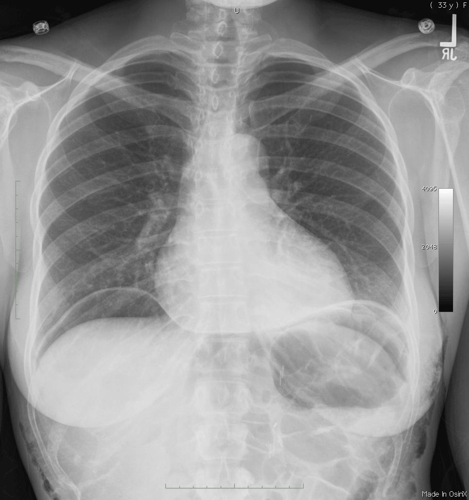
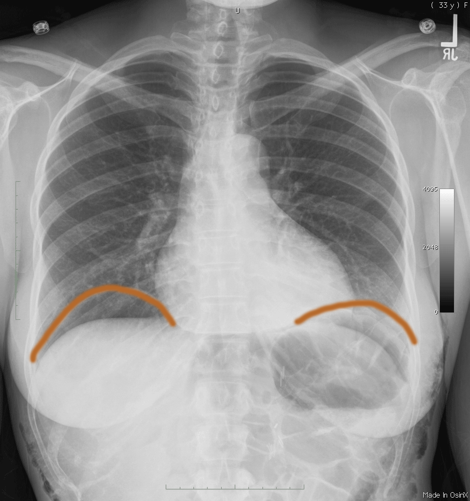
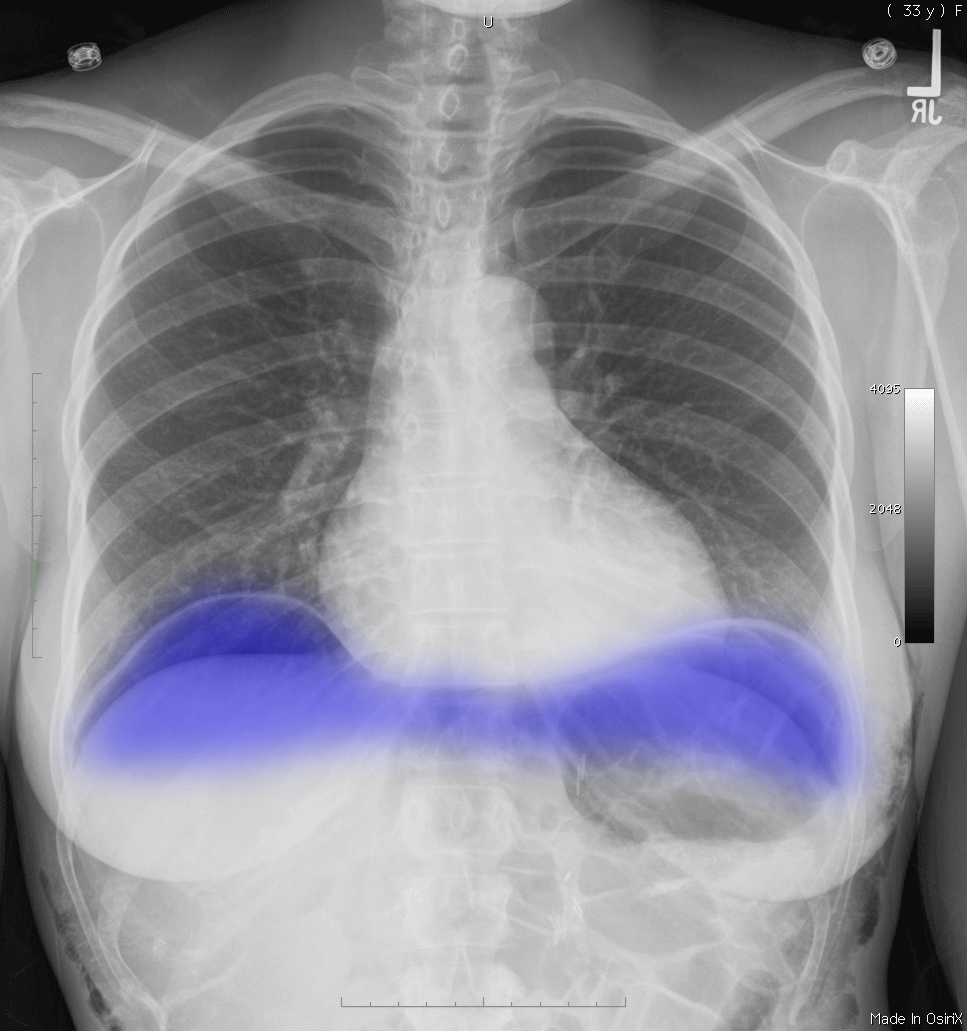
This patient has no support lines and their lungs and heart are relatively normal in appearance. This might suggest that they are upright. There is air under the diaphragm that outlines the normal thin diaphragms between the abnormal air and the normal lower aerated lung. This indicates that the patient was upright, as this will allow the air in the peritoneal cavity to rise to the highest position in the abdomen. Air in the peritoneal cavity could be normal in a patient ho recently had a laparoscopic procedure, but in most other circumstances this finding should suggest perforation of a viscus.
This patient has no support lines and their lungs and heart are relatively normal in appearance. This might suggest that they are upright. There is air under the diaphragm that outlines the normal thin diaphragms between the abnormal air and the normal lower aerated lung. This indicates that the patient was upright, as this will allow the air in the peritoneal cavity to rise to the highest position in the abdomen. Air in the peritoneal cavity could be normal in a patient ho recently had a laparoscopic procedure, but in most other circumstances this finding should suggest perforation of a viscus.
