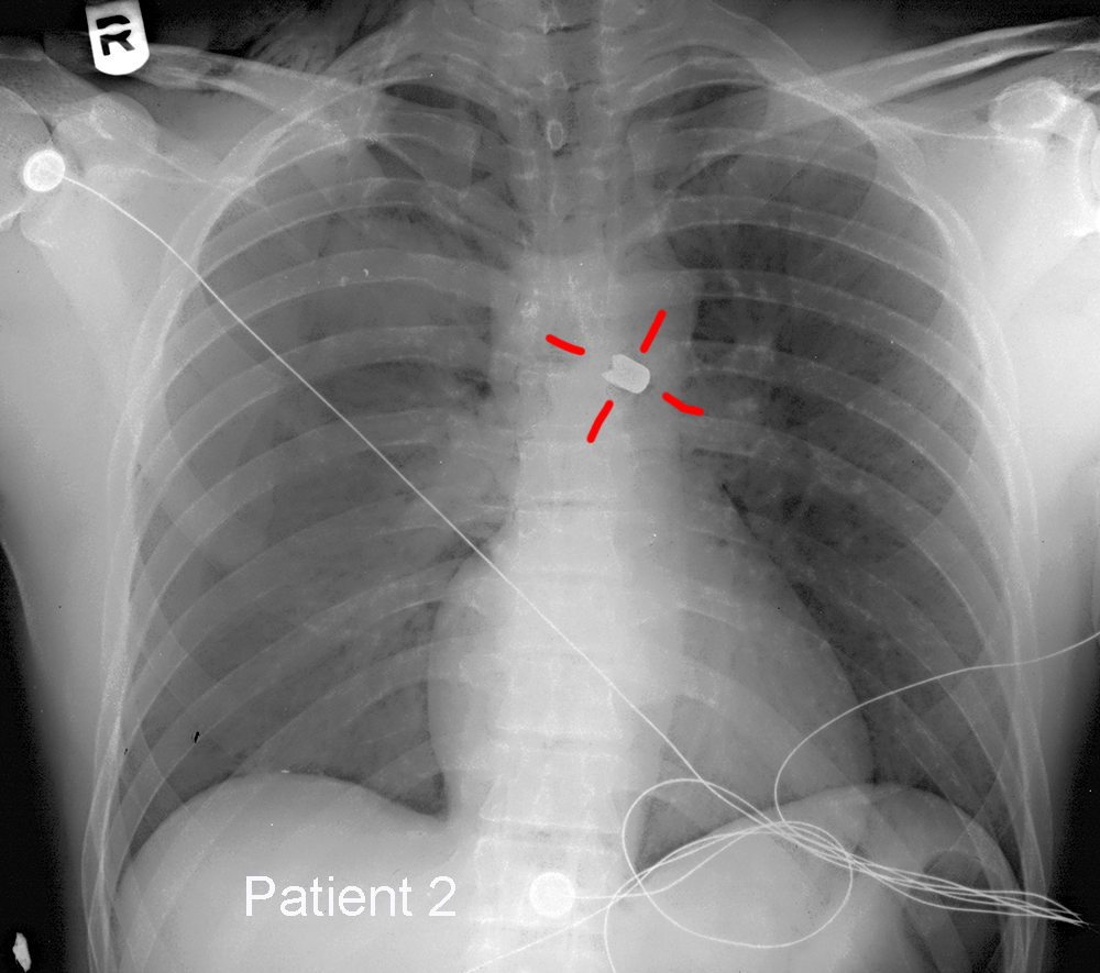


Case 5
The last thing on our pCXR checklist is E, which stands for everything else, but is mostly the lungs.
Question 1:
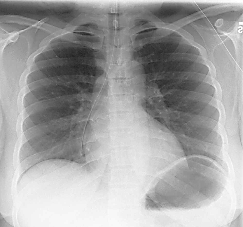
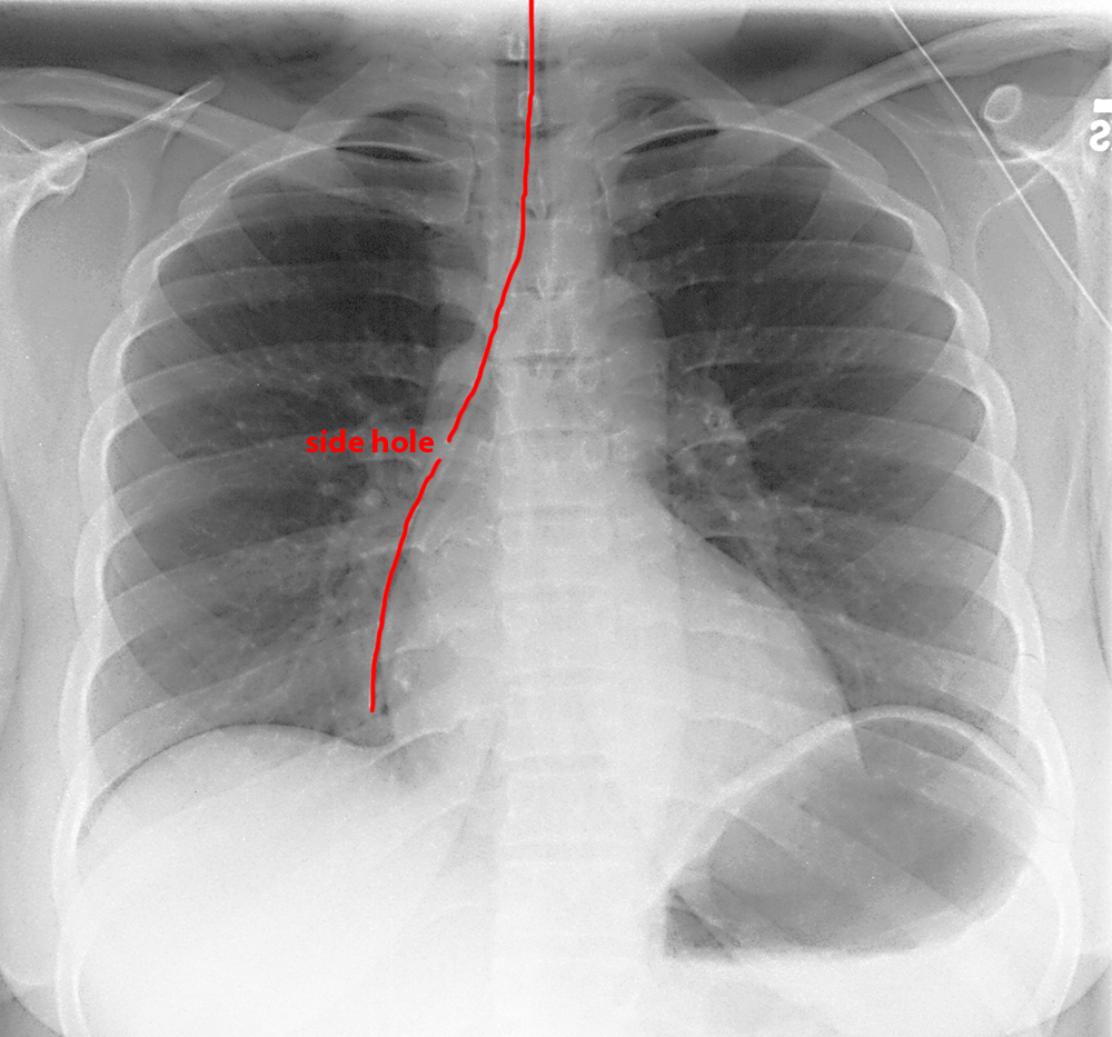
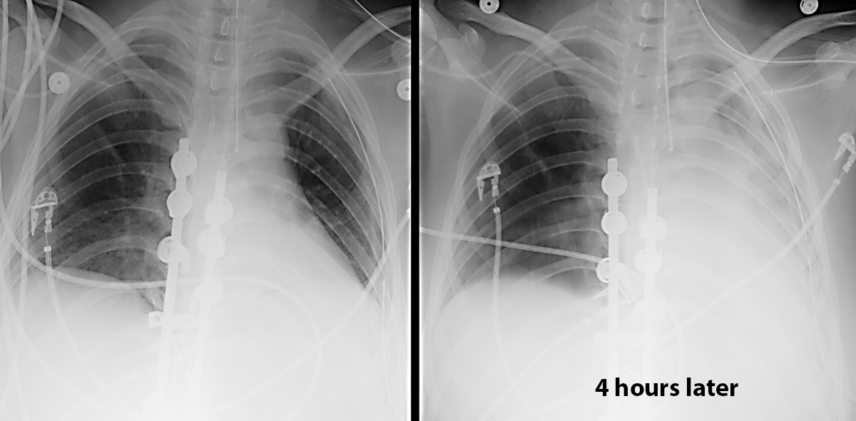
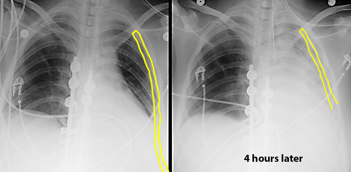
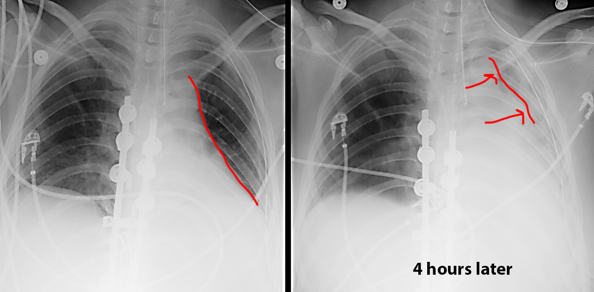
The second image was obtained four hours after the first in a patient with increasing shortness of breath. What do you think of the lungs, and what is the mystery device outlined below?
It is vital when lung opacities are seen on portable chest radiographs to consider the differential of effusion vs pneumonia vs collapse, since the treatment is different for each. In this case, there is new opacity on the left on the later image, with accompanying shift of the mediastinum toward the opacity. This should suggest collapse rather than consolidation or effusion. Most pneumonias do not produce shift of the mediastinum, and effusions should push the mediastinum away. The mystery device is a malpositioned NG tube, which you can identify by the presence of a side hole, which would not be present in an endotracheal tube. The tip of this NG tube is in the upper esophagus and should be below the diaphragms in the stomach.
Case 5
When looking at the lungs on a portable CXR, you should compare right to left for symmetry.
Question 2:
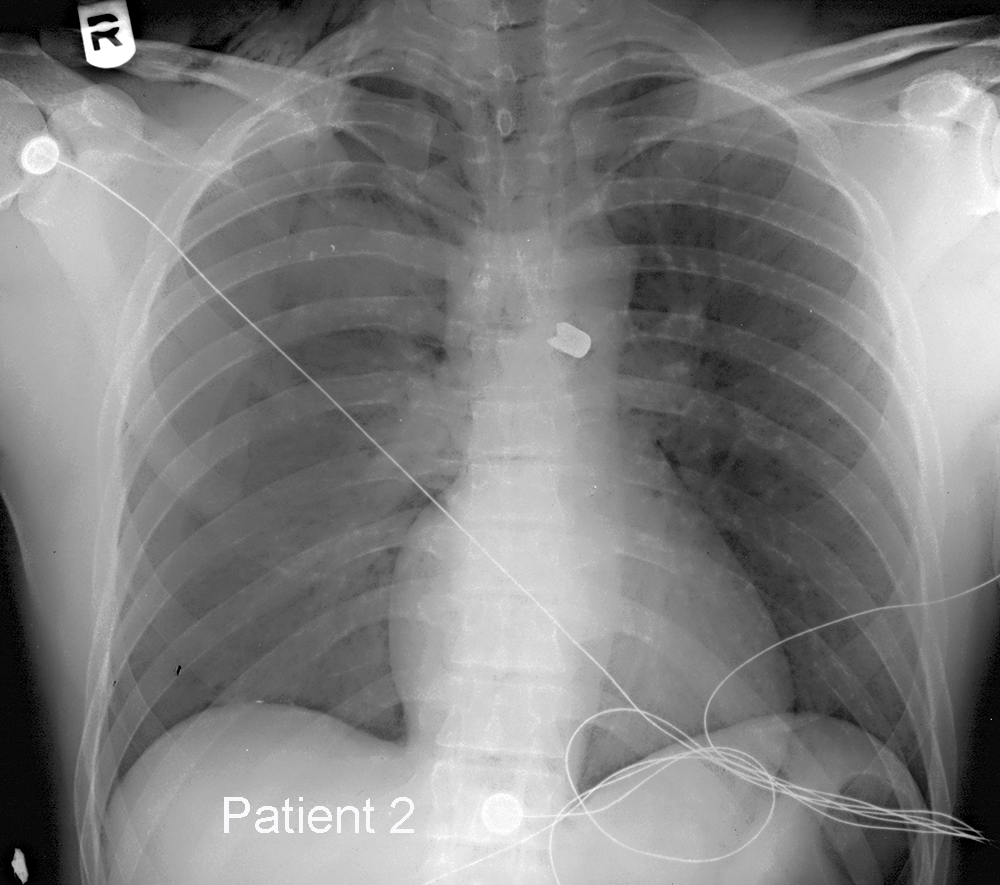
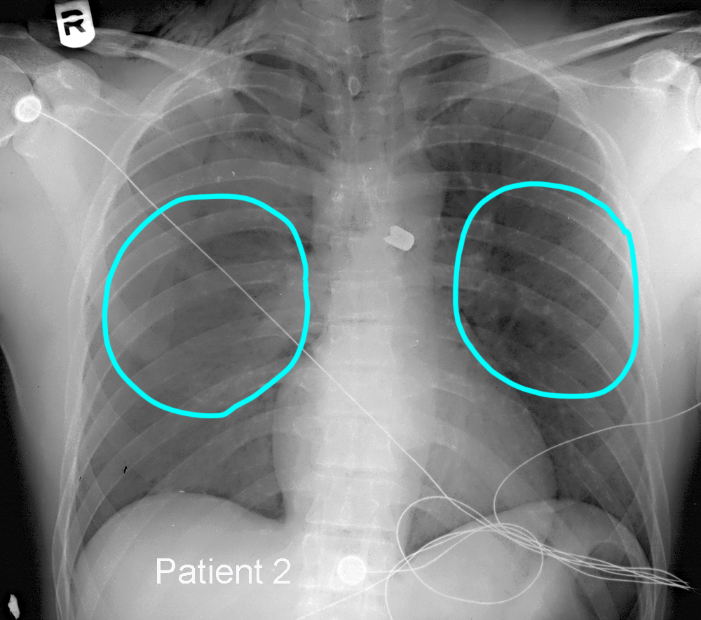
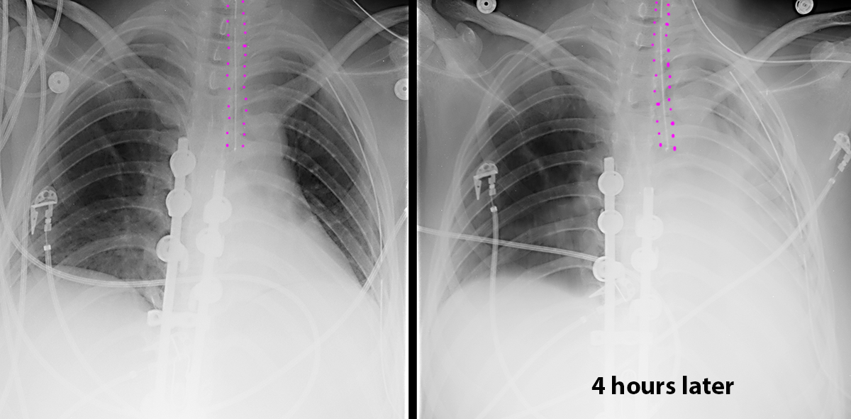
What do you think of this patient's right lung? Is there anything else on the image that should not be there? For patient 2, what do you think of the lungs, and do you see that might explain the findings?
This patient's right lung is clear. There is a tube that starts in the midline in the neck but with the tip projecting over the medial right lung. This is an NG tube that has entered the airway instead of the esophagus. This tube should NOT be used to administer anything, as it will go into the lung. For patient 2, the right lung looks overall denser than the left, but with no correspondence to any lobe and with no air bronchograms. This might suggest that the density could be in the pleural or extrapleural space instead of the lung. Detection of pleural effusions is more difficult on a supine image. The other finding of note is a bullet projecting over the mediastinum.
