

Case 1-esophageal disease
There are many ways to image the esophagus, but one of the most informative is a barium swallow.
Question 1:
a) What does a barium swallow show that is not visible on other imaging studies such as CT or MR?
A barium swallow is a real-time examination, meaning that imaging shows the actual motility and contraction of the esophagus. It is a relatively high radiation dose compared to a chest radiograph (approximately 10 times as high), but still less than a CT scan (which is about 10 times more).
b) What is the normal appearance of the esophageal mucosa on a barium swallow?
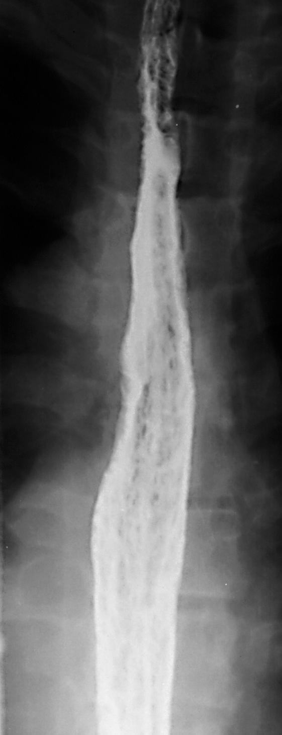
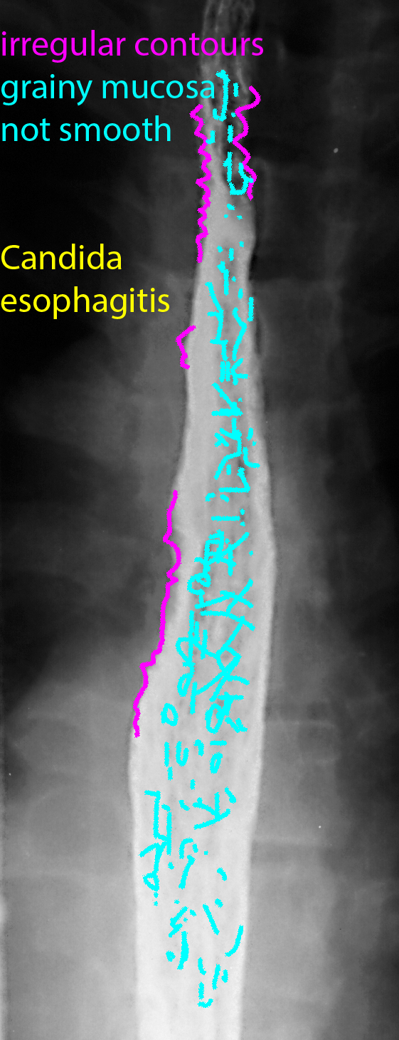
In a properly done barium swallow, which requires both expertise on the part of the operator and cooperation from the patient, images with barium coating the mucosa and gas in the lumen should show an entirely smooth mucosa with no folds and not bumpy or grainy in appearance. Frequent peristaltic waves are expected as the esophagus is very sensitive to stretch, leading to vigorous contraction. So timing is crucial to obtain images between peristaltic waves. You may also notice that the images shown are not straight AP, but a bit oblique. this is to move the esophagus off the spine, to get a better view of the organ.
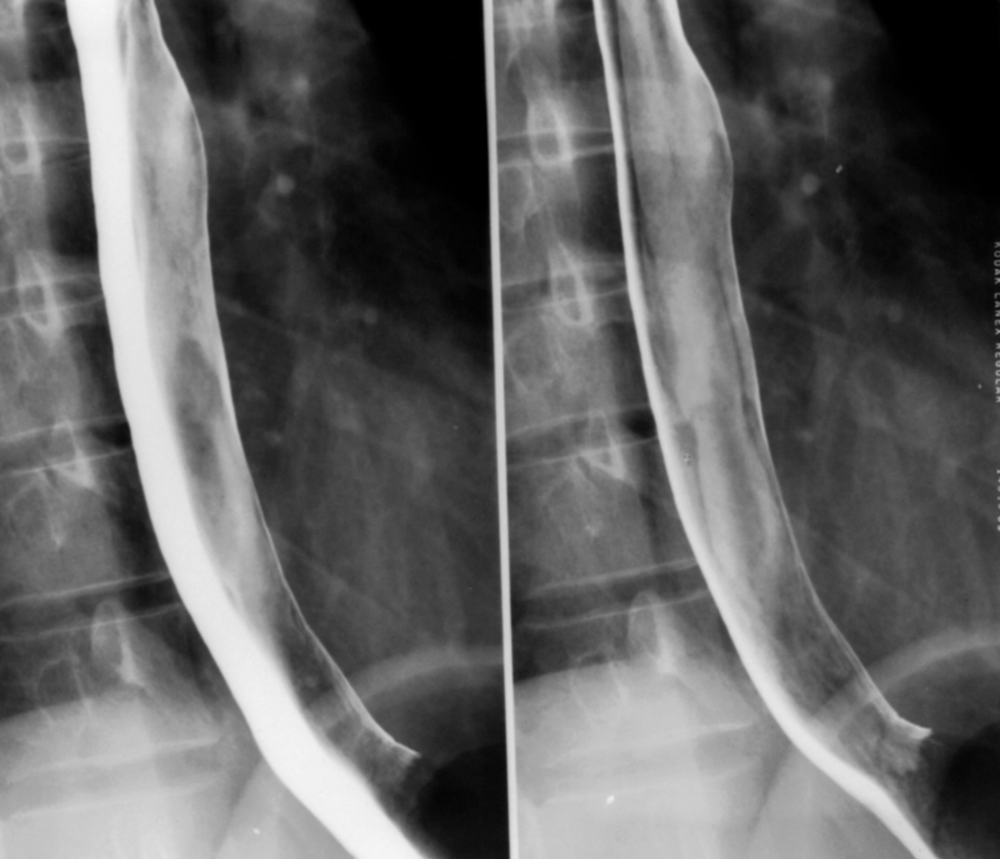
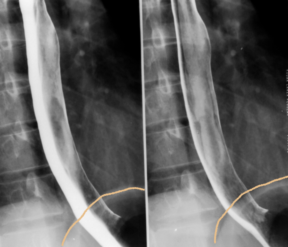
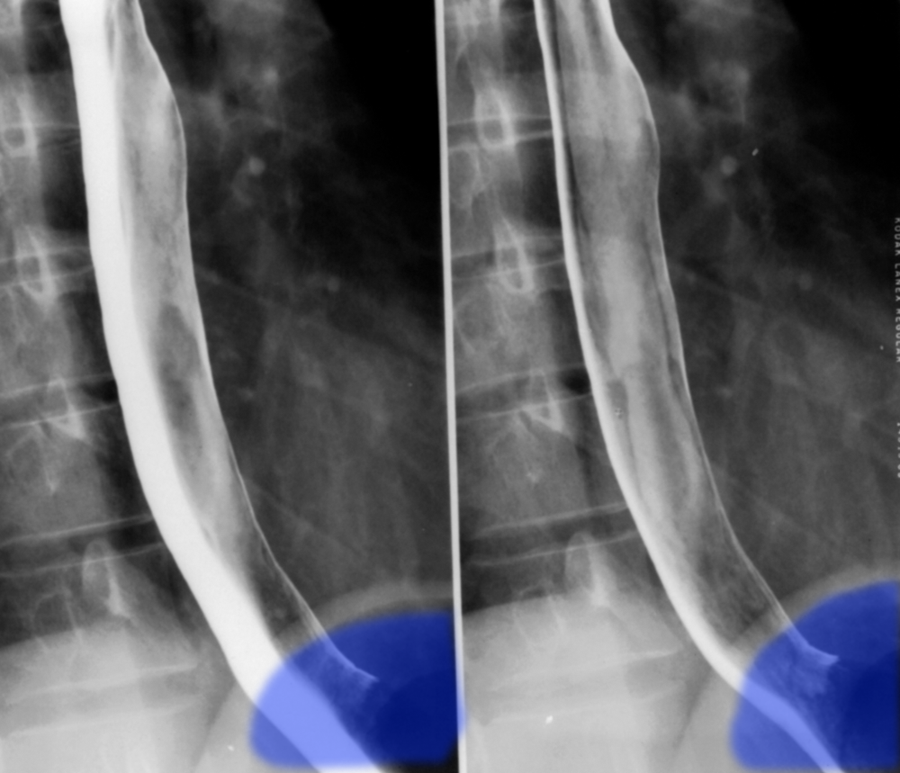
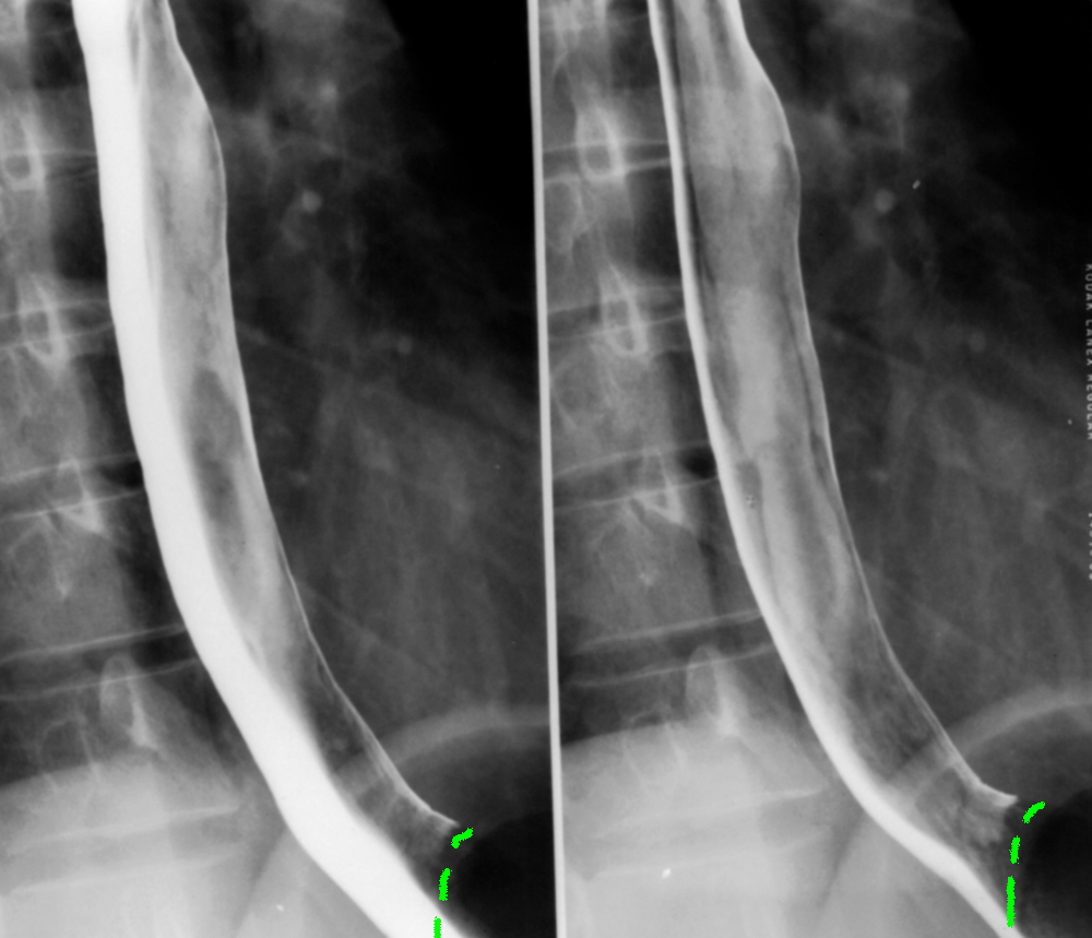
Case 1-esophageal disease
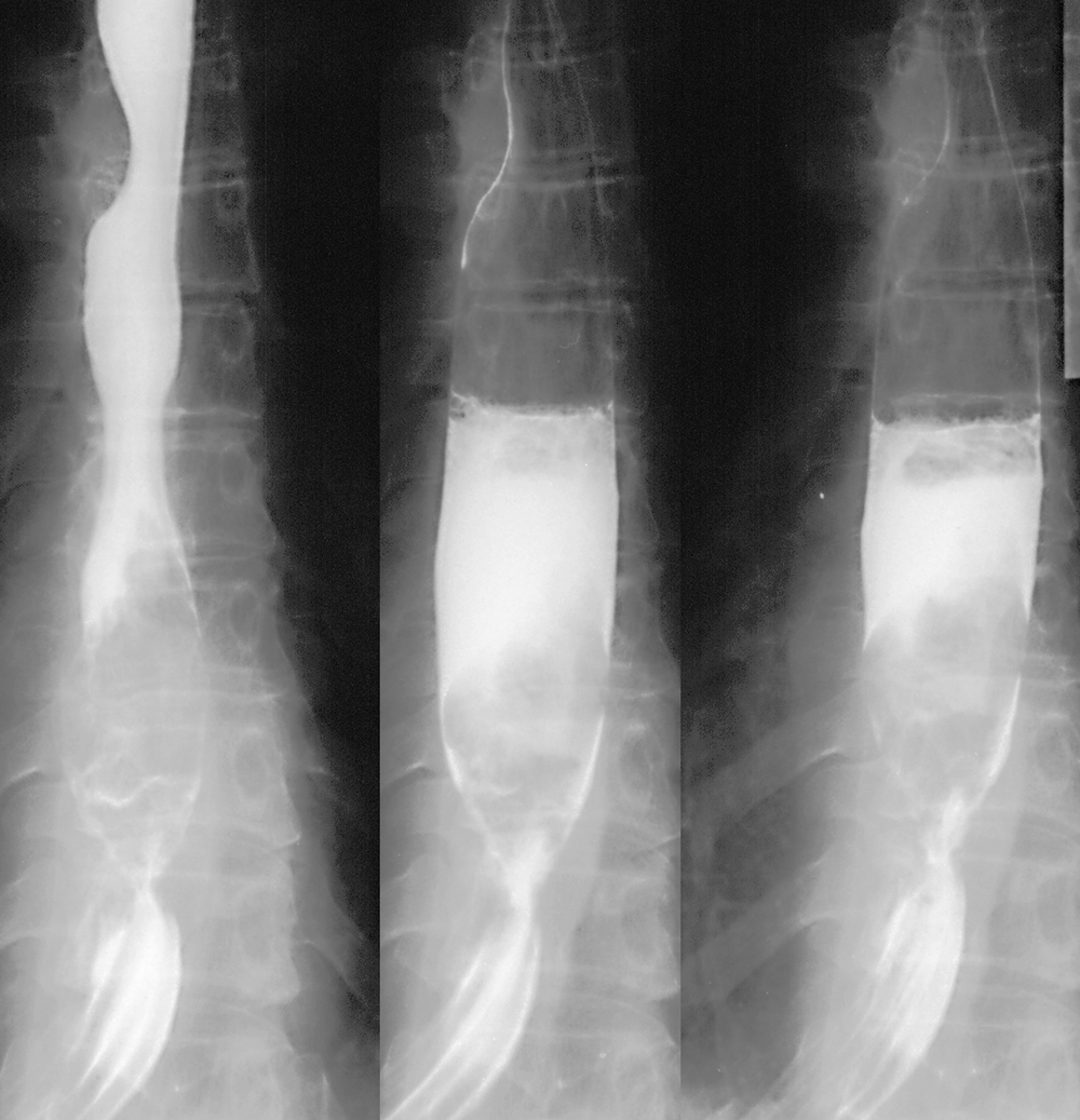
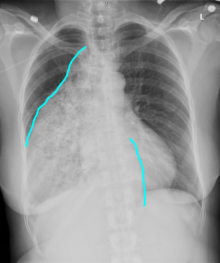
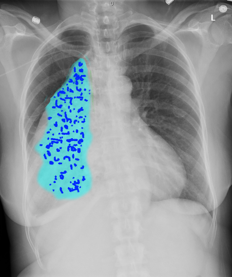
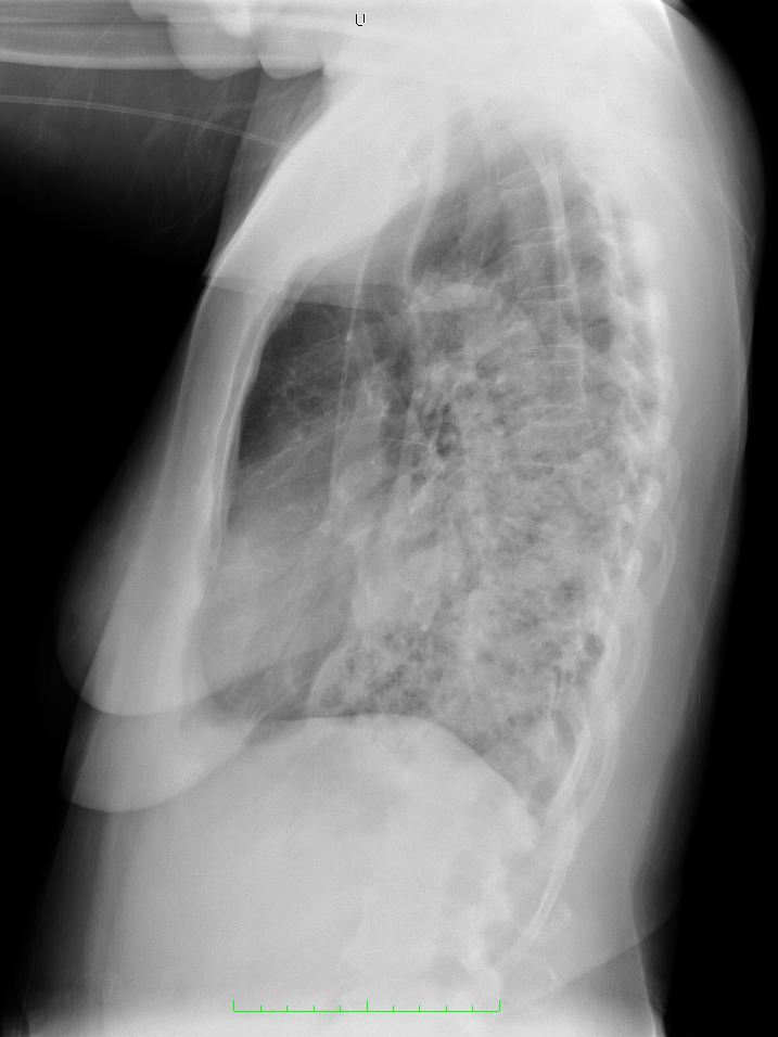
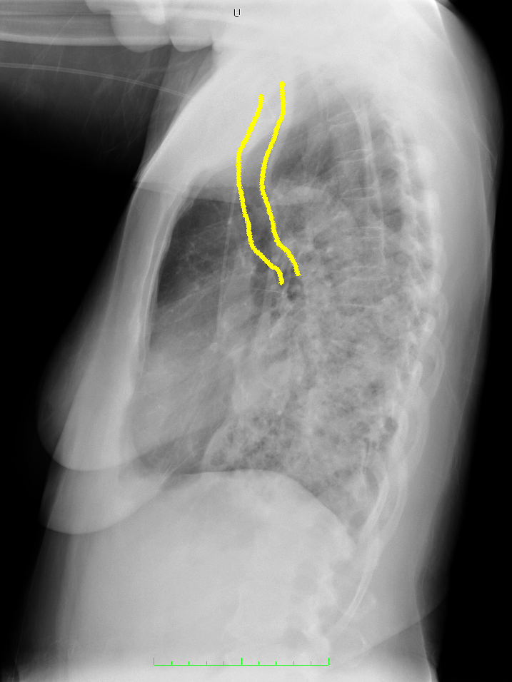
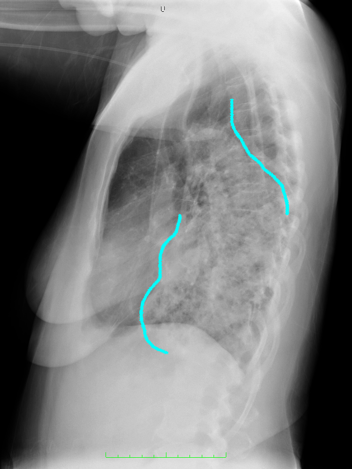
This patient has a long history of dysphagia.
Question 2:
What other study might be useful in this patient?
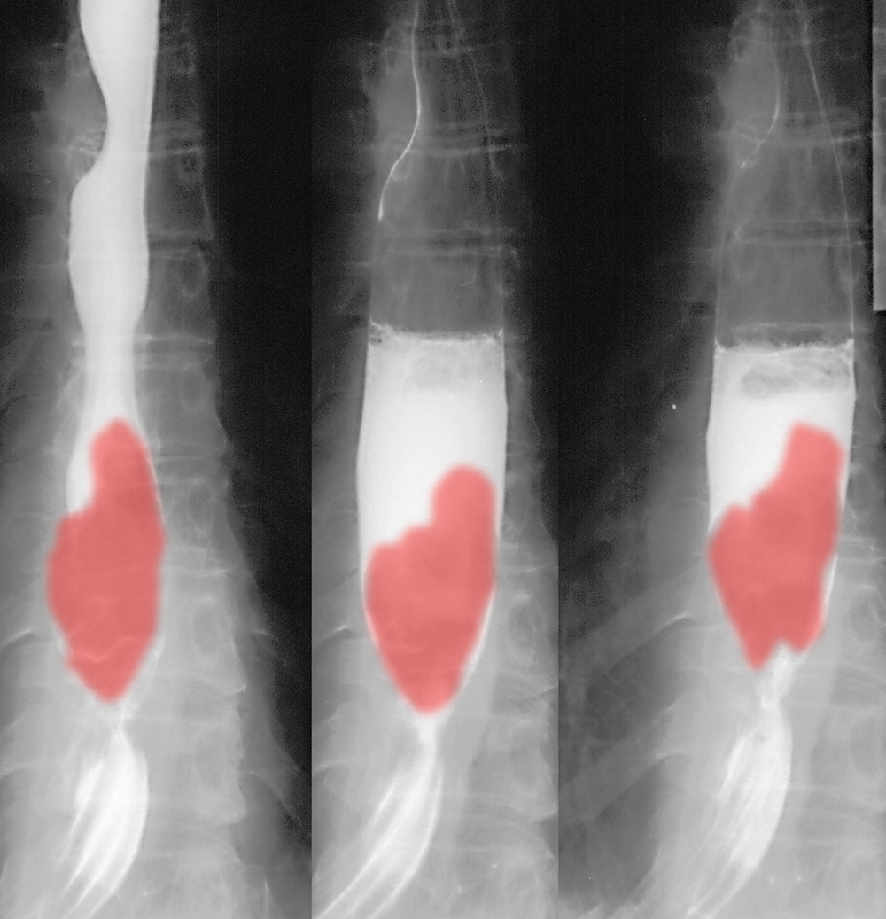
Barium swallow might be performed, but likely a CT scan will better demonstrate the associated findings. This is a patient with long-standing achalasia, a motility disorder of the lower esophageal sphincter. The speckled appearance is produced by chewed food and debris collecting in the severely dilated esophagus.

Case 1-esophageal disease
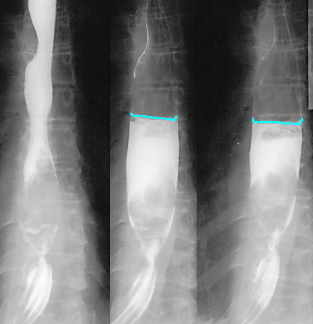
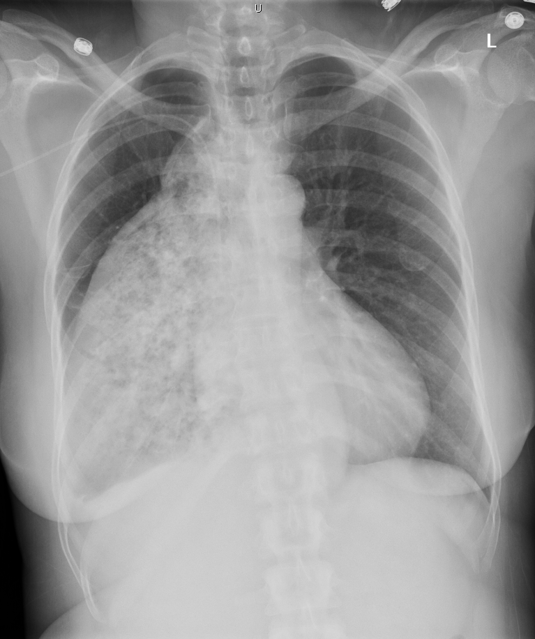
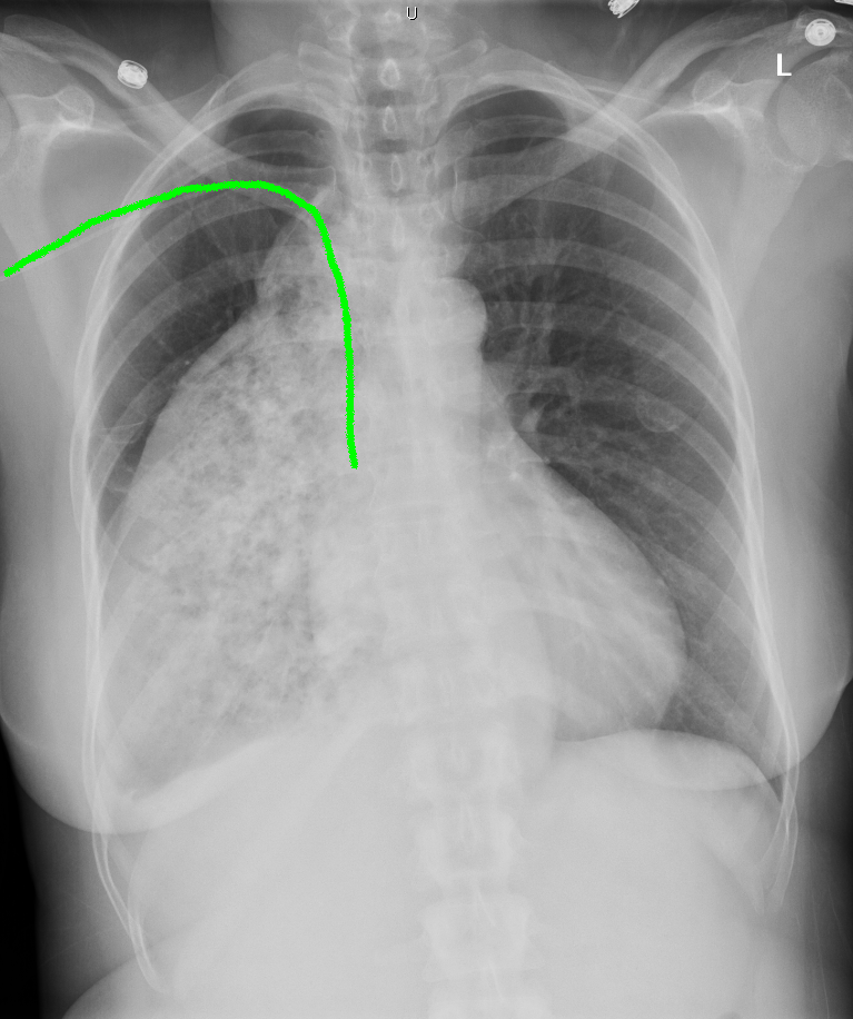
This is another patient with dysphagia.
Question 3:
a) Is an air fluid level normal to see in the esophagus?
No, an air fluid level usually indicates some degree of obstruction. While air and fluid are often swallowed, the high rate of peristalsis usually prevents formation of an air fluid level. Normally material in the esophagus is stripped out into the stomach quickly.
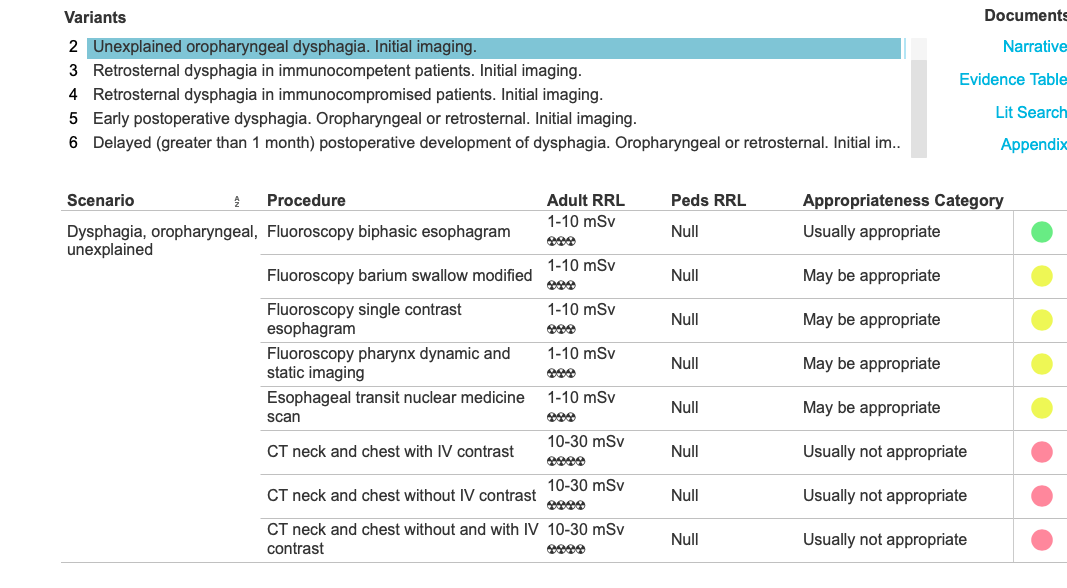
b) What other imaging can be done for dysphagia?
There are many variations on the barium swallow, some focusing on the pharynx with videotaping of the actual swallowing mechanism at the level of the larynx. As noted in the ACR document link, some sort of barium swallow is often done before other types of studies, such as cross-sectional imaging.
