

Case 2-peptic ulcer and stomach disease
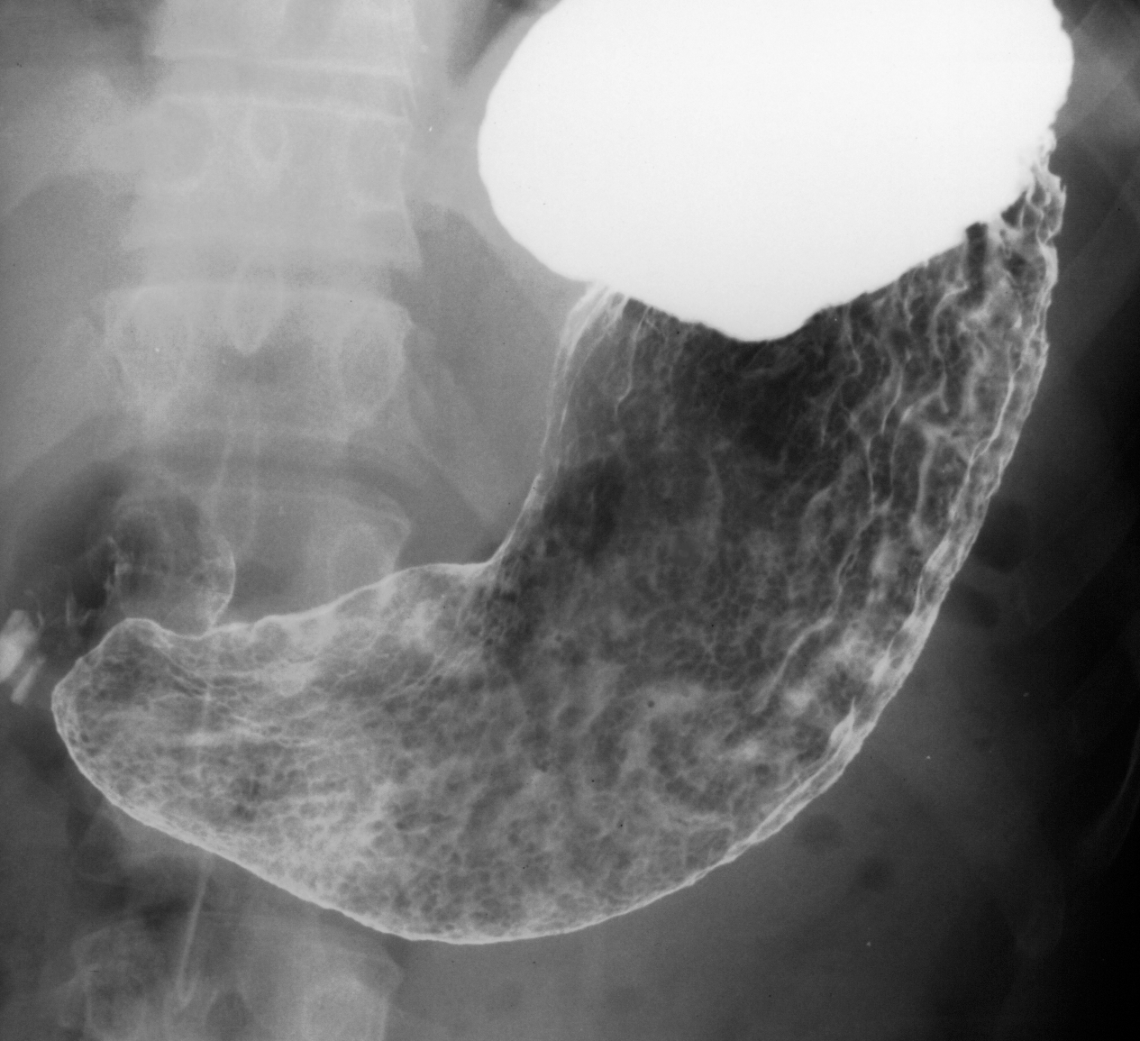
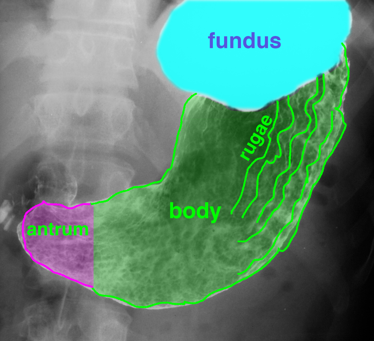
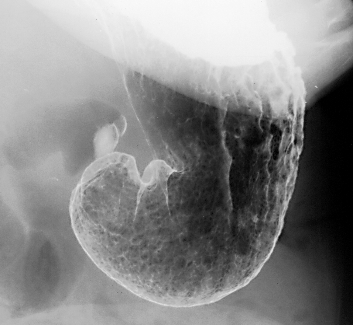
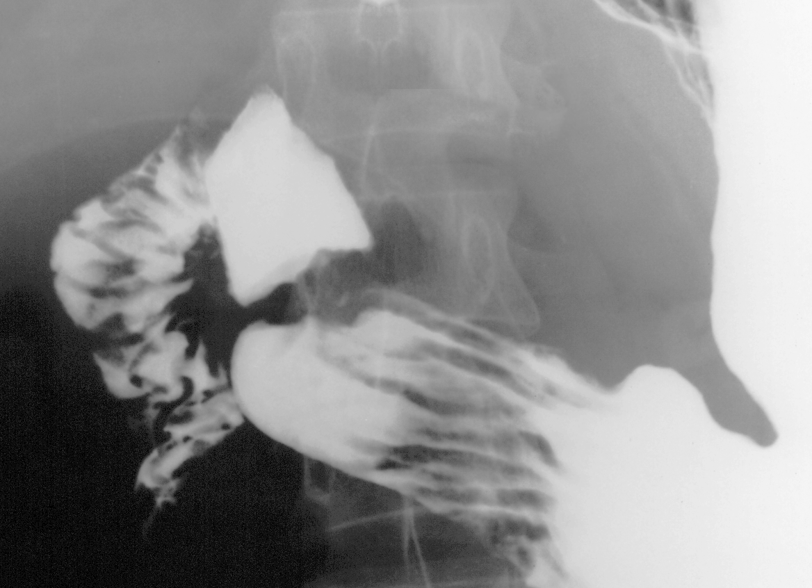
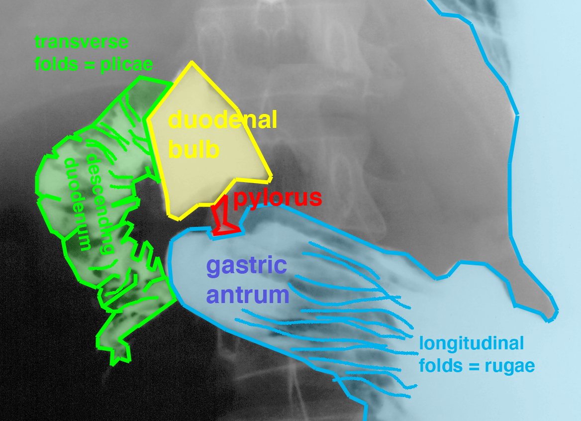
This is a barium fluoroscopic study of the stomach, called an upper GI.
Question 1:
How can you tell if a barium study is single or double contrast?
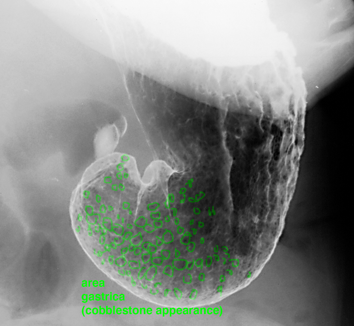
Double contrast means that something dense and something lucent are used to give a 'see-through' view of the GI structures. Usually the dense material is barium and the lucent material is gas. For patients who are more acutely ill, sometimes only barium is used, which will give a dense outline of the organ, but you will not be able to see through it very easily. This can show gross abnormalities of shape or positions as well as leakage, but will not show subtle mucosal abnormalities as well. But single contrast studies are faster to perform and do not require as much cooperation from patients in terms of positioning. Notice that the stomach mucosa is NOT smooth like the esophagus but has a cobblestone appearance due to slightly raised areas, called 'area gastric'.
Case 2-peptic ulcer and stomach disease
This patient presents with indigestion and upper abdominal pain.
Question 2:
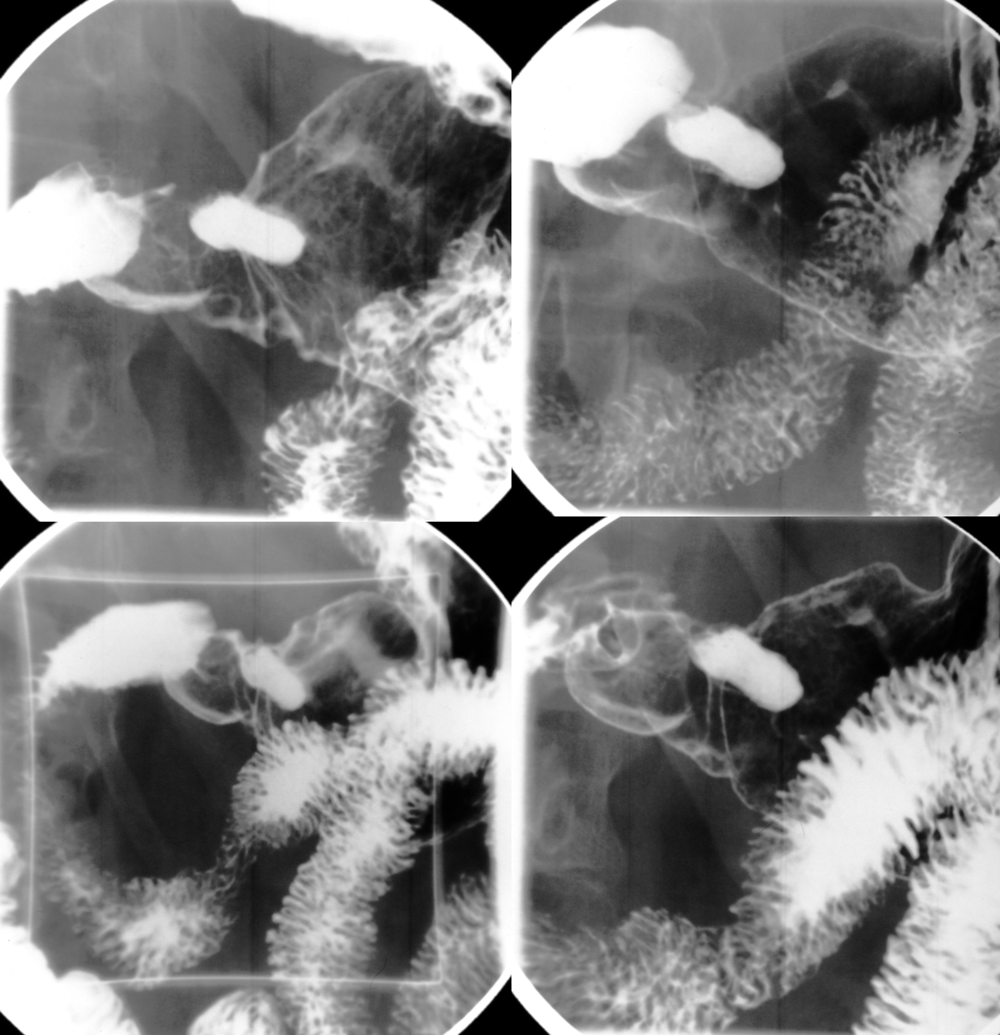
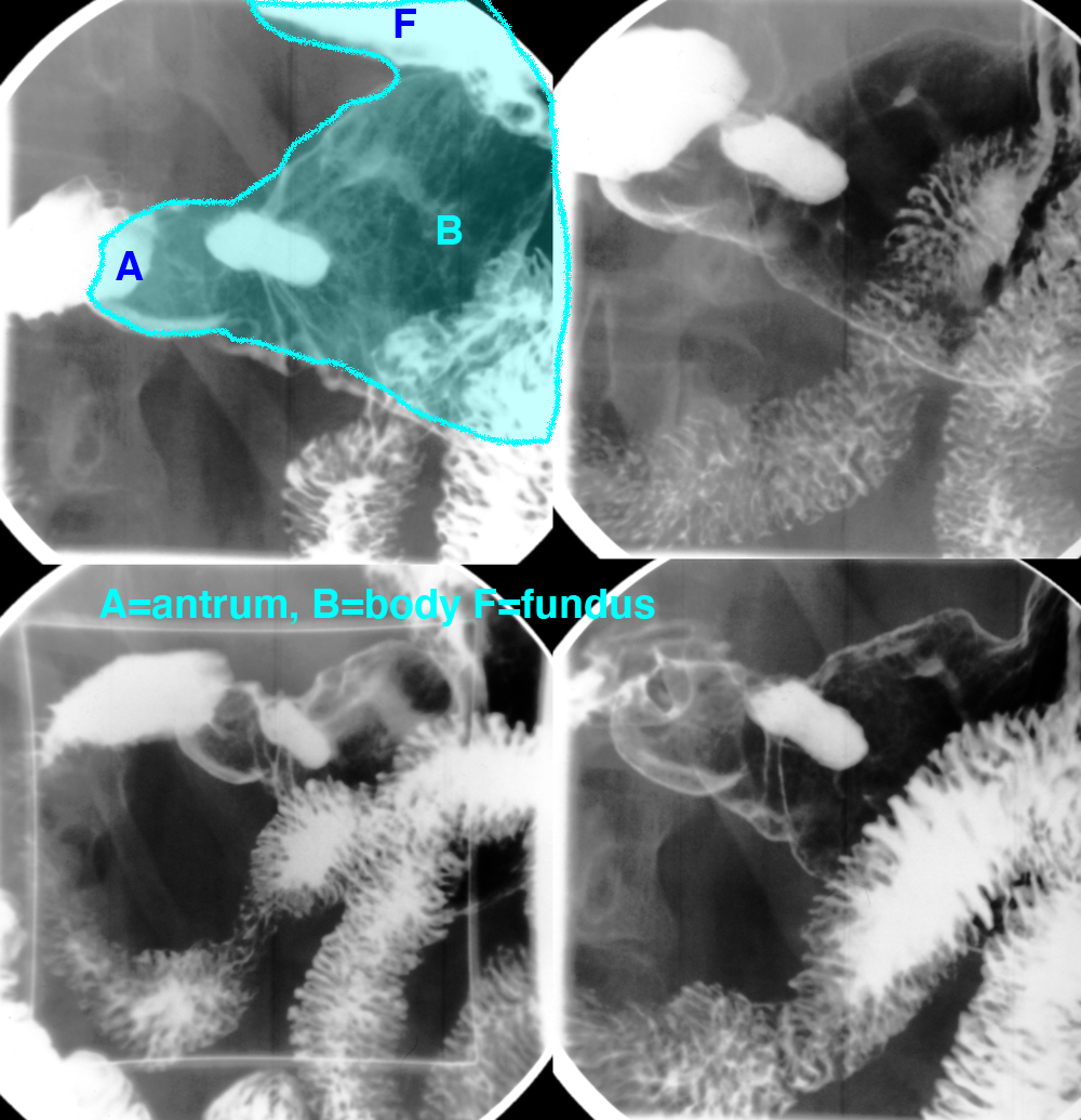
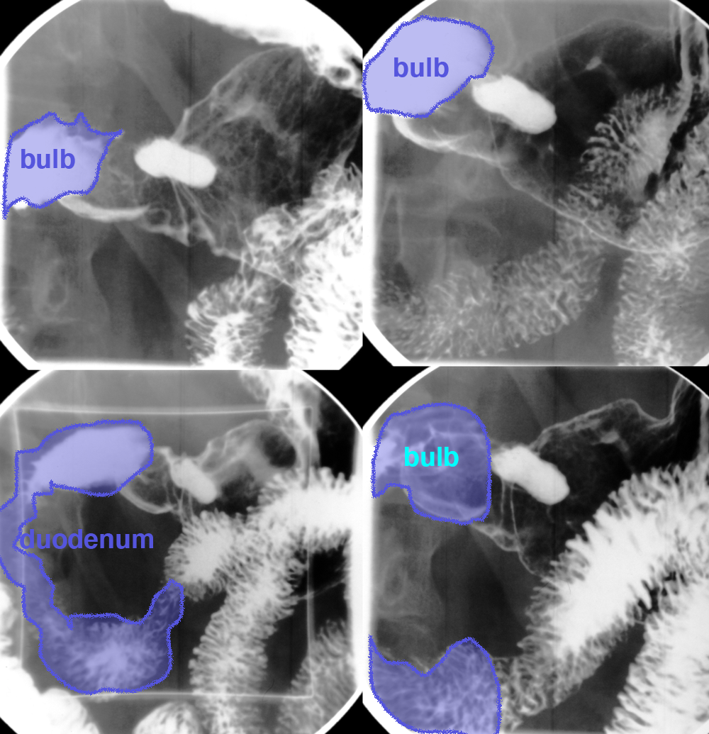
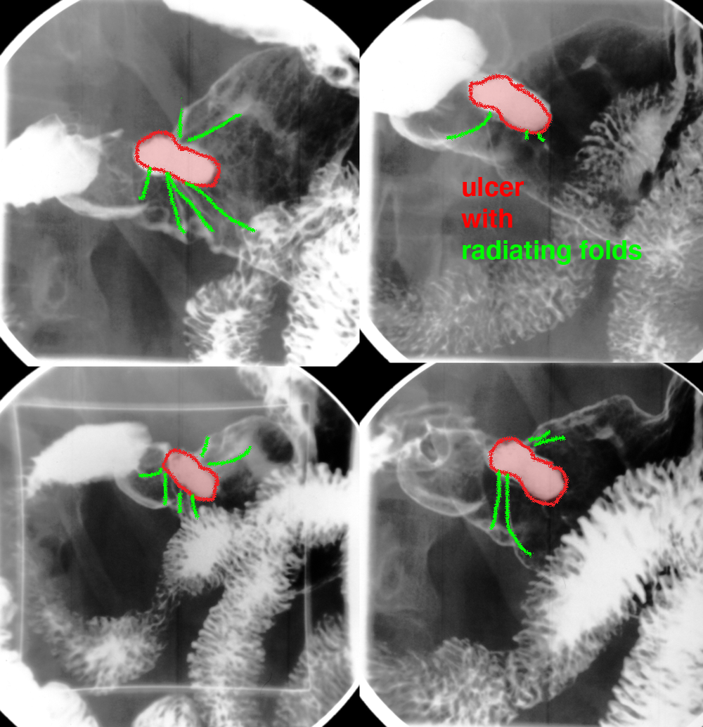
a) What are these images? How can you tell the ulcer from the duodenal bulb?
These are 'spot' views from an upper GI study, double contrast with barium and air. They are like snapshots of what is seen in real time with fluoroscopy. The ulcer retains contrast (appears unchanging and white) on various different views with the patient turned in different directions. On some of the images, the duodenal bulb also appears white (when gravity fills it with barium) but on others the bulb is filled with gas. An ulcer typically fills with barium that stays relatively unchanging as the patient moves around, as the ulcer crater captures and holds the barium.
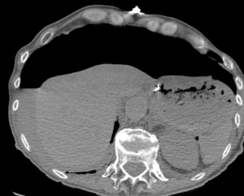
b) What complication of gastric ulcer is shown on the CT image?
An ulcer can perforate through the wall of the stomach, and let gastric contents and air enter the peritoneal cavity. The CT image shows a large amount of air in the peritoneal cavity, with a small amount of fluid in a dependent position along the liver. The high attenuation (white) area in the stomach is the tip of an NG tube.
Case 2-peptic ulcer and stomach disease
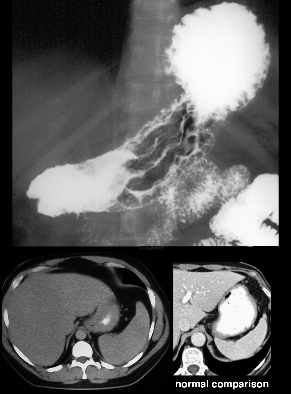
Patient with vague epigastric pain and weight loss.
Question 3:
What is the term for a stomach that has lost distensibility, as in this case?
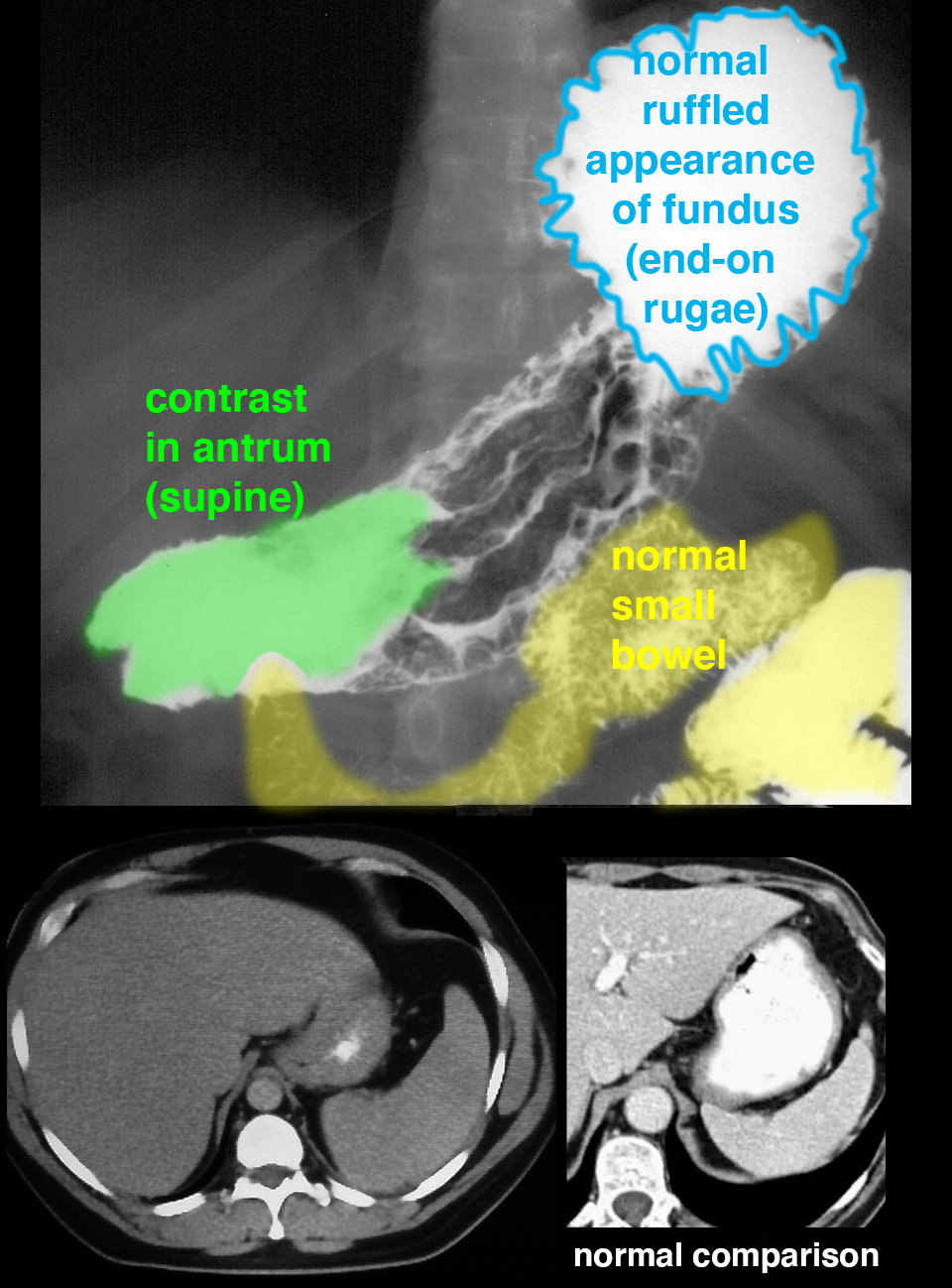
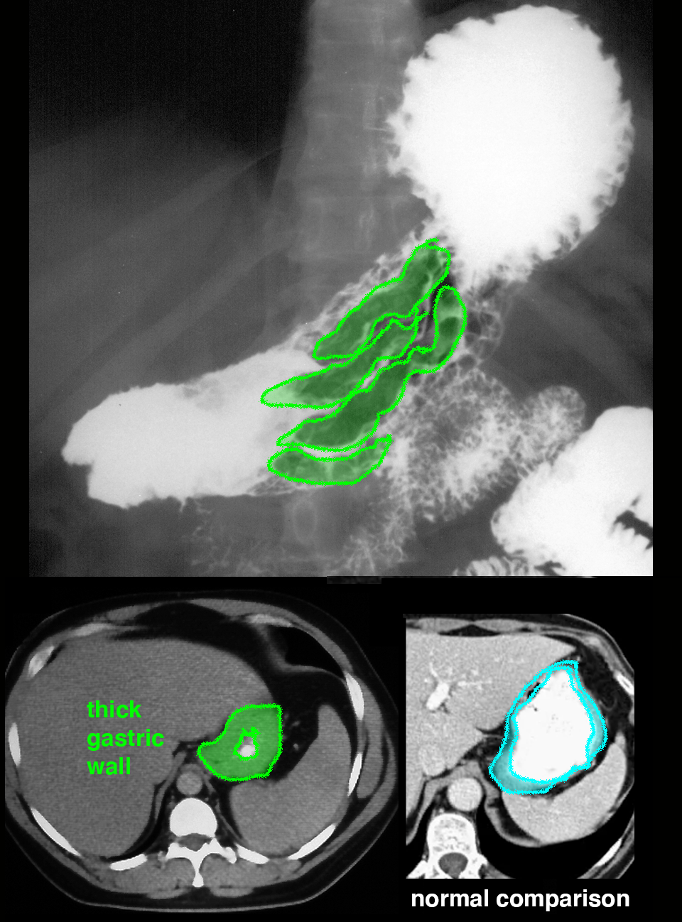
This stomach has very thick rugae and does not distend as much as normal in this upper GI image. The accompanying CT image also shows diffuse thickening of the gastric wall, compared to the adjacent normal image. This stiff stomach is termed linitis plastica and can be seen due to involvement by a type of adenocarcinoma of the stomach with an infiltrative pattern of growth. There is pooling of contrast in the fundus and antrum on this supine view. The ruffled appearance of the fundus is normal, due to rugal folds seen end-on.
