

Case 3-inflammatory bowel disease
This image is from another type of contrast study of the GI tract, called a barium enema.
Question 1:
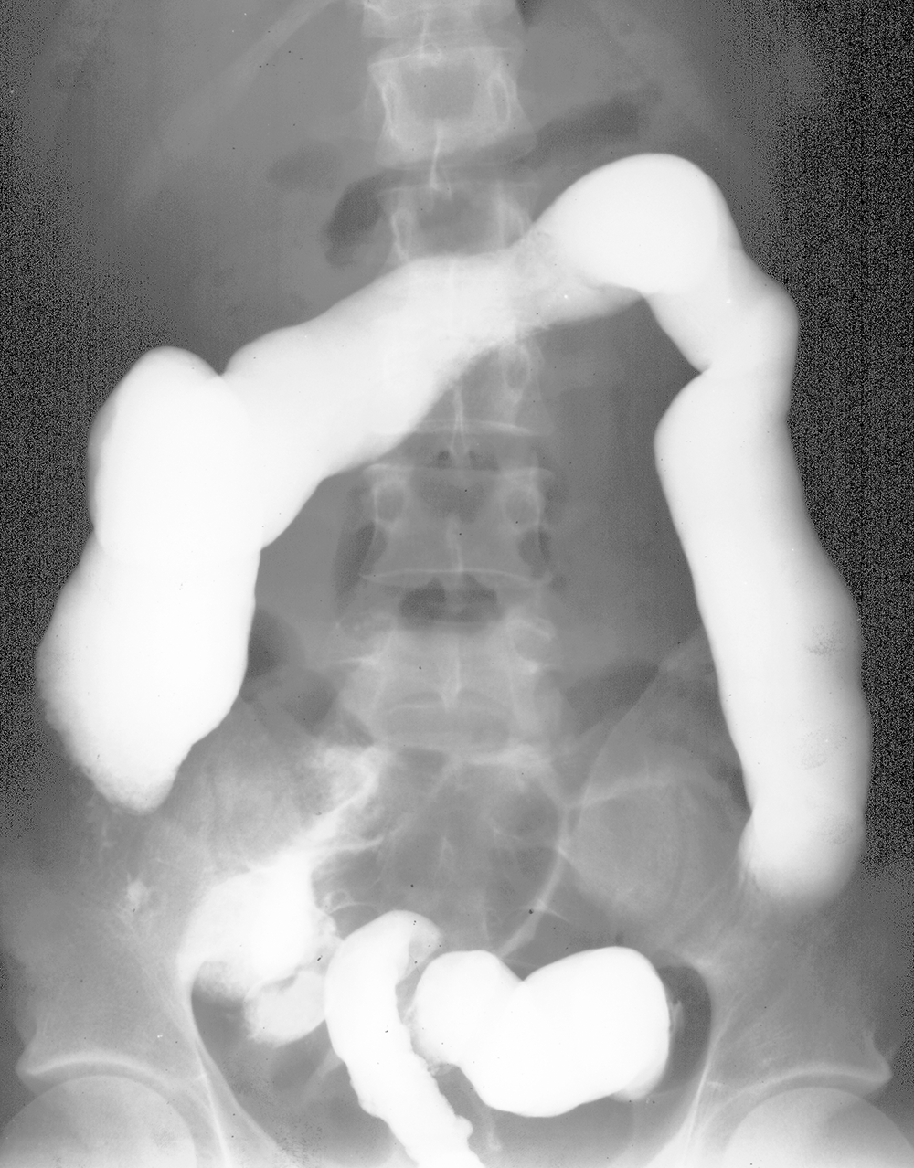
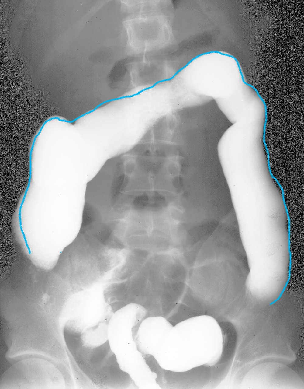
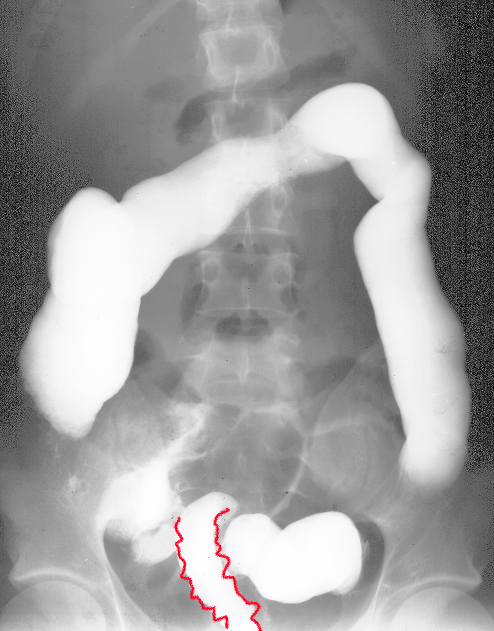
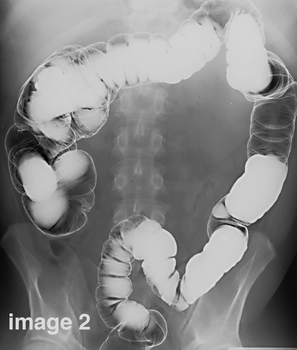
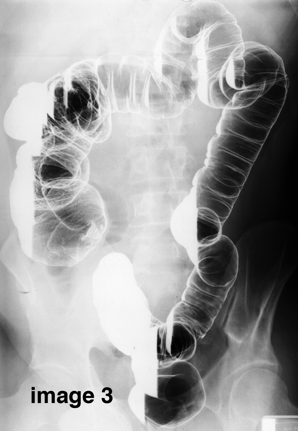
Try to determine the patient's position for each of the three images shown from this double-contrast barium enema.
×
Answer:
Image 1 is supine. You can tell this because most of the heavy barium is located in the most posterior parts of the colon--the ascending and descending portions, which are secondarily retroperitoneal. Image 2 is prone. You can tell this because the barium has moved into the most anterior part of the cooling--the transverse colon. Image 3 is decubitus with the patient's right side down and with barium pooling in the right parts of the colon.
Image 1 is supine. You can tell this because most of the heavy barium is located in the most posterior parts of the colon--the ascending and descending portions, which are secondarily retroperitoneal. Image 2 is prone. You can tell this because the barium has moved into the most anterior part of the cooling--the transverse colon. Image 3 is decubitus with the patient's right side down and with barium pooling in the right parts of the colon.
Case 3-inflammatory bowel disease
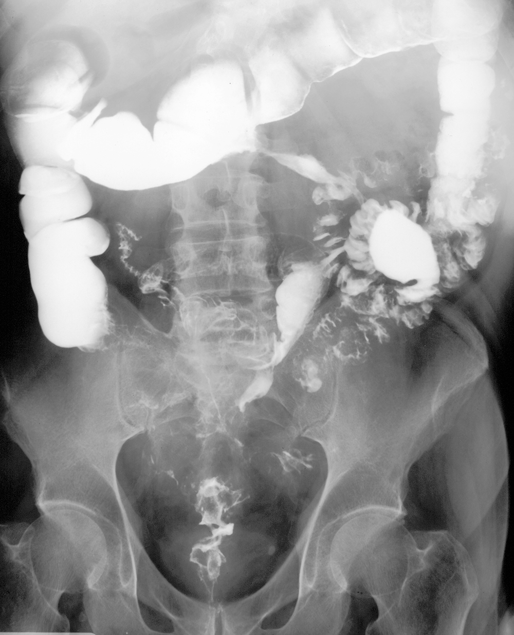
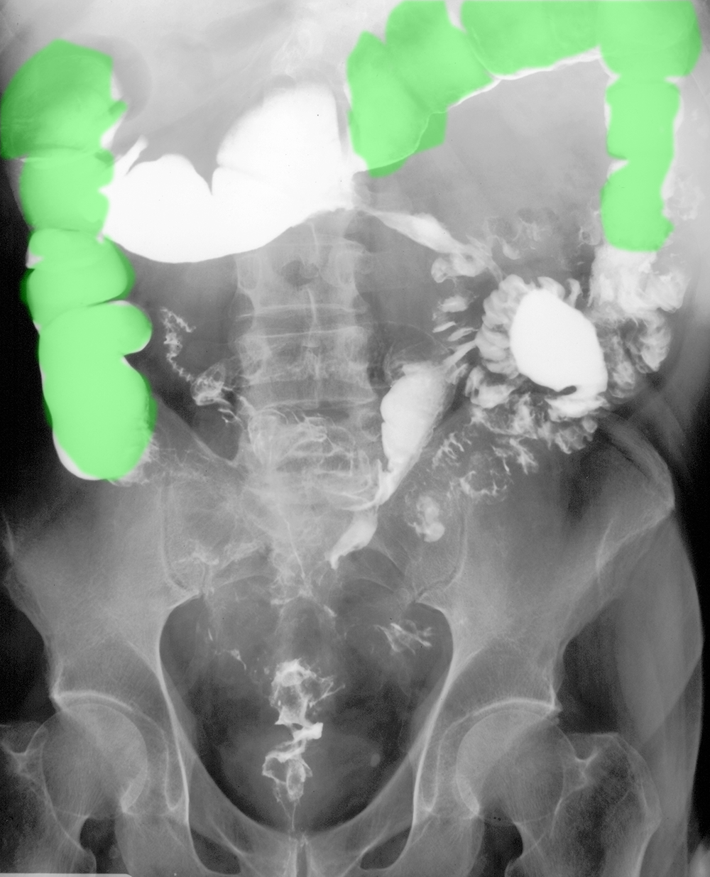
This is a study of a patient with long-standing diarrhea and abdominal pain.
Question 2:
What is this study? What imaging features favor Crohn's disease?
×
Answer:
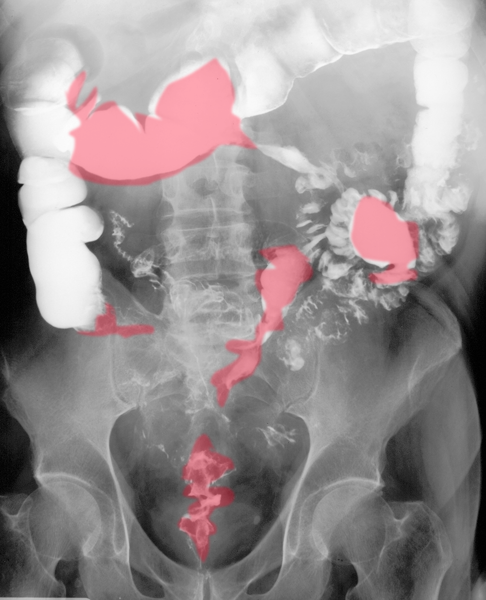
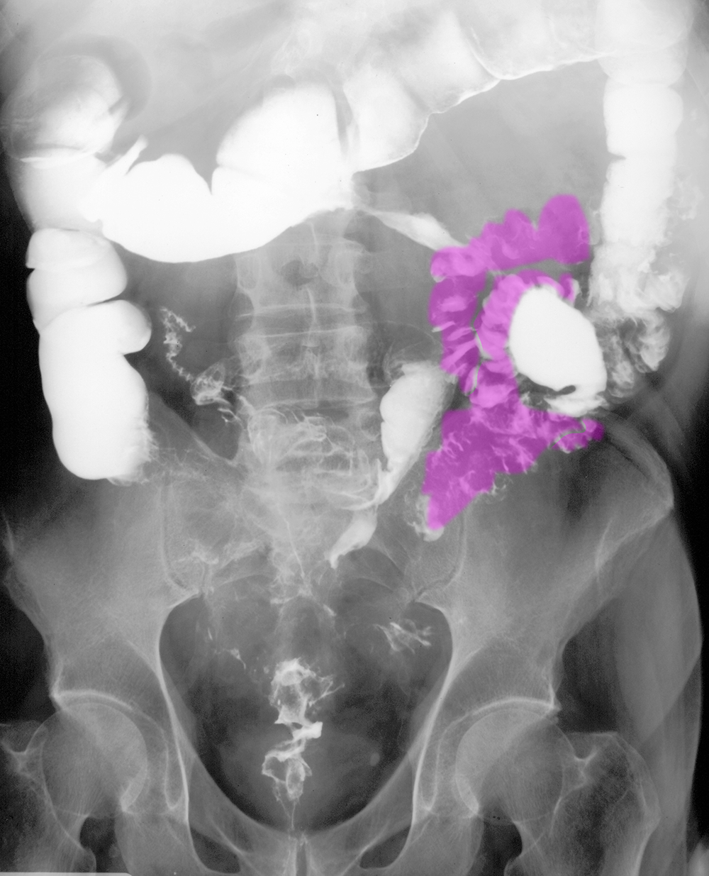
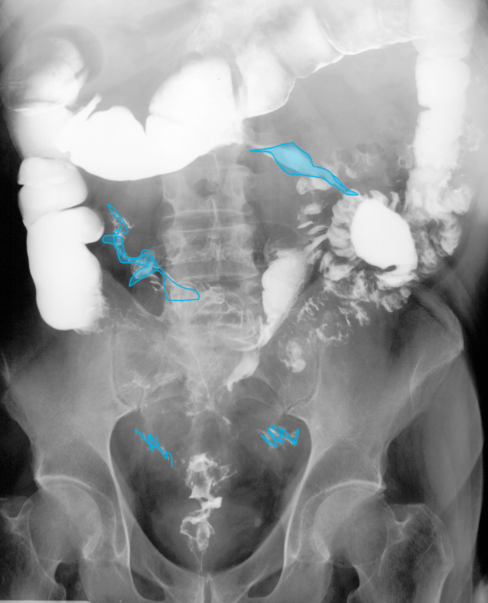
This is a single contrast barium enema. There is only barium primarily in the colon with very little gas. As shown with the labels, there are segments of normal appearing colon and very abnormal segments. The involvement of the disease is discontinuous. There are also fistulae (abnormal channels) connecting various areas, with contrast in the left small bowel loops that filled from contrast in fistulae.
This is a single contrast barium enema. There is only barium primarily in the colon with very little gas. As shown with the labels, there are segments of normal appearing colon and very abnormal segments. The involvement of the disease is discontinuous. There are also fistulae (abnormal channels) connecting various areas, with contrast in the left small bowel loops that filled from contrast in fistulae.
Case 3-inflammatory bowel disease
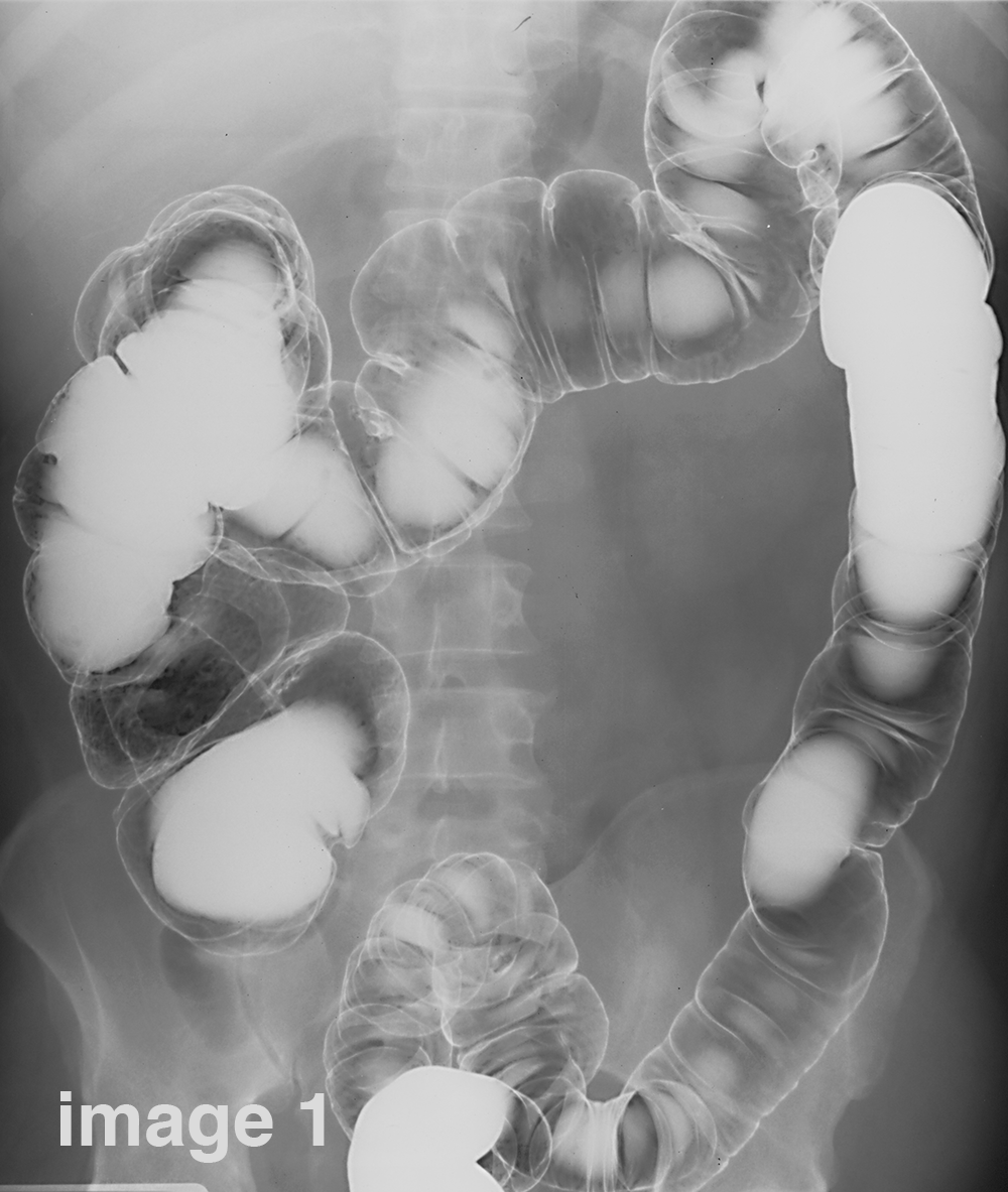
Patient with chronic diarrhea and weight loss.
Question 3:
How would you describe the appearance of the colon on this study?
×
Answer:
This is again a barium enema. The entire colon is unusually smooth in contour with no normal haustra. The overall caliber seems a bit small especially in the sigmoid and the margins of the colon in this region appear shaggy, likely representing ulceration. The continuous involvement without skip areas is more typical of UC than Crohn's.
This is again a barium enema. The entire colon is unusually smooth in contour with no normal haustra. The overall caliber seems a bit small especially in the sigmoid and the margins of the colon in this region appear shaggy, likely representing ulceration. The continuous involvement without skip areas is more typical of UC than Crohn's.
