


Case 4-GI parasites
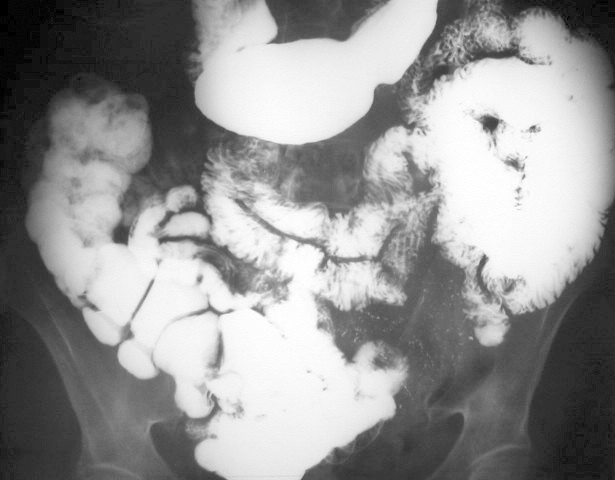
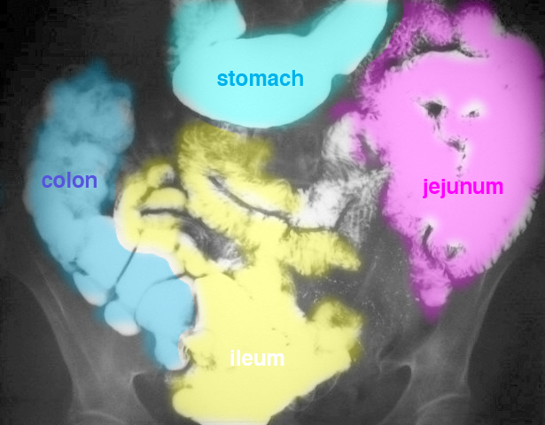
This is another normal study in a patient with abdominal pain.
Question 1:
What is this study and how is it done?
×
Answer:
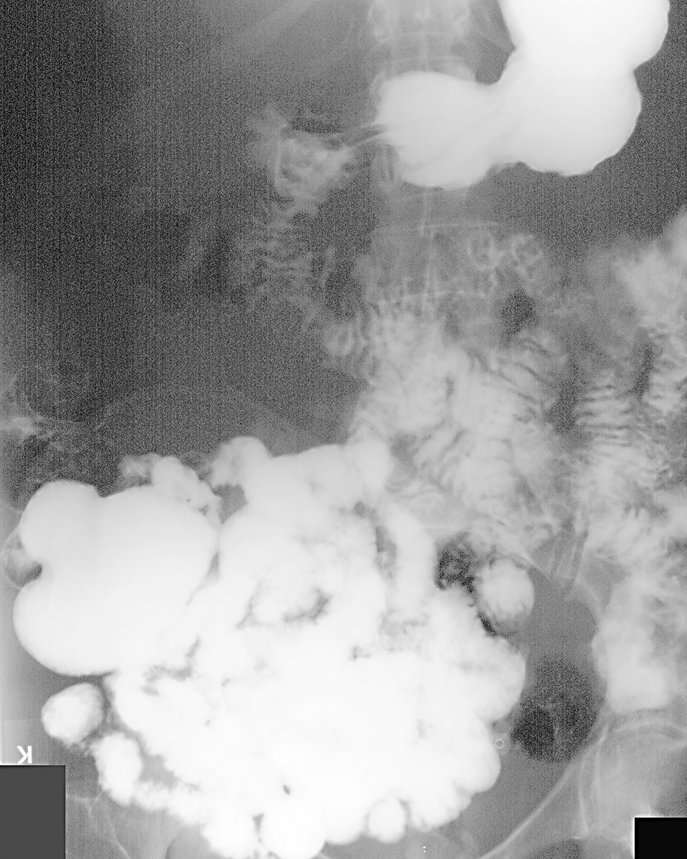
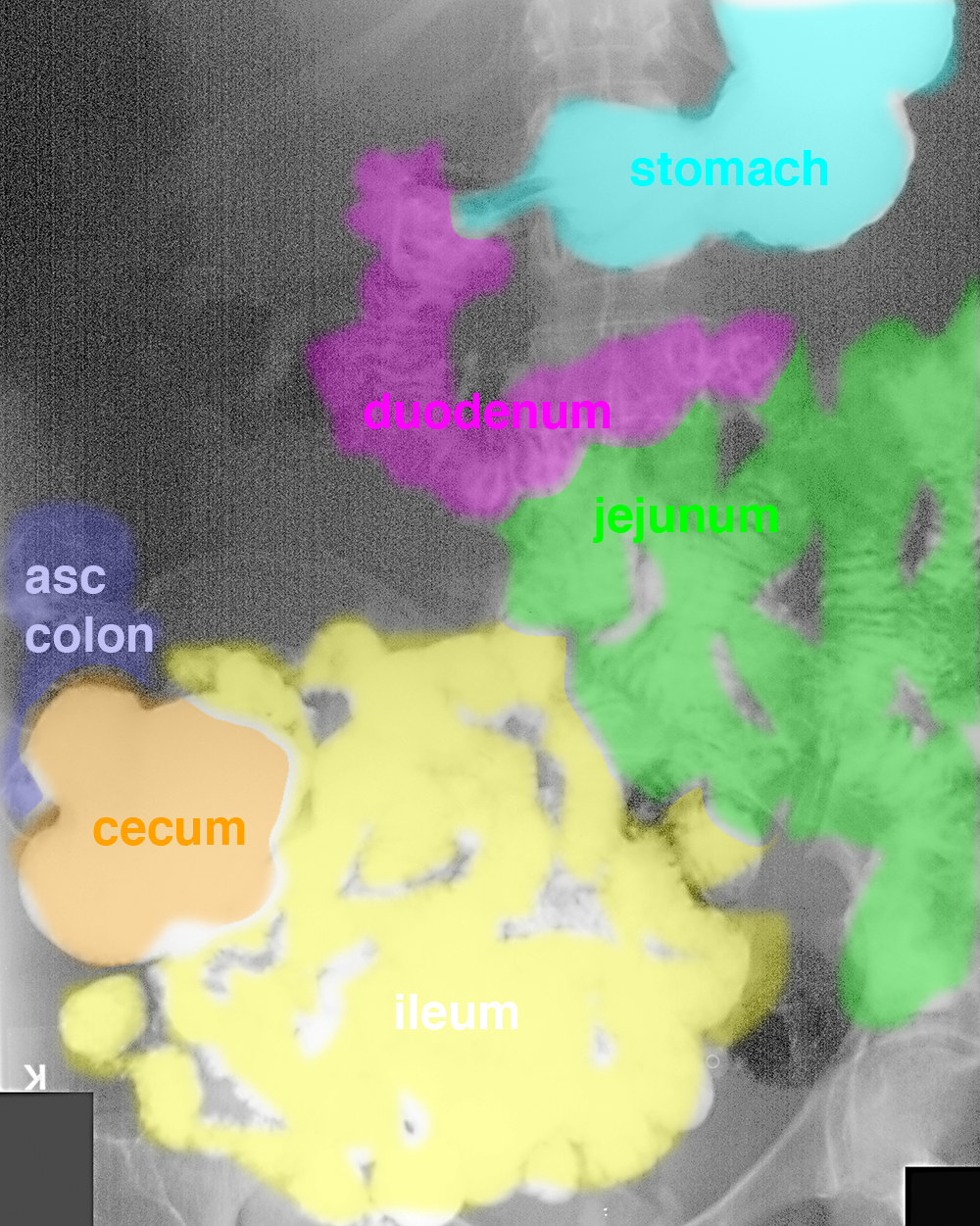
This is a small bowel follow-through. In this study, a patient is gradually given oral barium to drink over time, and serial fluoroscopy with spot films are obtained as the contrast moves through the small bowel. It is not a double contrast study, since only barium is used. There is often a lot of overlap of loops making it hard to detect small or focal abnormalities.
This is a small bowel follow-through. In this study, a patient is gradually given oral barium to drink over time, and serial fluoroscopy with spot films are obtained as the contrast moves through the small bowel. It is not a double contrast study, since only barium is used. There is often a lot of overlap of loops making it hard to detect small or focal abnormalities.
Case 4-GI parasites
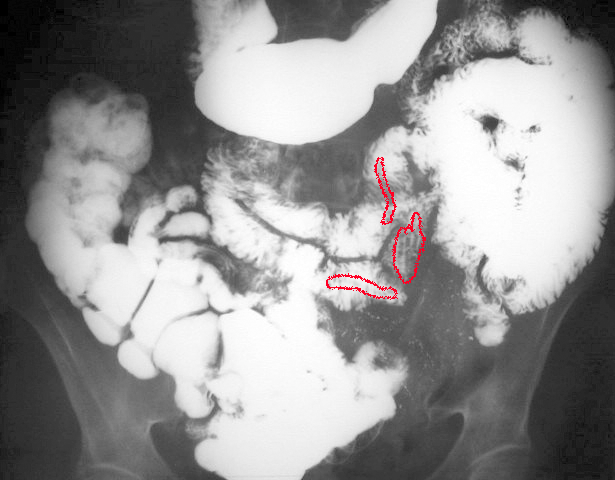
Patient with abdominal pain.
Question 2:
What is this study?
×
Answer:
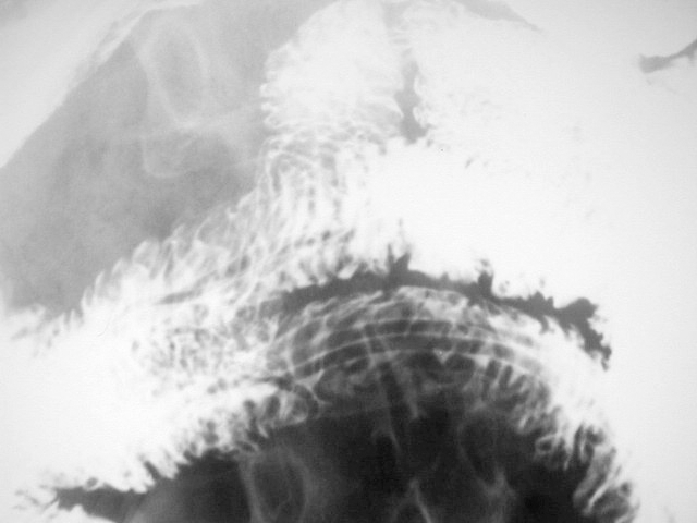
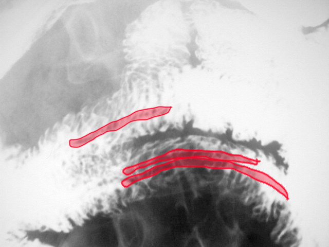
This is another small bowel follow through. It is a single contrast study with lots of overlap. During the study, closeup views are often obtained with gentle abdominal compression to move loops around and get a better look at them. You are also shown one of these closeup views from the central part of the abdomen.
This is another small bowel follow through. It is a single contrast study with lots of overlap. During the study, closeup views are often obtained with gentle abdominal compression to move loops around and get a better look at them. You are also shown one of these closeup views from the central part of the abdomen.
Case 4-GI parasites
Screening mammogram on a patient from Nigeria.
Question 3:
What are these views? What do they show?
×
Answer:
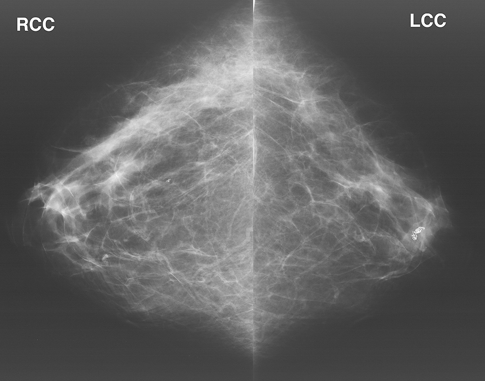
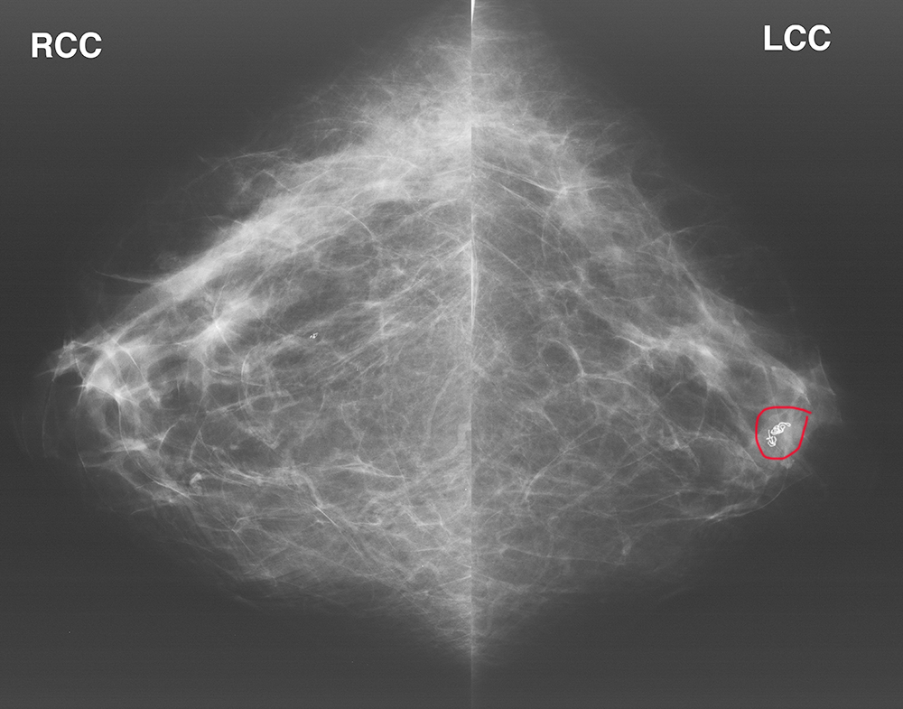
These are CC views, meaning cranio-caudal. That indicates that the beam is coming top-down through the compressed breasts. We typically show the images back-to-back, which helps to identify differences from one side to the other. One of the things we look for is calcifications. Try to find a small white abnormality before checking the label.
These are CC views, meaning cranio-caudal. That indicates that the beam is coming top-down through the compressed breasts. We typically show the images back-to-back, which helps to identify differences from one side to the other. One of the things we look for is calcifications. Try to find a small white abnormality before checking the label.

Case 4-GI parasites
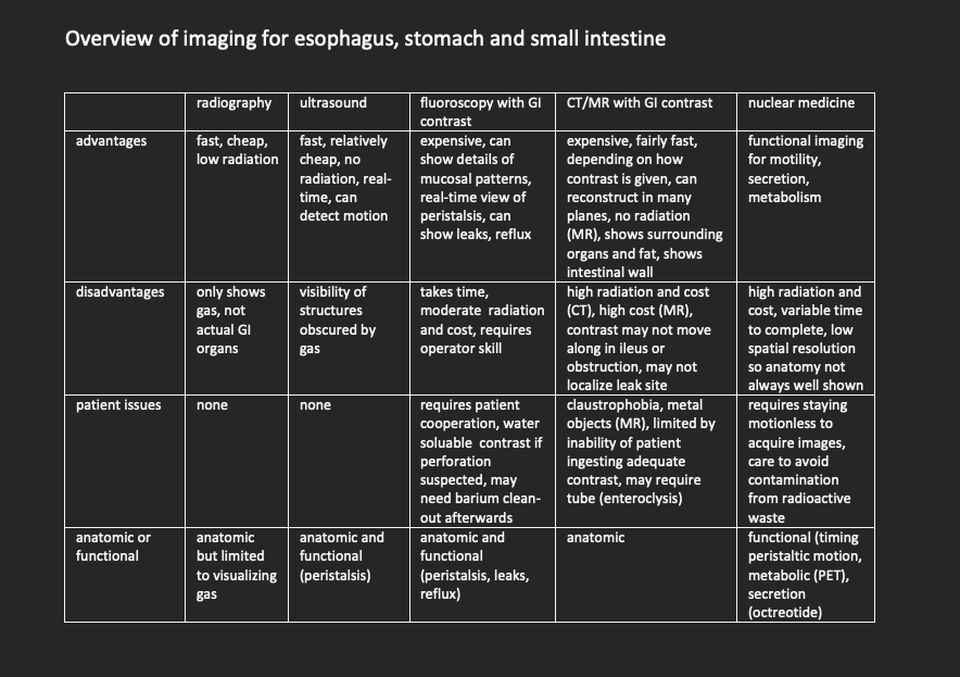
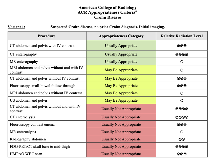
This table shows an overview of various types of imaging for the esophagus, stomach and small intestine. Click the link to see a scenario from the ACR Appropriateness Criteria for imaging Crohn's disease.
Further Explanation: