

Question 1:
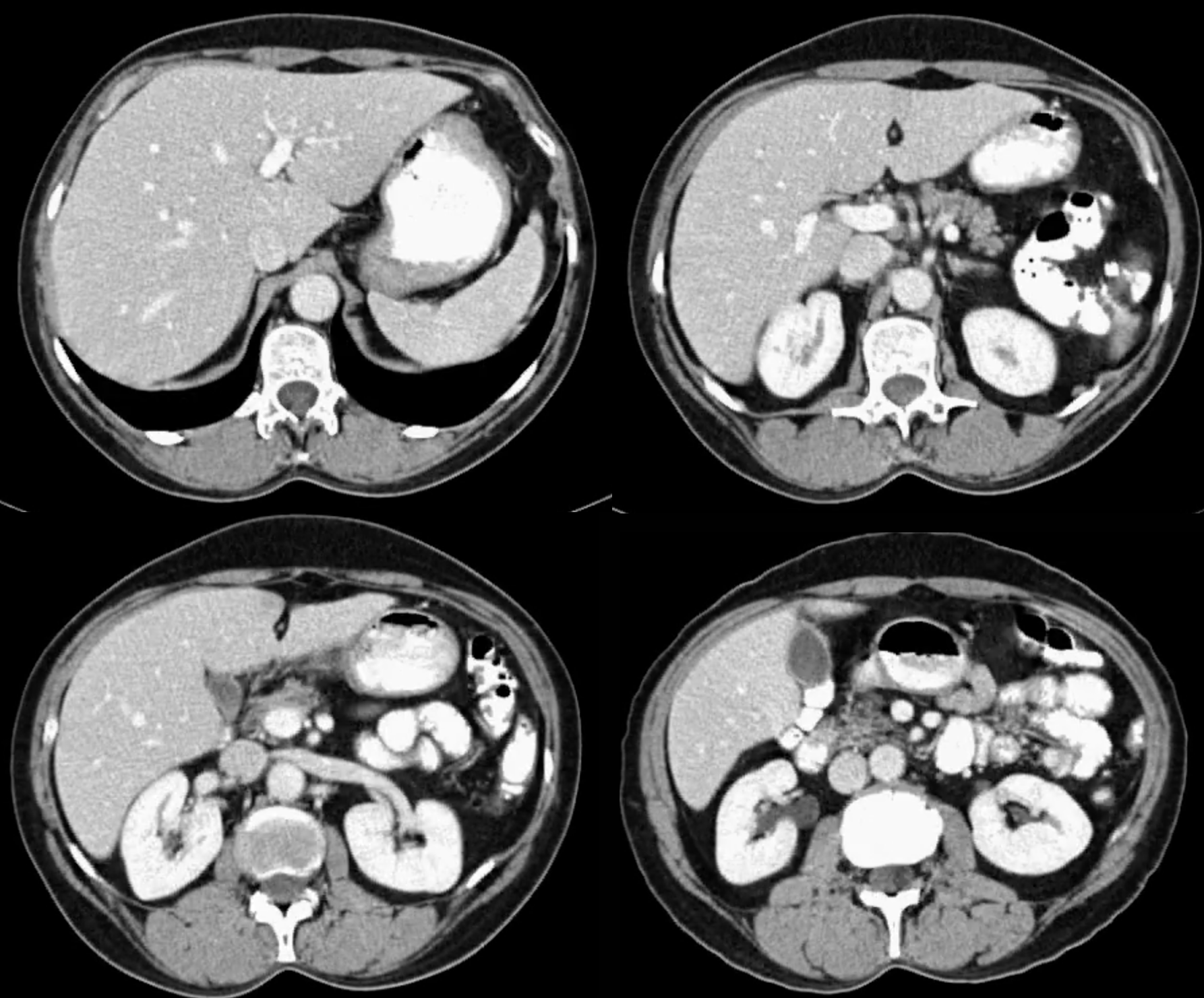
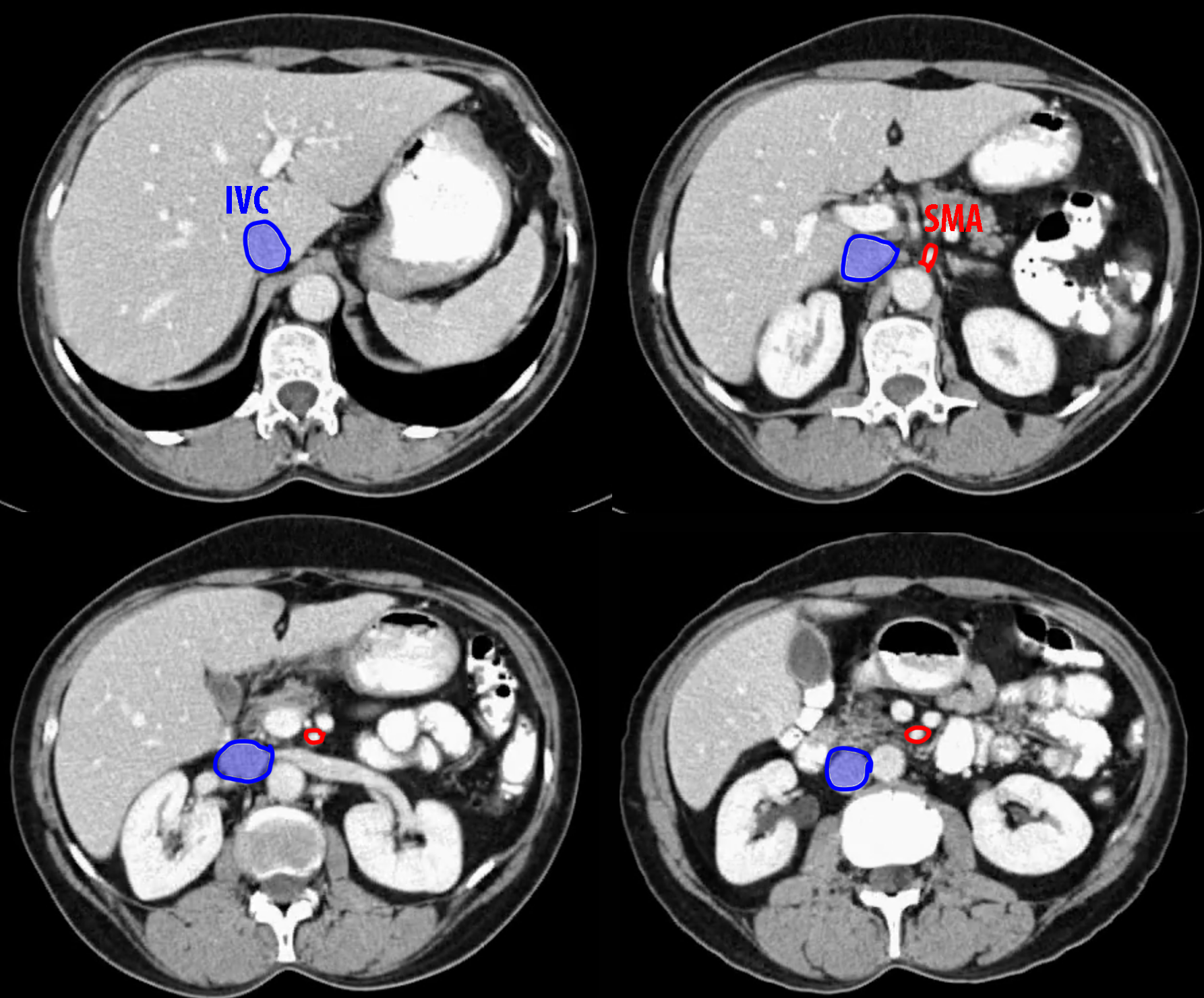
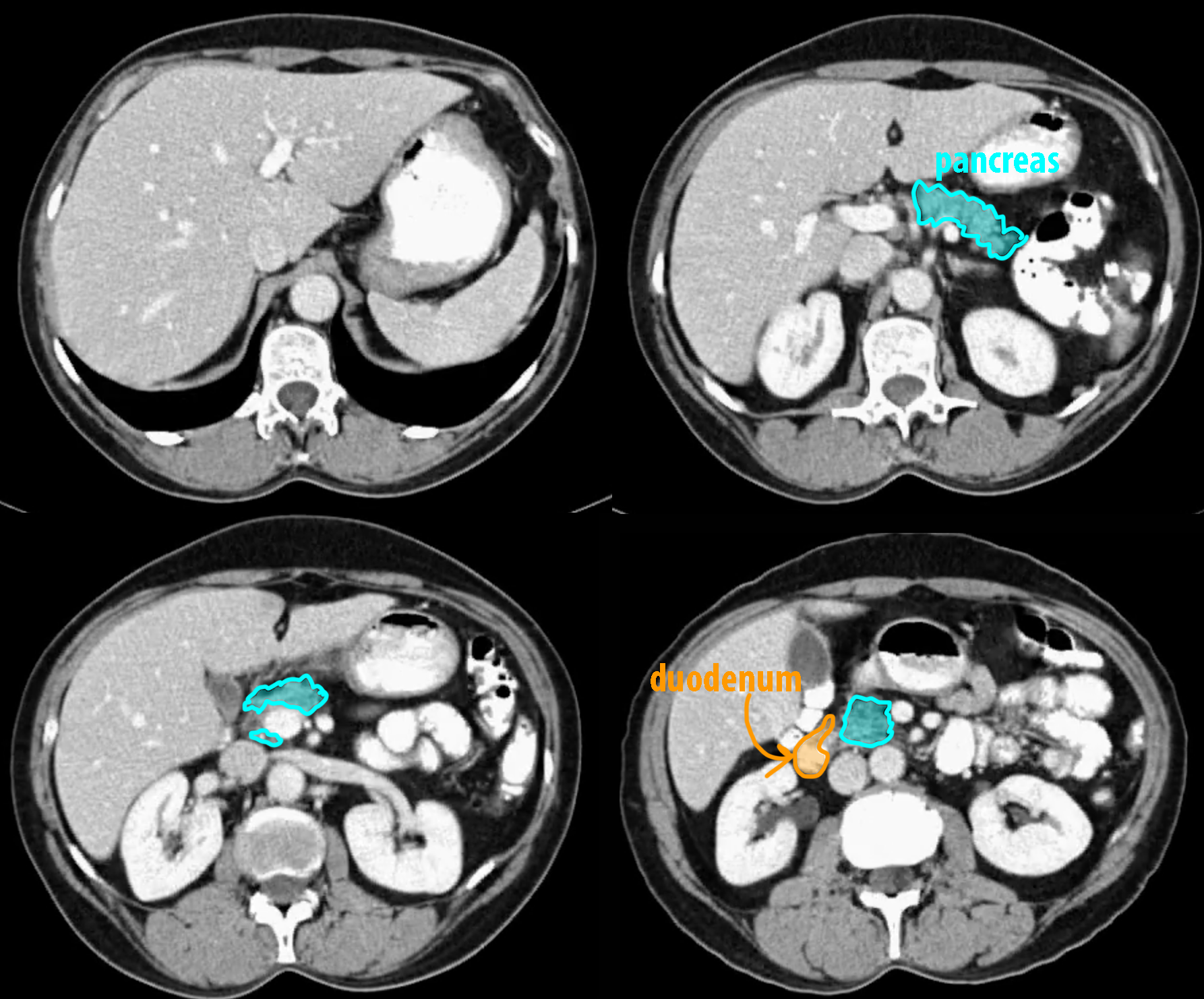
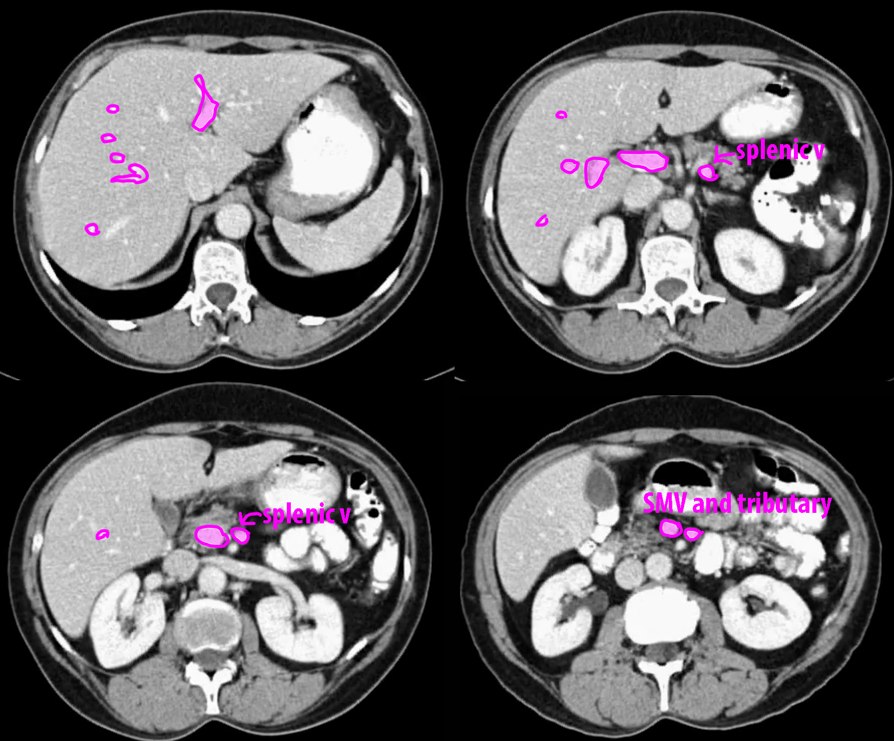
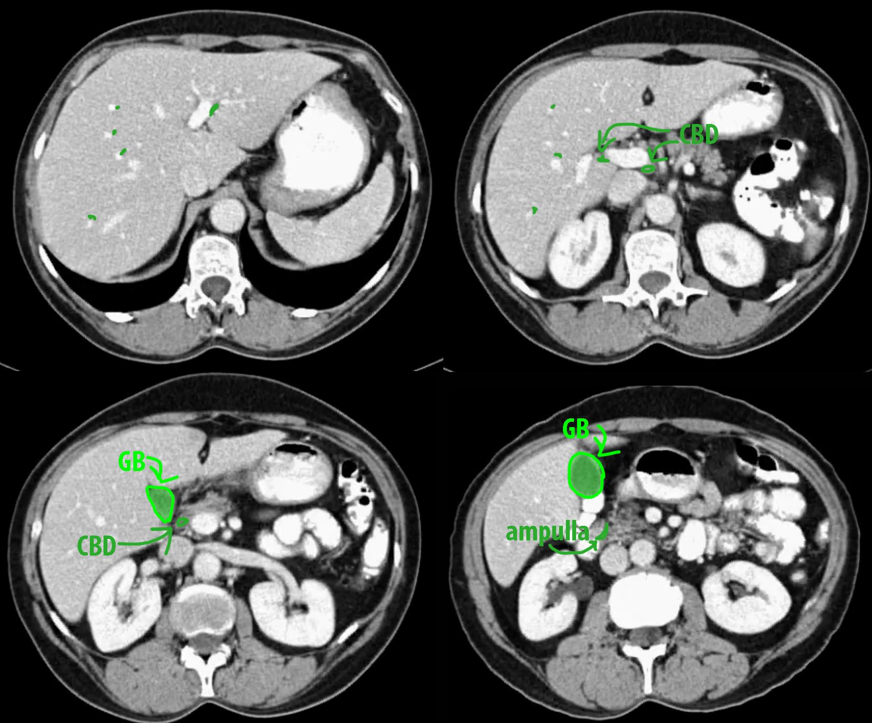
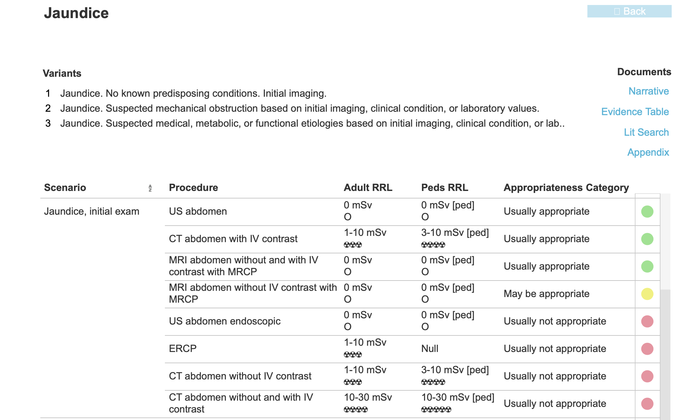
On these four normal abdominal CT images try to identify structures before clicking on labels. What density is bile on CT, and what are the two presented mystery studies? The link AC jaundice shows the Appropriateness Criteria for a patient with jaundice.
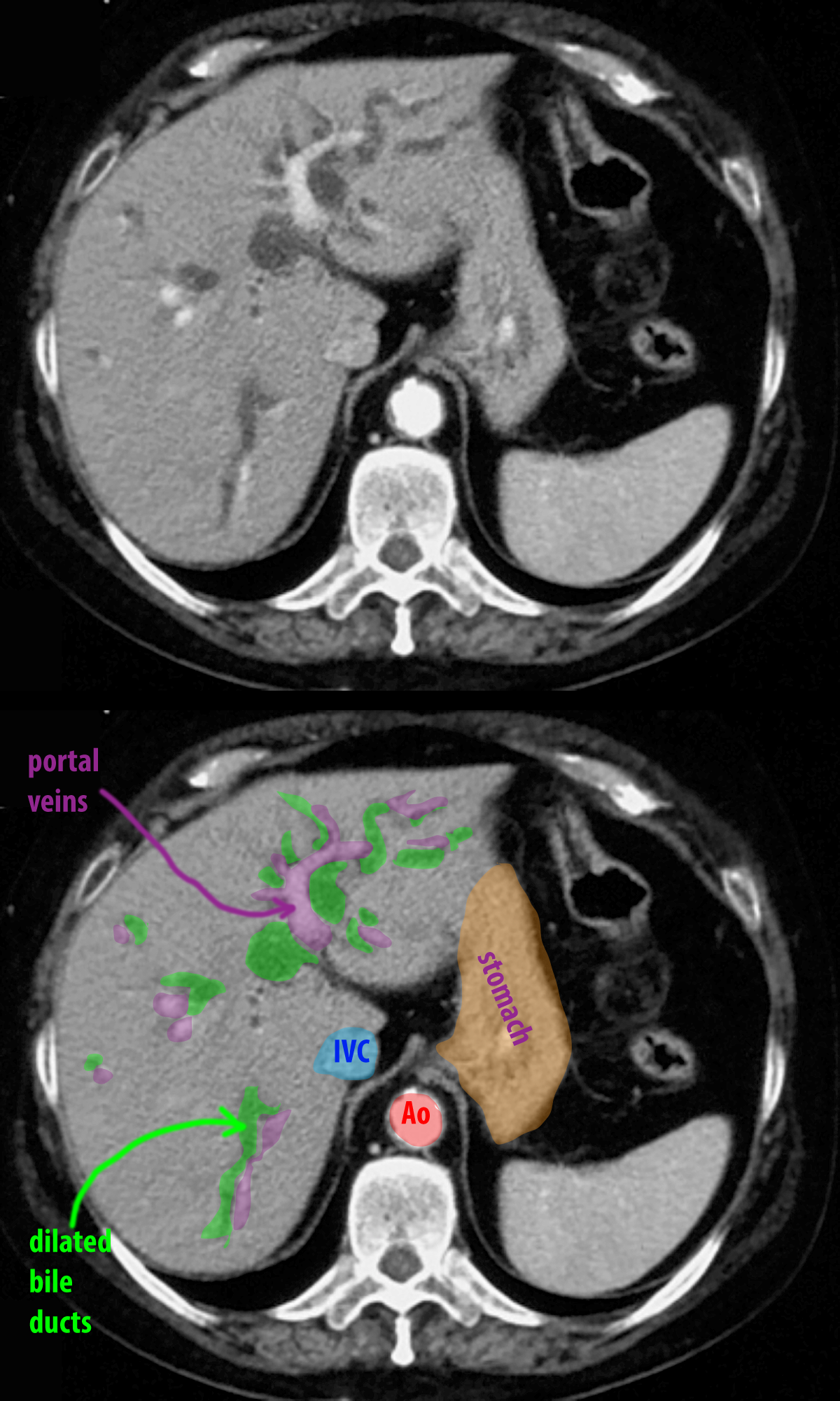
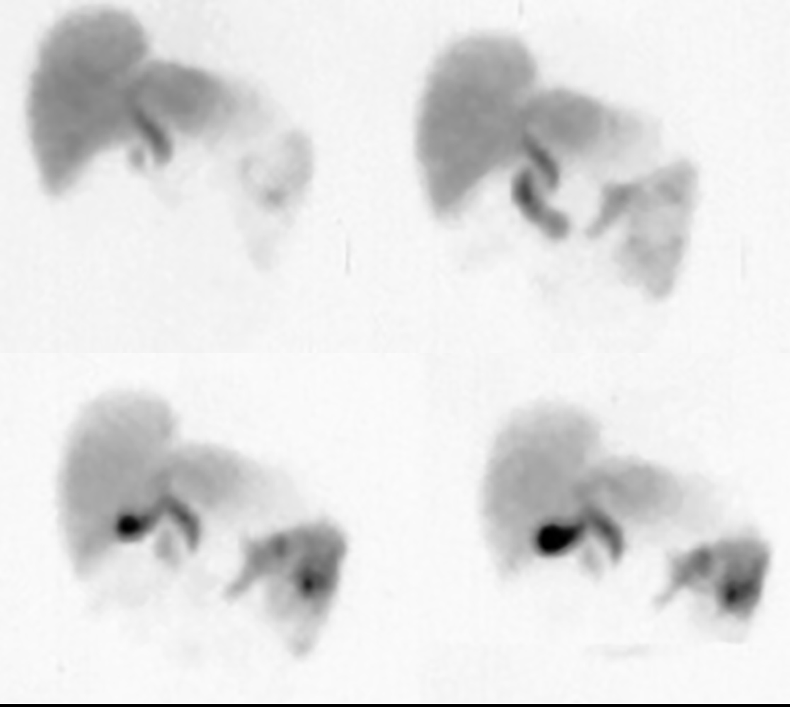
Bile is the same density as water on CT, around 0 Hounsfield units. The normal intrahepatic ducts are very small and barely visible, running adjacent to the portal vessels (which are white on this contrast enhanced study performed in the portal phase). When you look at the dilated bile ducts example, you can see that there are water-density tubular structures throughout the liver. You can use the gallbladder as an example of water density. Mystery study 1 is a HIDA scan, or hepatobiliary scan, using a radio tracer that is taken up by hepatocytes and excreted into the bile. The four images are at different points in time, and you can gradually see the common bile duct and gallbladder, and contrast moving into the small intestine. Mystery study 2 is an MR done with T2 weighting and fat saturation (so only water will be white). This highlights the biliary tree, and is called an MRCP. You will also see the renal collecting systems and ureters on this study, since they are also filled with fluid.
Question 2:
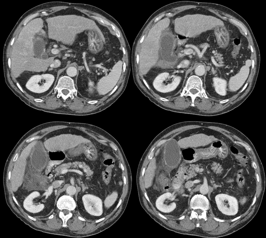
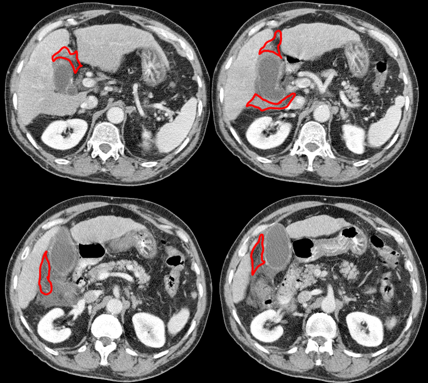
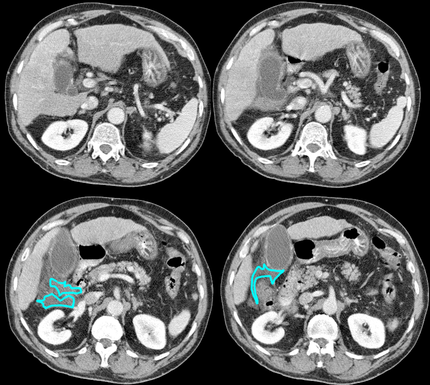
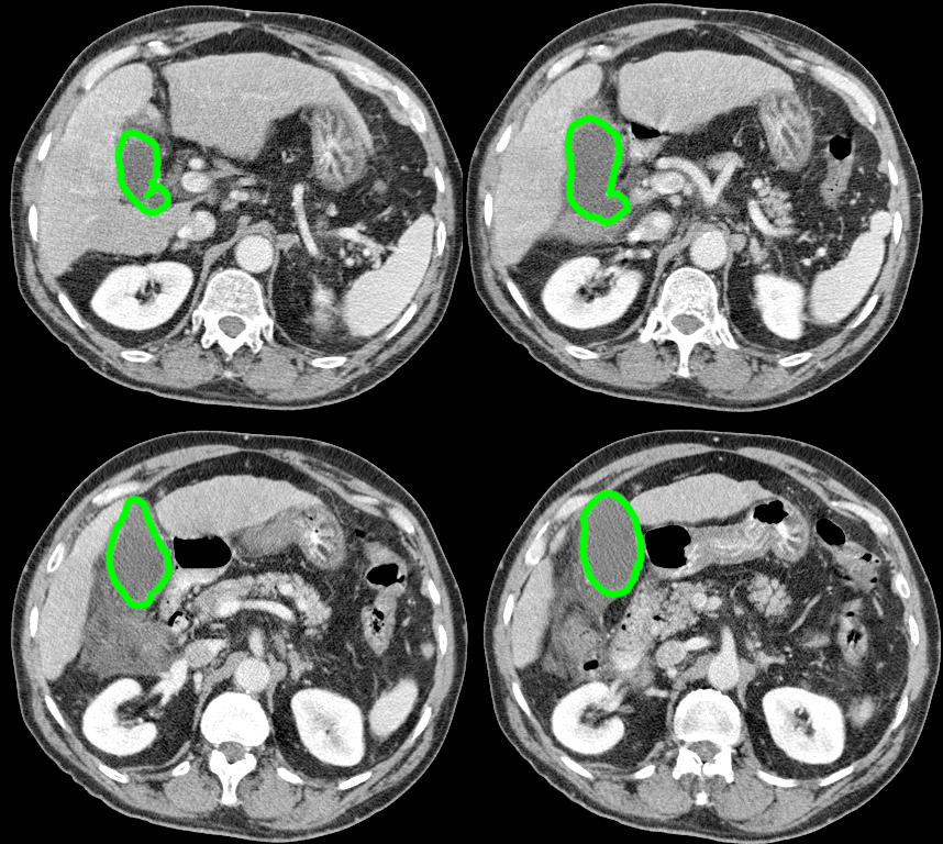
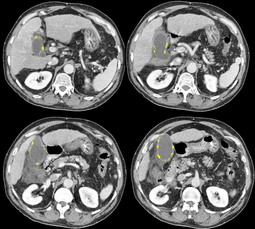
Focus attention on the liver and gallbladder on this CT of a patient with right upper quadrant pain. What diagnosis is most likely and why?
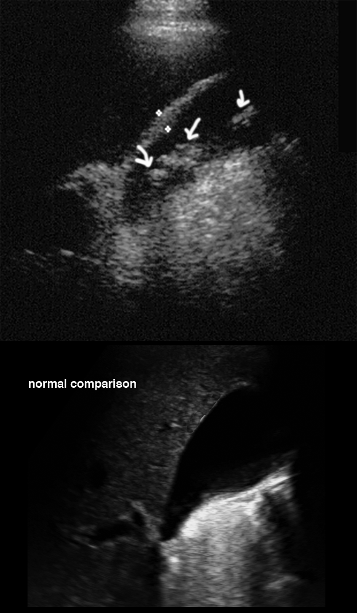
As noted on the labels, the gallbladder is distended, with a mildly thickened wall and some bright foci in the wall that are either calcifications or contrast enhancement. There is fluid around the gallbladder as well as fat stranding, which can be seen in many infiltrative processes in the abdomen, but most often indicate inflammation. These findings are most consistent with acute cholecystitis. On the normal GB CT movie, there is no fluid or stranding in the area and the gallbladder wall is very thin, almost inapparent. The top US image shows the thickened appearance of the GB wall in a different patient cholecystitis, who also has gallstones, indicated by the white arrows. A normal US of the gallbladder is shown in the bottom image from a different patient.
Question 3:
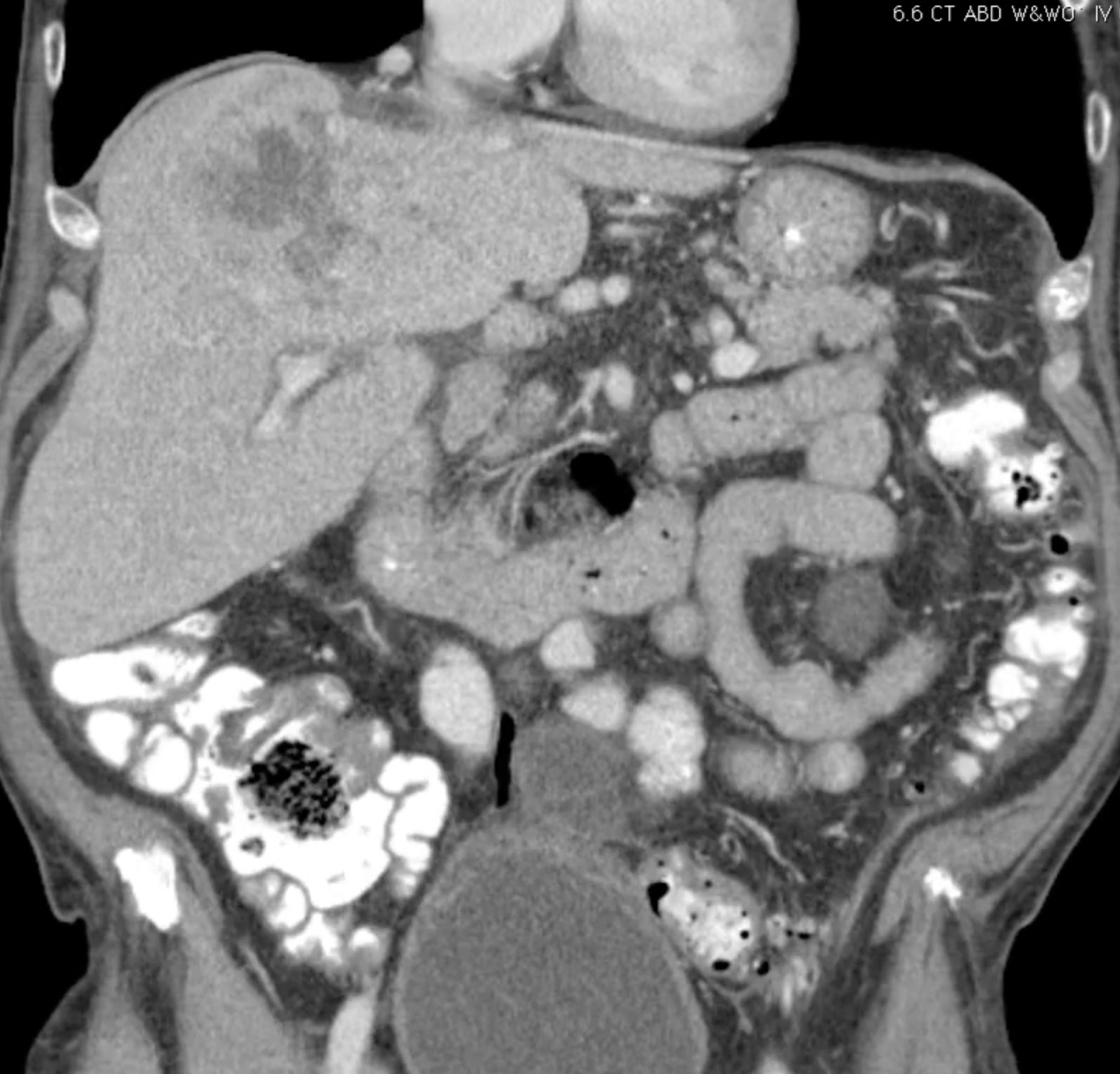
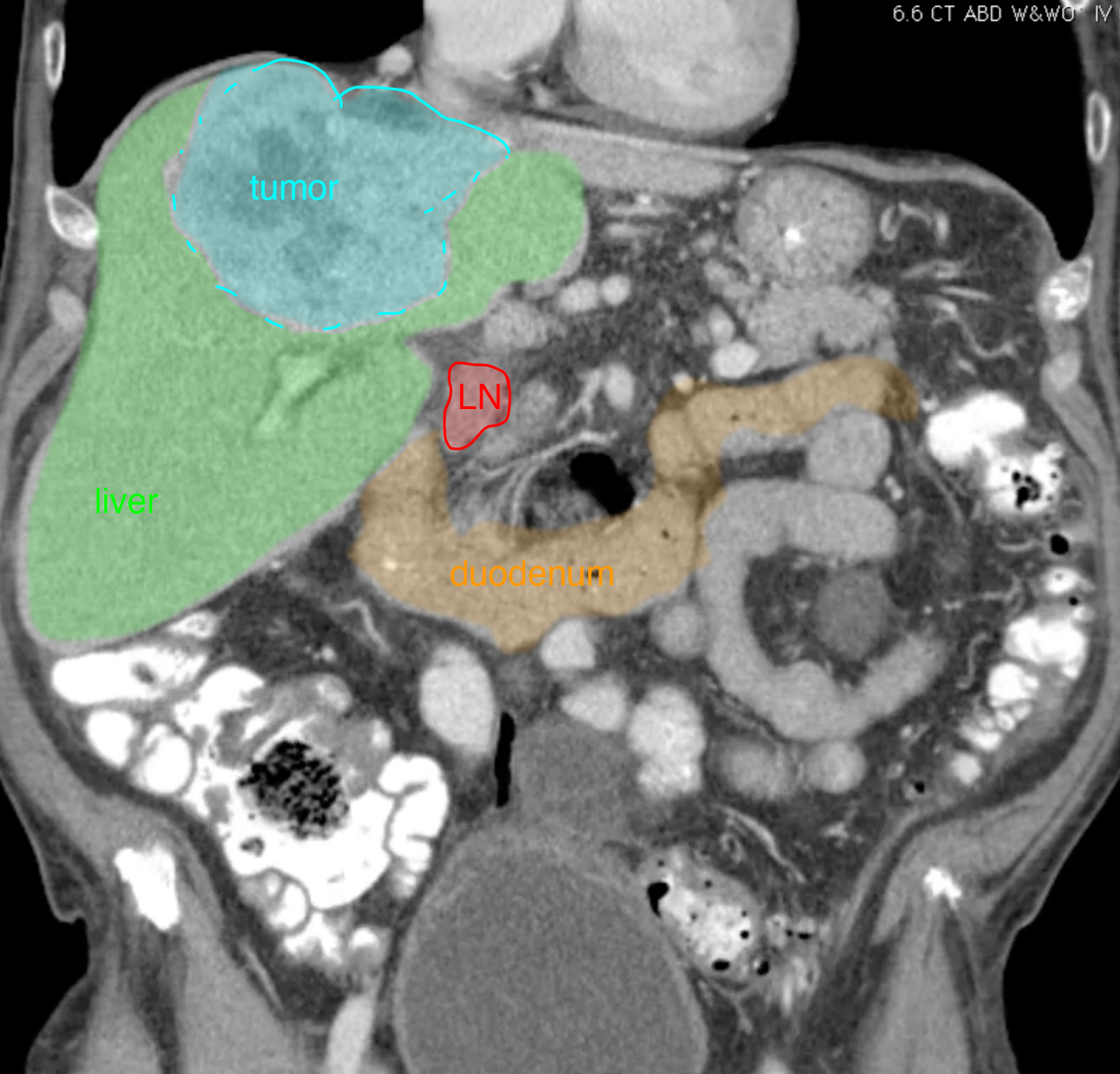
These CT images are from another patient with right upper quadrant pain. In what phase of contrast enhancement were these images obtained, and what do you see in the liver? Is there biliary dilatation?
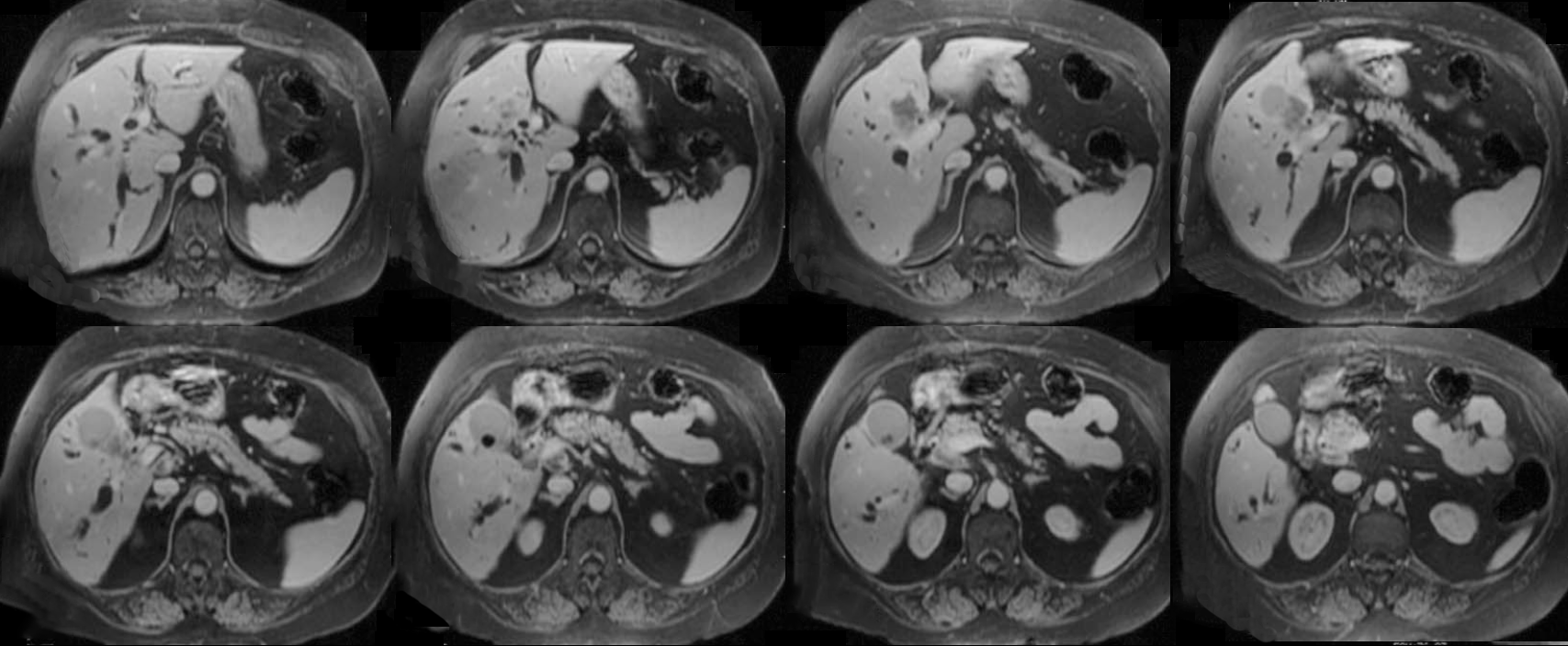
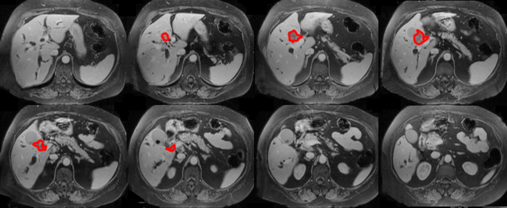
This CT sequence shows bright contrast in the abdominal aorta and its visceral branches, but not in the IVC or portal veins, so this is in the arterial phase, early after contrast injection. When evaluating liver and pancreatic tumors, the phase of contrast is important to note, and studies are often done at multiple points in time (which increases the radiation dose for the study). There is no biliary dilatation and there is a large heterogeneously enhancing upper central liver mass. The mass has low attenuation centrally suggesting necrosis. The main considerations for diagnosis here would be hepatocellular carcinoma or cholangiocarcinoma. The lack of biliary dilatation might favor HCC, but given the relatively peripheral position of the mass in the liver (best seen on the coronal image), even a cholangiocarcinoma (which is what this is) would not produce much biliary dilatation. If you look at the study on the other patient, there is extensive biliary dilatation on the MR images (dark tubular structures in the liver on this T1-weighted image) despite a very small tumor (shown in red on the labeled images, which was also a choriocarcinoma). Biopsy is generally required to establish the type of tumor present as there is considerable overlap in the imaging features of cholangiocarcinoma and other tumors.
