

Question 1:
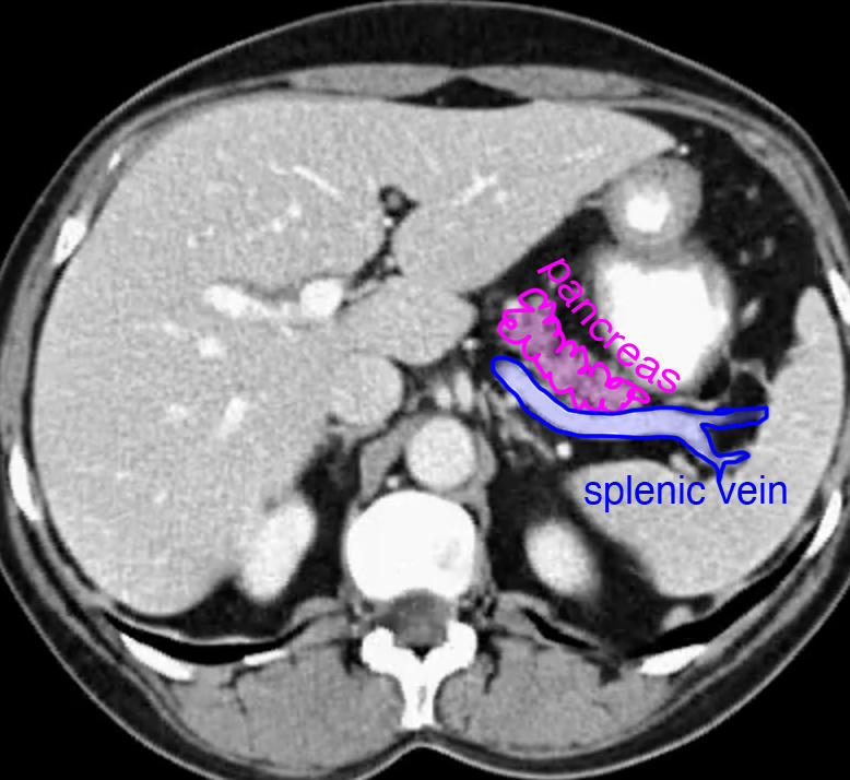
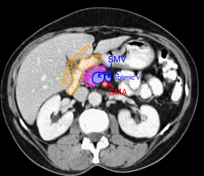
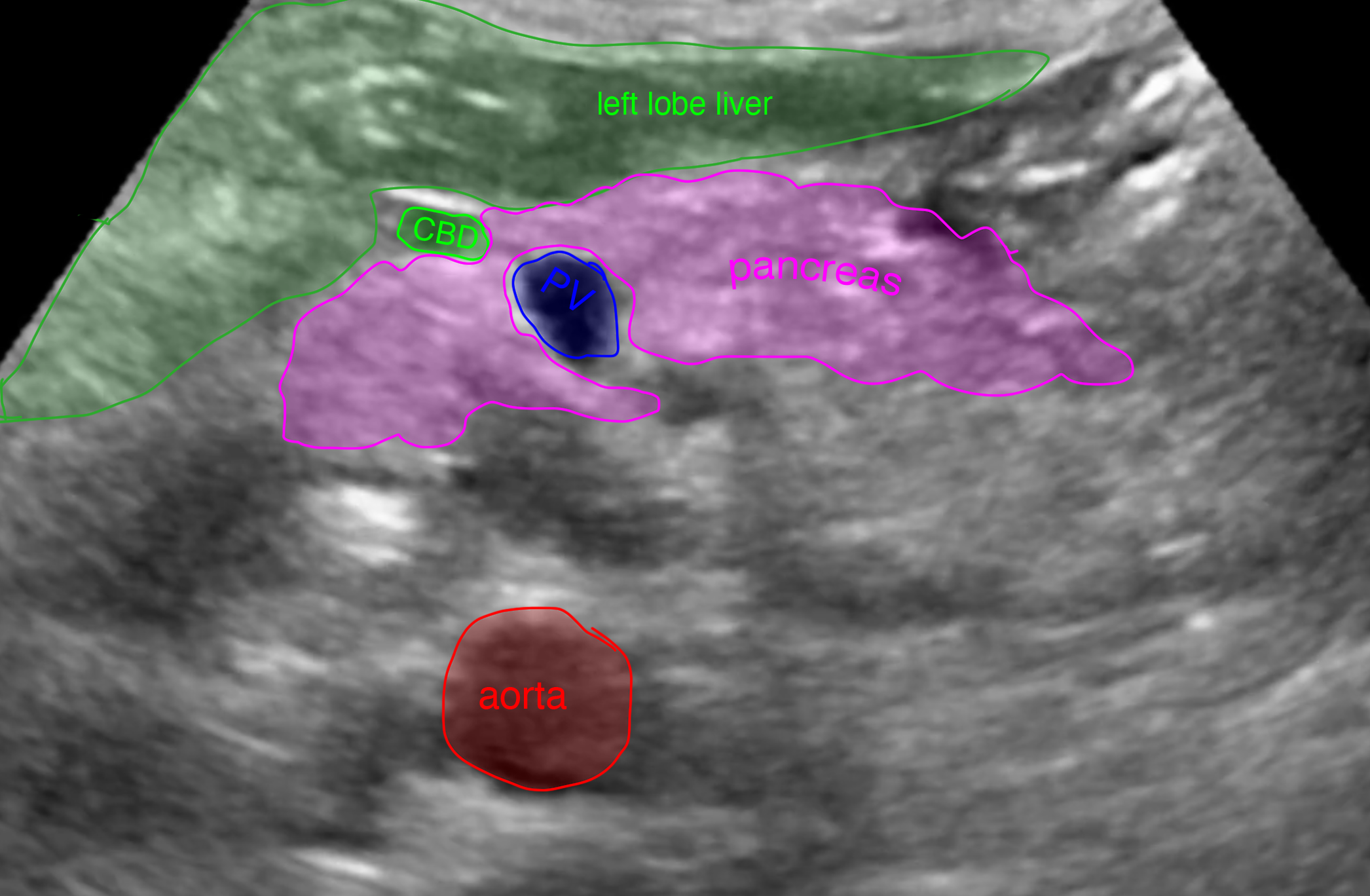
This is another normal CT. What phase of contrast is displayed? Try to identify the pancreas before clicking on the selected images, which have normal structures labeled. What often makes the pancreas difficult to visualize on ultrasound?
This CT was obtained in the portal phase. You can see that there is dense contrast in the portal veins, a bit brighter than what is in the aorta. The pancreas often moves in and out of the plane of axial scans, so you need to look at several images to visualize the entire organ. The splenic vein has a straight course along the deep surface of the pancreas. The pancreas is often difficult to visualize on ultrasound, due to overlying bowel gas. you can see from the CT how many gas-filled loops are lying anterior to the pancreas.
Question 2:
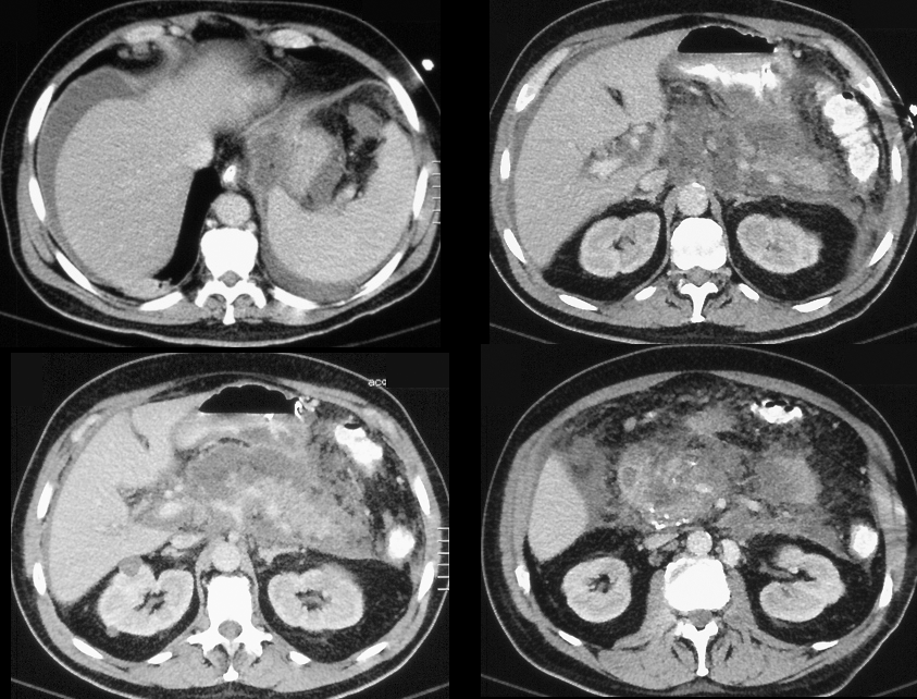
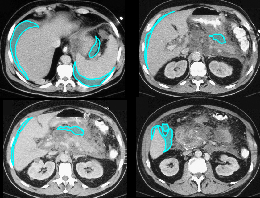
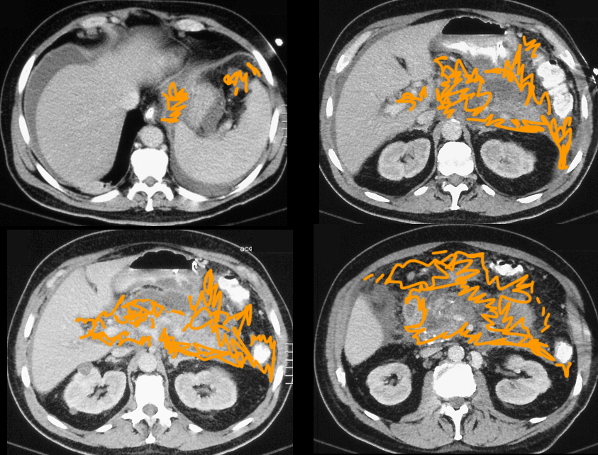
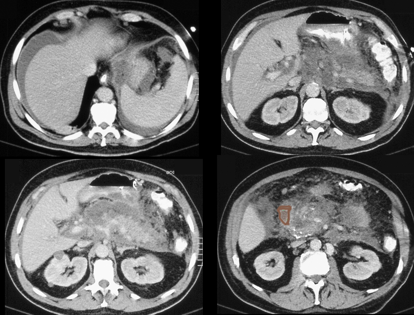
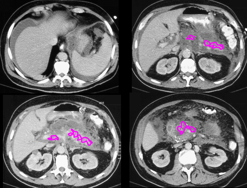
This patient has severe abdominal pain. What imaging features do you see that are similar to those in the patient with cholecystitis?
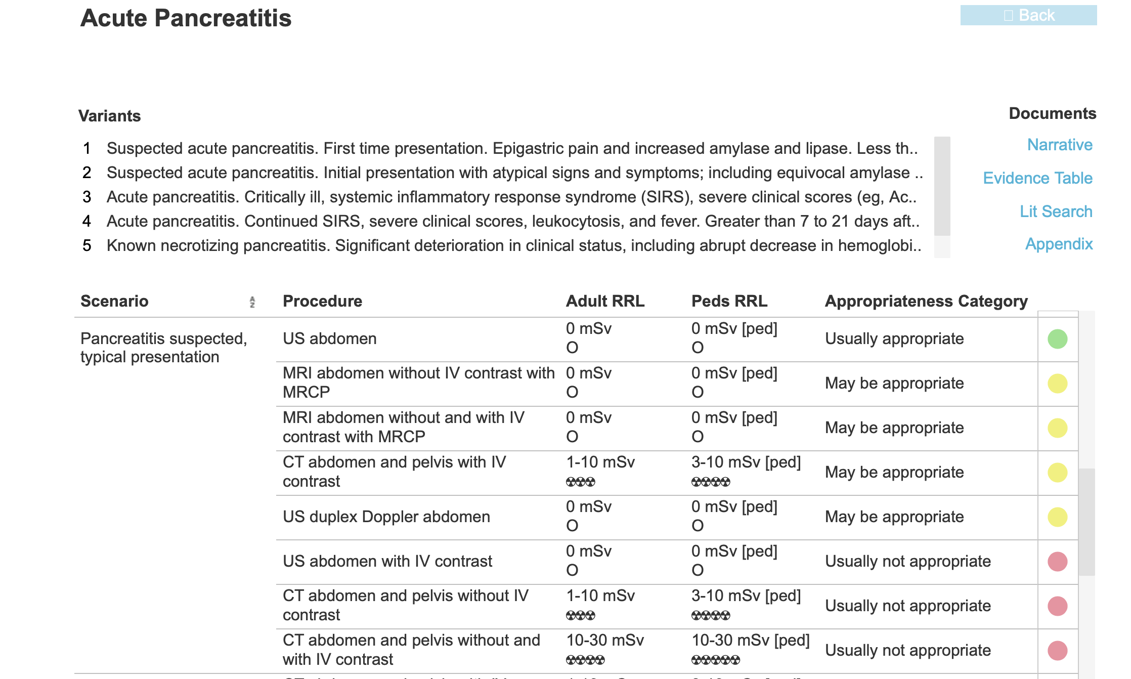
Fluid and fat stranding are both present surrounding the enflamed pancreas, similar to the case of acute cholecystitis. The pancreas is nearly inapparent, as it has been extensively damaged by the inflammatory process and release of digestive enzymes. You are also shown the ACR Appropriateness criteria for pancreatitis.
Question 3:
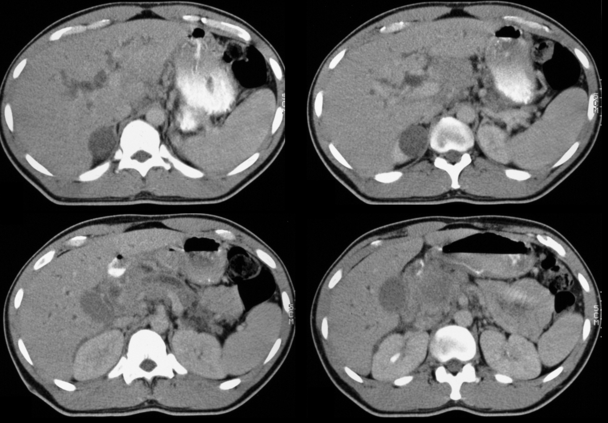
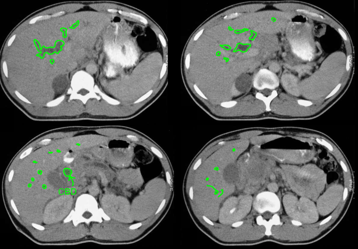
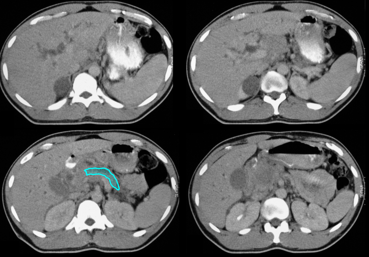
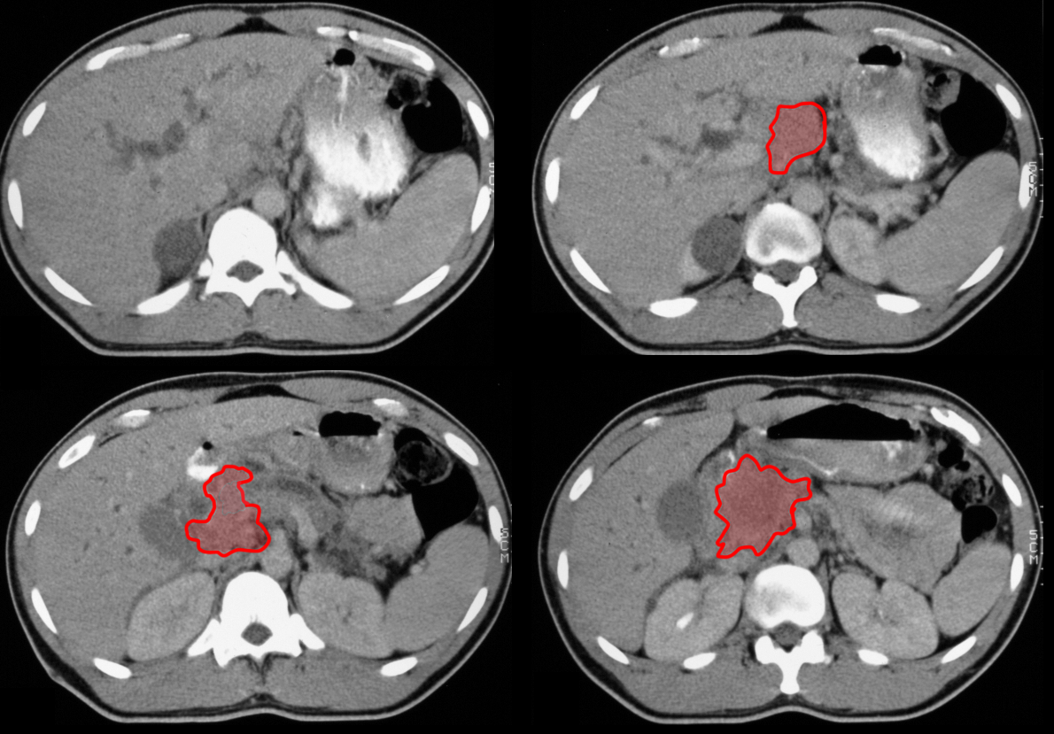
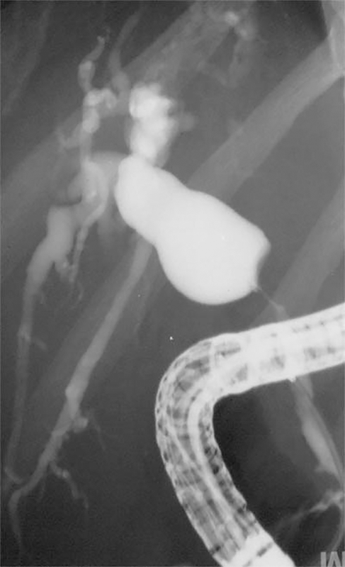
You are shown four CT images on another patient with more chronic upper abdominal pain. Is there biliary dilatation? What do you think of the pancreatic duct? You are shown another study in this patient--what is this additional study?
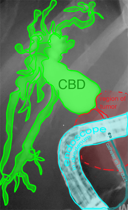
This CT was done very late after contrast injection, with very little contrast in the vascular structures. There is an incidental right kidney upper pole cyst, a common benign finding with no relationship to the patient's symptoms. There is extensive biliary dilatation, and the pancreatic duct is also dilated. This suggests an obstructive process somewhere in the region of the ampulla of Vater. This could arise from an exophytic liver mass, a tumor of the distal bile duct or the ampulla itself, or a pancreatic head mass, which is present in this case. The other study done on this patient is an ERCP, or endoscopic study with direct introduction of contrast into the common bile duct. The ERCP shows the marked narrowing of the duct in the region of the pancreatic head tumor, and the marked dilatation of the intrahepatic ducts and hepatic duct.
