

Question 1:
You are shown three normal angiographic studies that might be done in a patient with GI bleeding. What are these studies? What bleeding sources would be likely for each study? What are some other ways to identify the source of GI bleeding?
Study 1 in a celiac angiogram. You can see that the catheter is coming up from the bottom right part of the patient, so access was via the right femoral artery. Celiac angiograms might show sources of bleeding from the stomach and duodenum. Study 2 is a superior mesenteric angiogram. This study might show bleeding from the duodenum, small intestine and right colon. Study 3 is an inferior mesenteric angiogram, and would show bleeding sources in the distal colon. Angiograms are invasive with risks related to vascular access. GI bleeding can sometimes be seen on less invasive studies like CT (if the bleeding is brisk enough), or using nuclear medicine imaging with tagged RBC's, with sequential imaging over time to show the source of bleeding through accumulation of the radioactive RBC's in a portion of the GI tract.
Question 2:
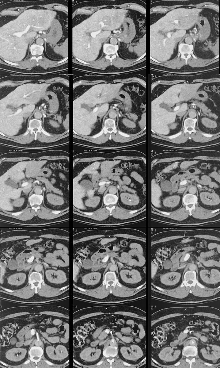
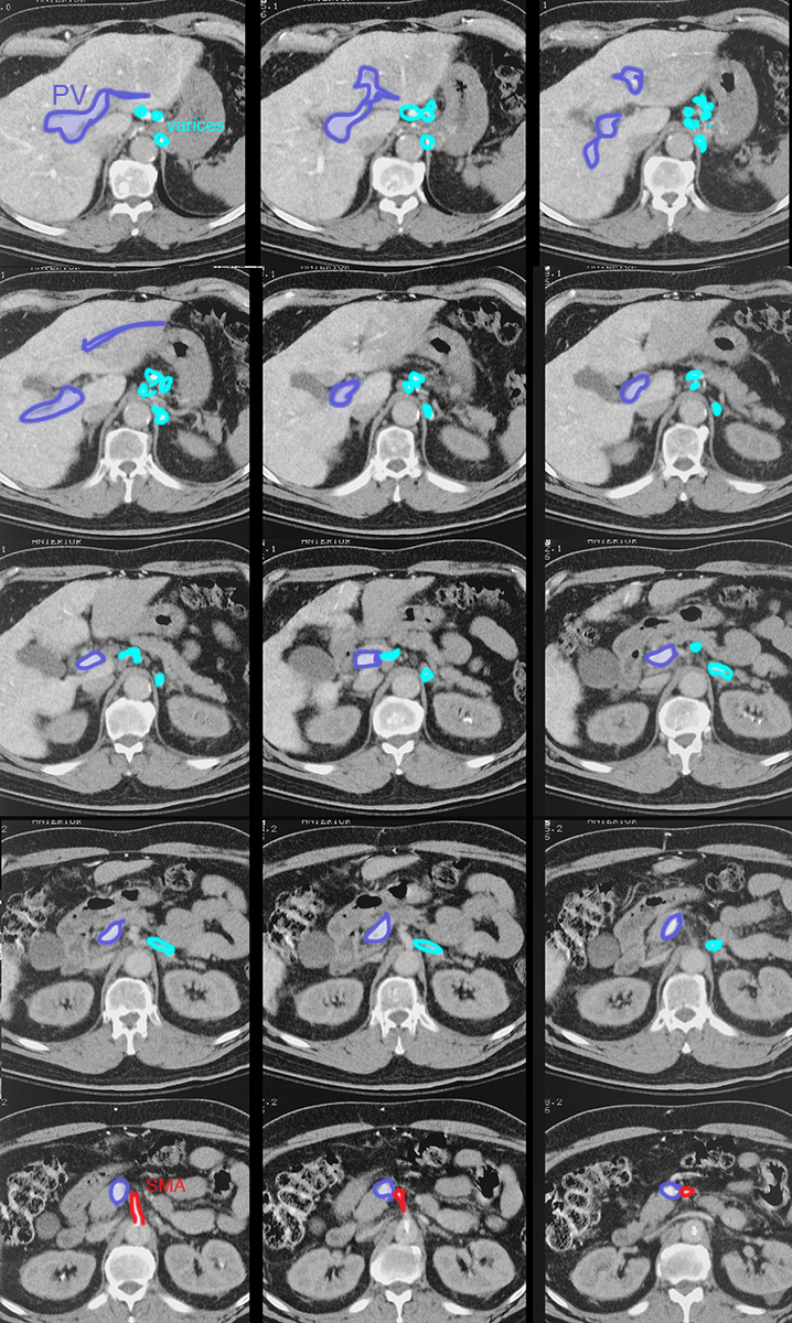
One possible source of GI bleeding is the distal esophagus. What condition would predispose a patient to bleeding of this type? Can you identify such an abnormality on these CT images? How was the contrast administered in this case?
Portal hypertension can produce portal-systemic shunts from the right and left gastric veins (which usually drain into the liver), in the opposite direction into para-esophageal veins. These veins can dilate (varices), and are located just deep to the esophageal wall, which can lead to bleeding. The labeled image shows the vessels of the portal system in dark blue and the dilated and tortuous esophageal varices in light blue. There is virtually no contrast in any other vessels on this study. The contrast was actually delivered through a catheter in the superior mesenteric artery, which would produce very bright contrast in the portal vein and all of its connections. This is not a common way to administer contrast for CT images.
Question 3:
This patient had black tarry stools, upper abdominal pain, which was worse several hours after eating, and anemia. What is this study? Does it involve radiation? Does it involve iodinated contrast material? What is an advantage of this study over a CTA?
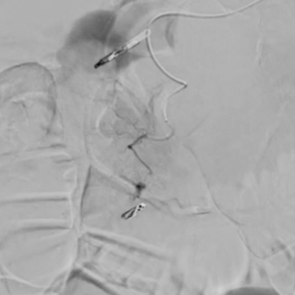
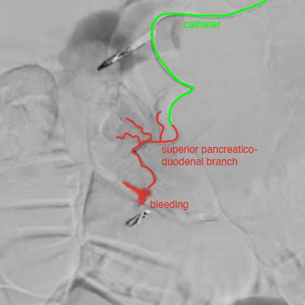
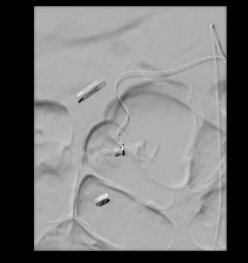
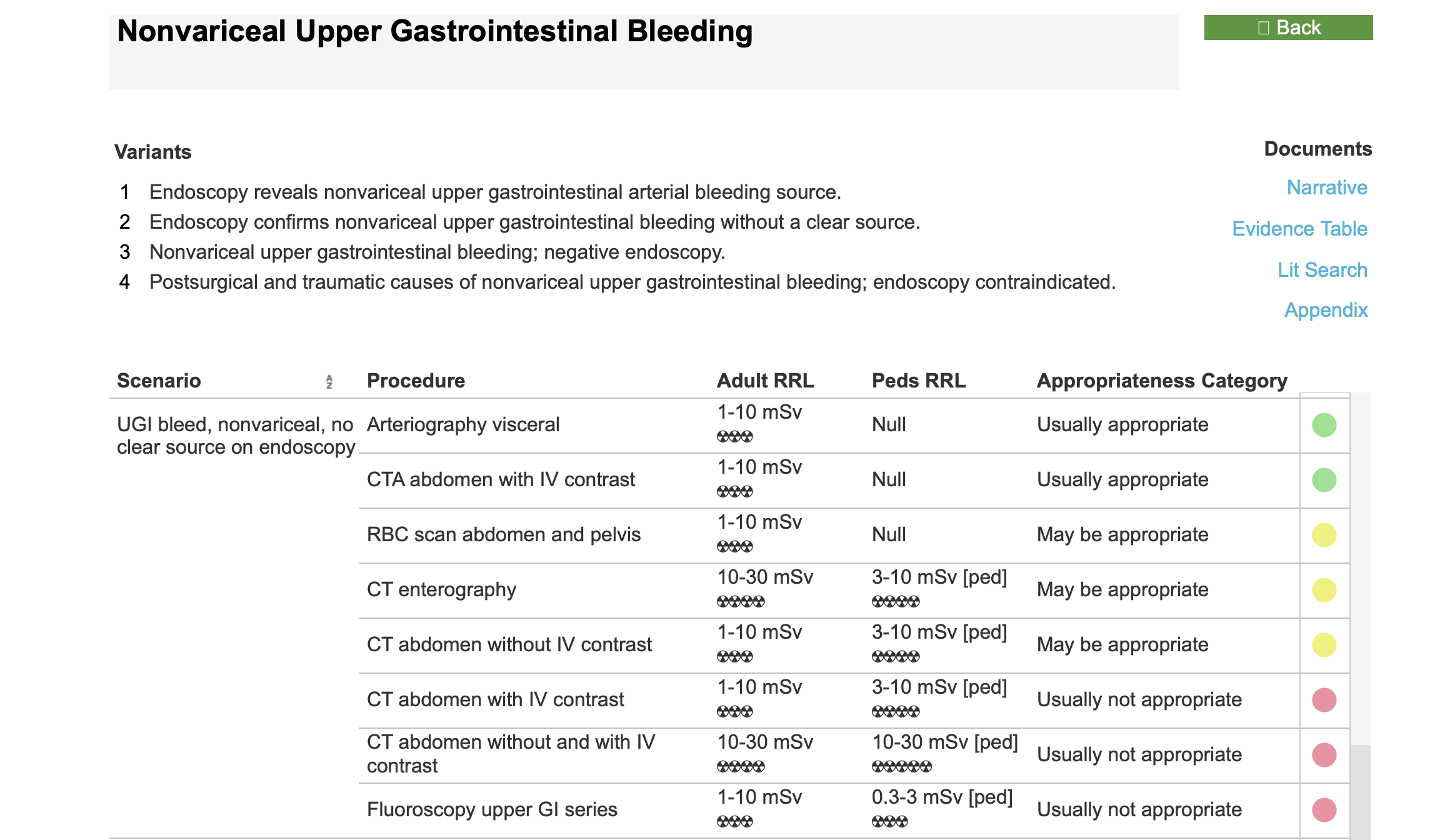
This study is a selective angiogram of a vessel supplying the duodenum. The patient had several angiograms of other vessels first, in particular the celiac artery and superior mesenteric artery. Once a site of bleeding was identified, a catheter was directed into the closest vessel. This study does involve a relatively high radiation dose, particularly as many series of images must generally be obtained to find the vessel that is bleeding. It also involves use of a relatively high dose of iodinated contrast, which can lead to renal problems. The main advantage is that once the bleeding vessel is identified, coils can be deployed to occlude the vessel and stop the bleeding. The coil image below shows coils in place occluding the bleeding site. You are also shown the Appropriateness Criteria recommendations for imaging in the clinical setting of upper GI bleeding.
