

Case 1-normal MSK imaging
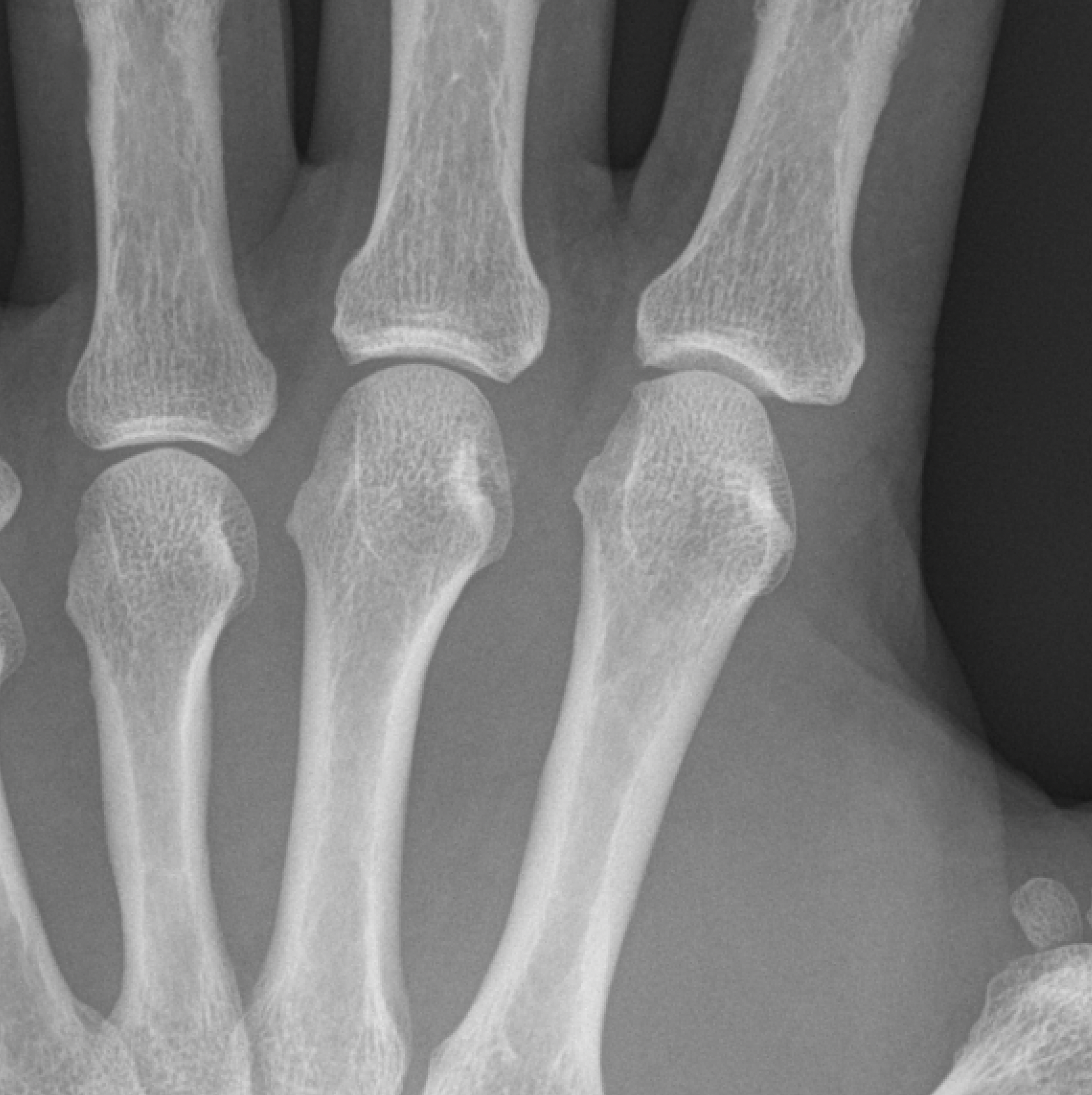
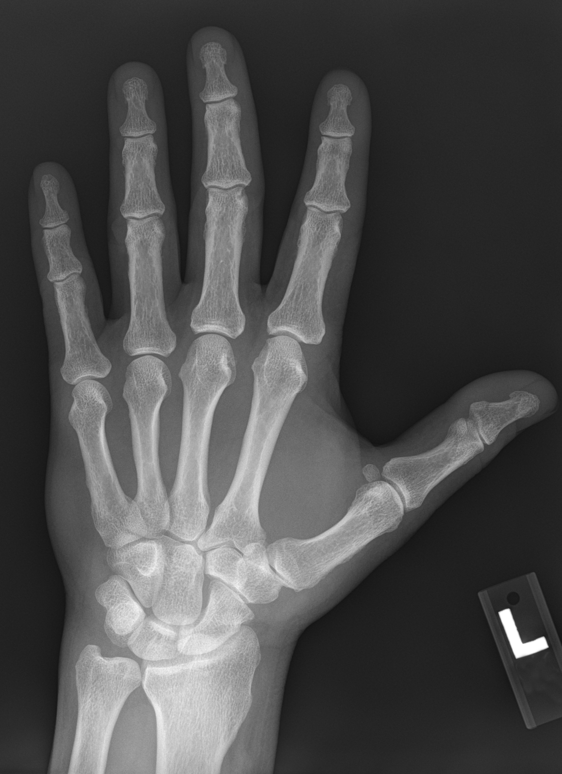
This case will demonstrate the normal appearance of various parts of the musculoskeletal system on different types of imaging. We will start with the hand, since it is involved in many rheumatologic disorders.
Question 1:
a) What is the commonest first imaging performed on patients with MSK complaints, overall?
Radiography, which is readily available, quick, cheap, with low radiation dose, is often the first imaging performed for patients with MSK complaints.
b) What other types of imaging could be considered for MSK complaints?
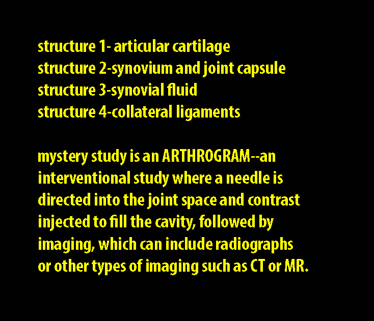
Other types of imaging include ultrasound, CT, MR and nuclear medicine. Contrast can also be introduced into a joint space (arthrography) prior to CT or MR to give more detailed examination of the intra-articular features of a joint. Depending on the clinical question and the joint involved, any of these modalities could be considered. To help in deciding what is appropriate, there is an online resource available to all from the American College of Radiology that lists recommendations based on clinical presentation.
Link to ACR Appropriateness Criteria for all topic areas:
If you click in the Panels field, you can select Musculoskeletal from the list, and click Search to bring up the specific recommendations for common MSK complaints.
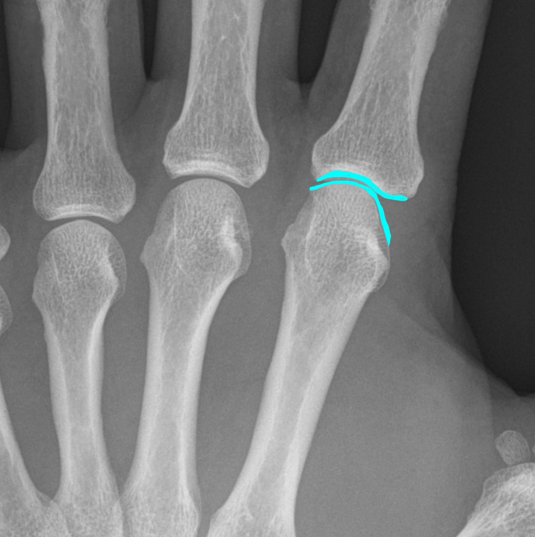
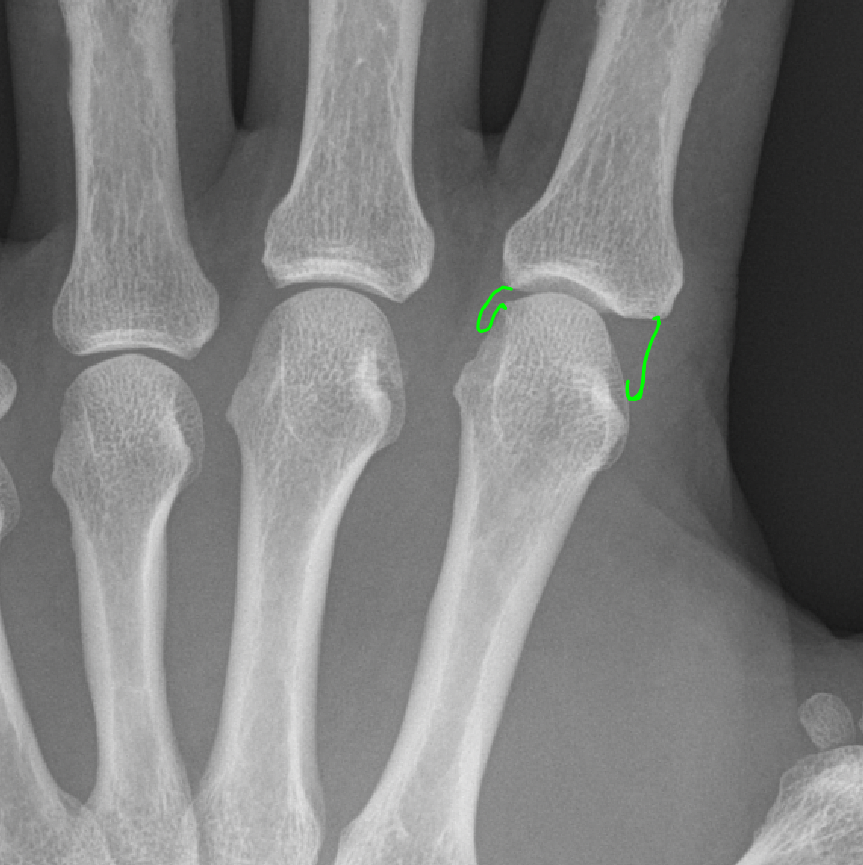
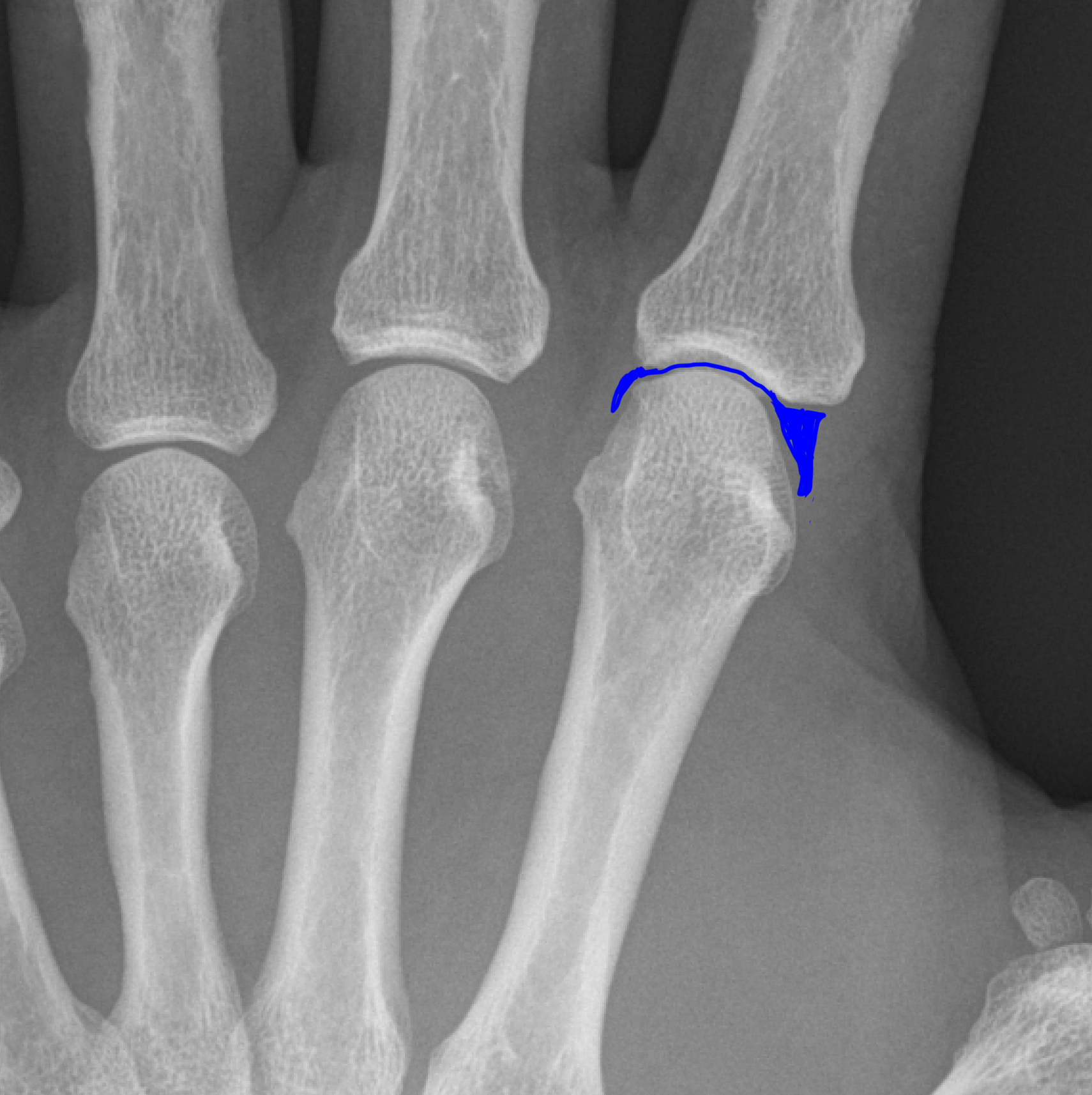
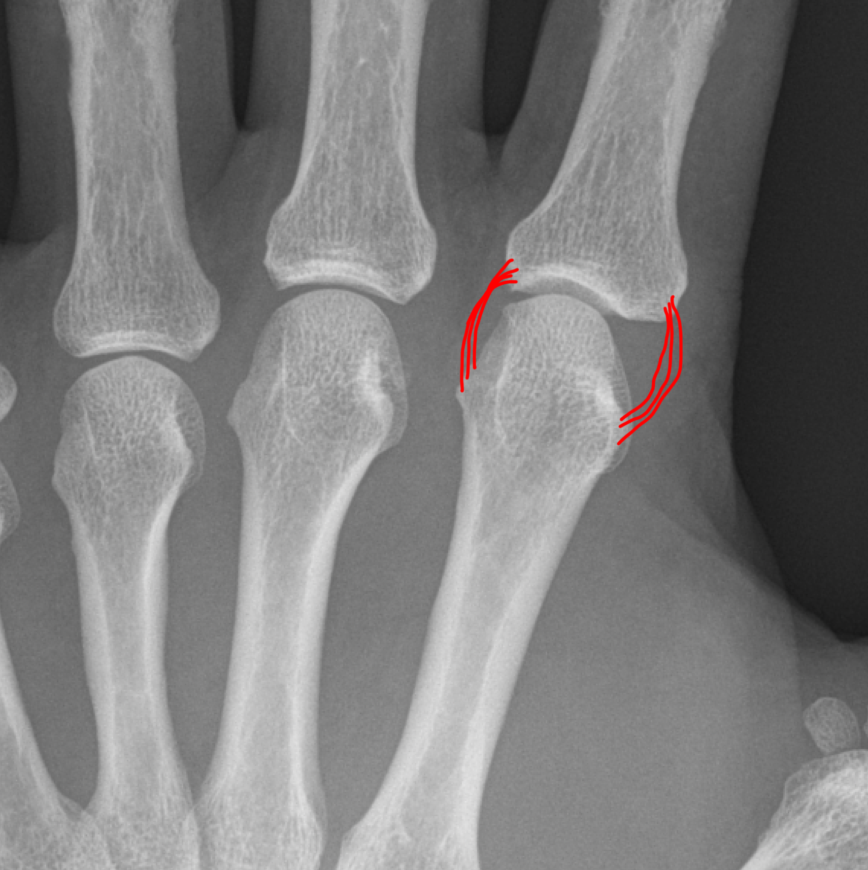
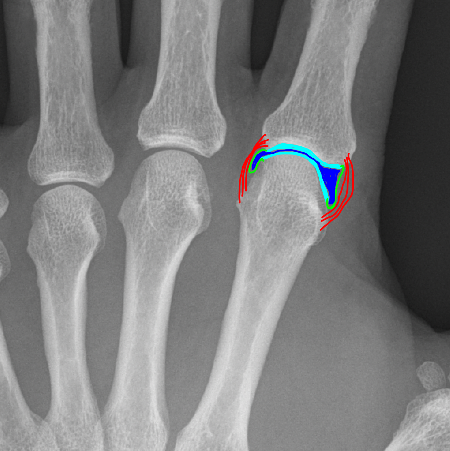
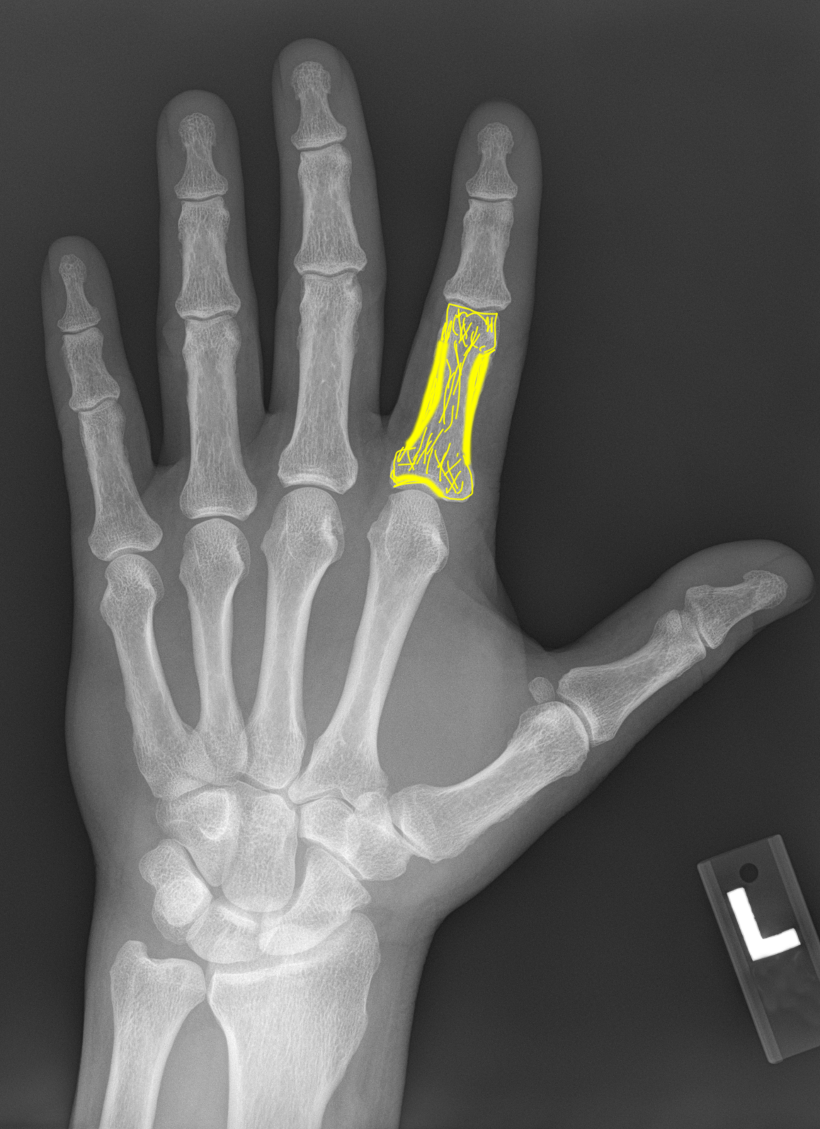
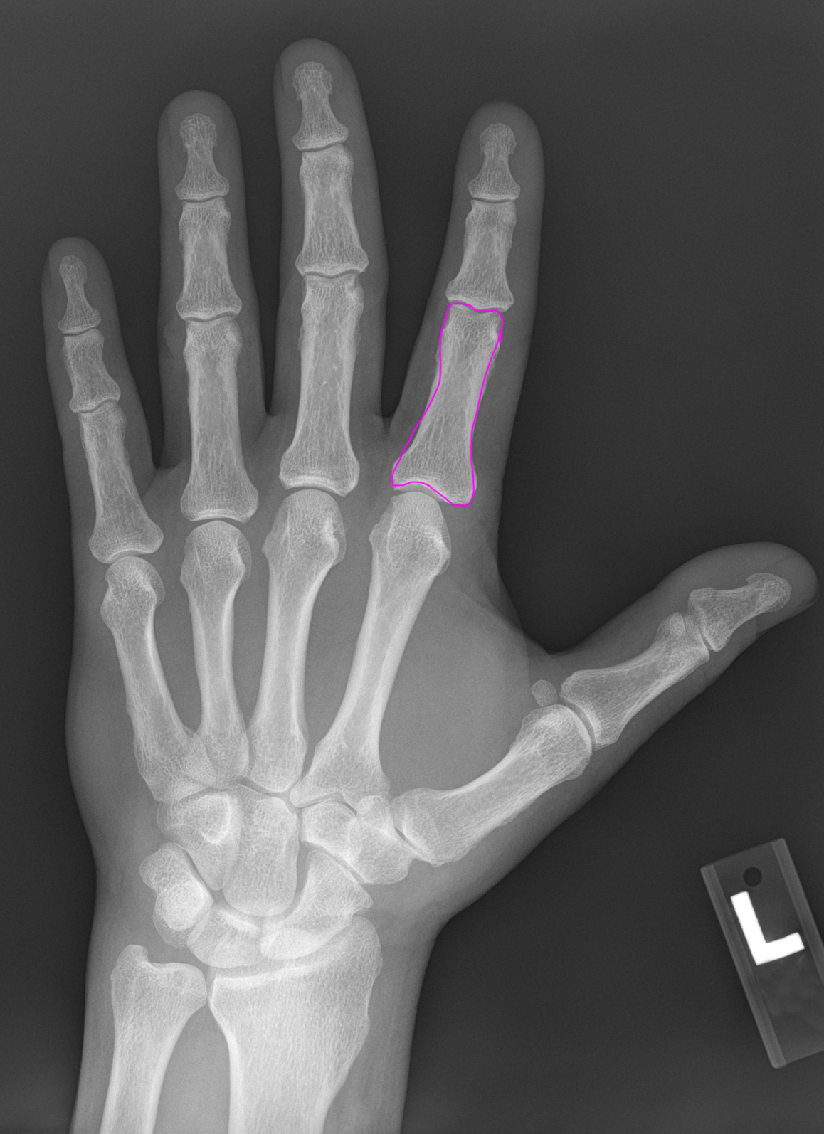
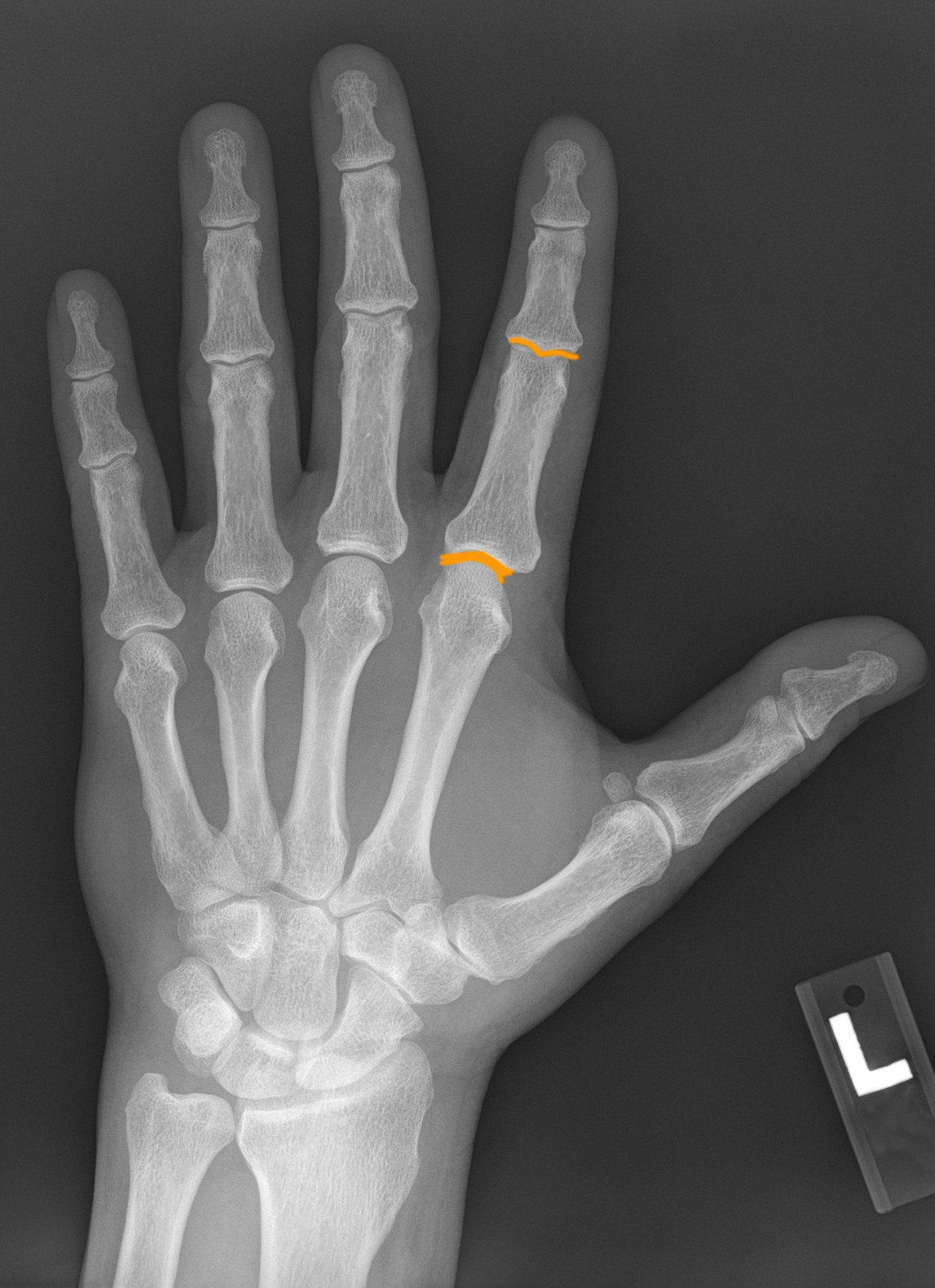
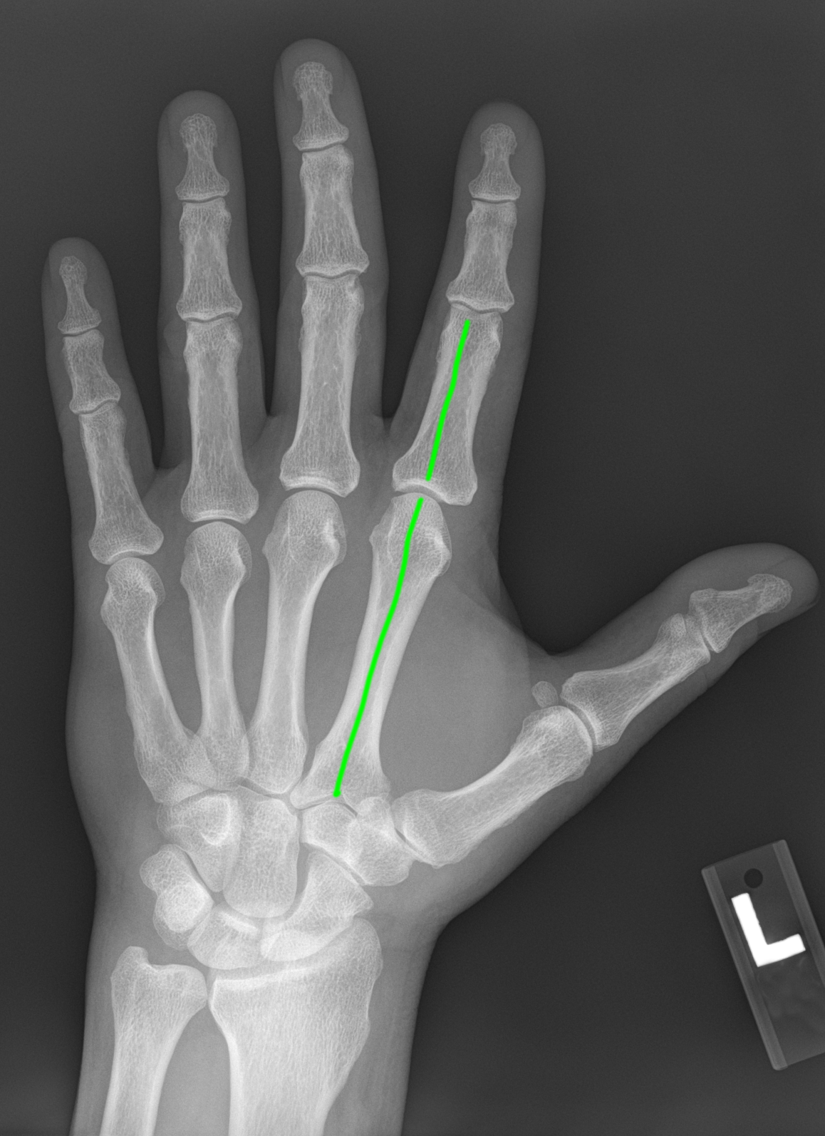
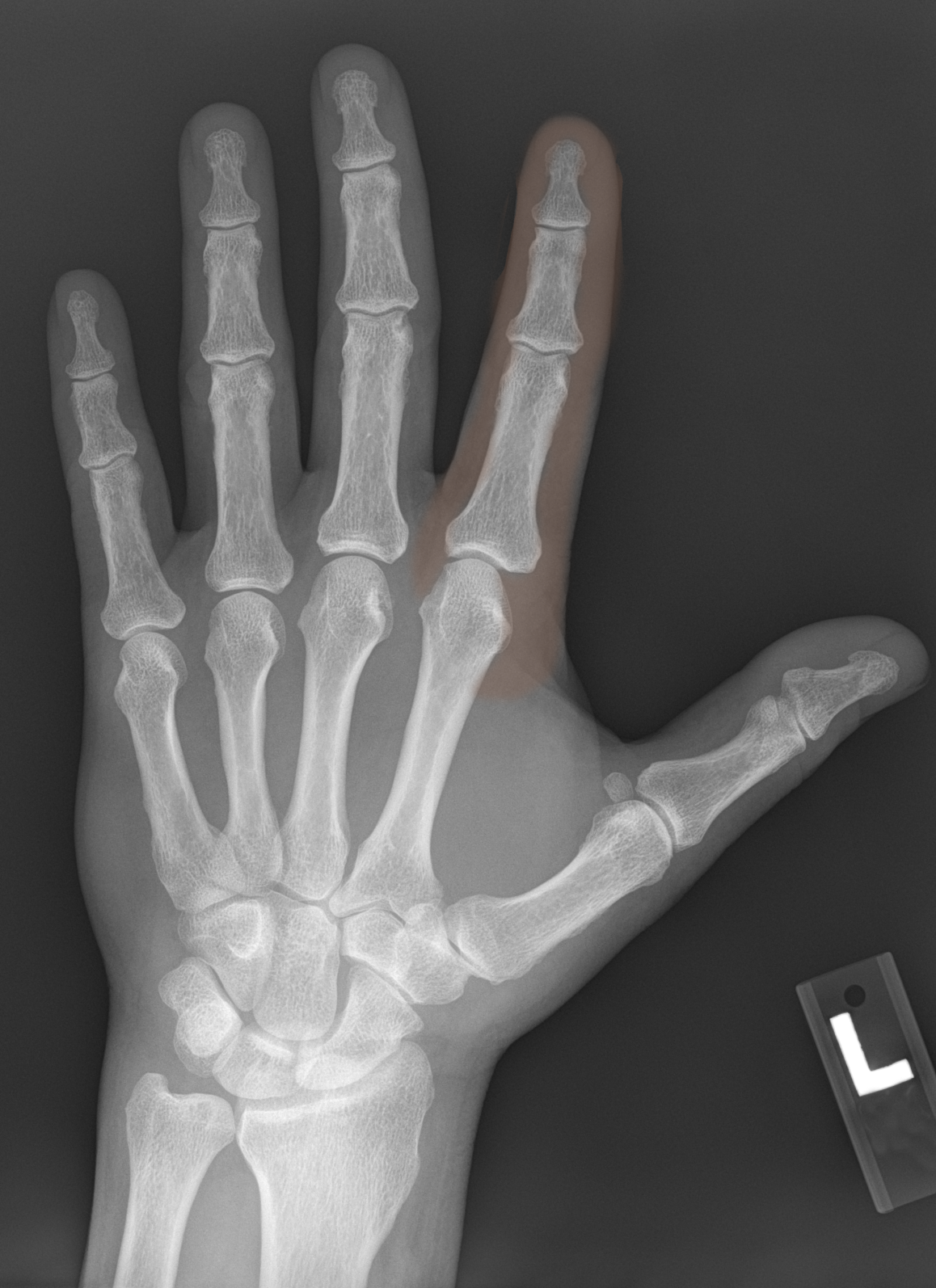
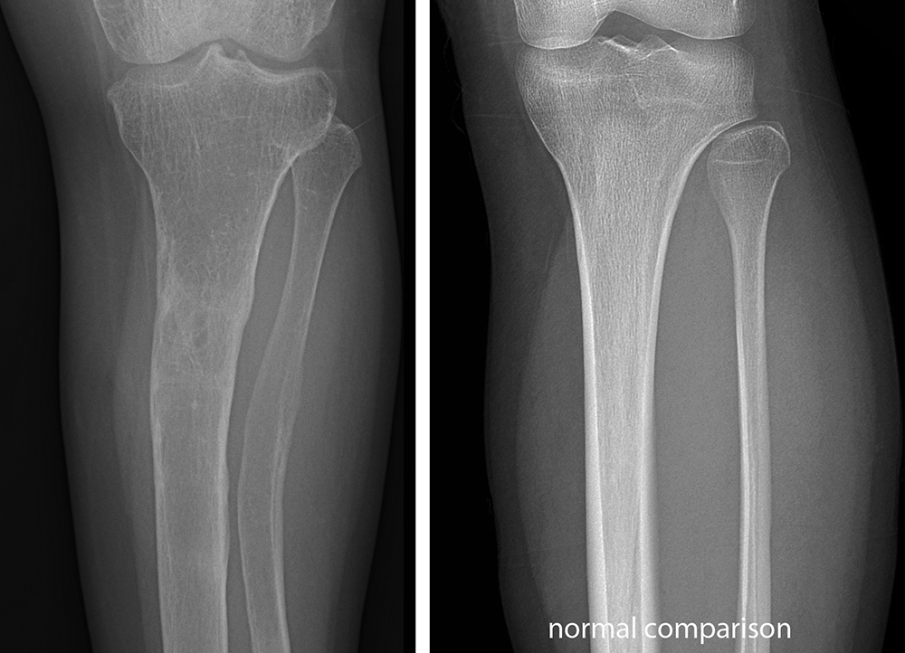
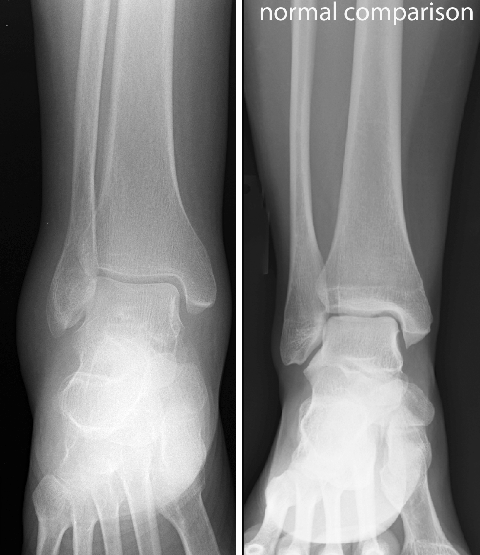
When examining a radiograph of a bony structure, check the links below to see some of the features that should be explored.
Further Explanation:


Case 1-normal MSK imaging
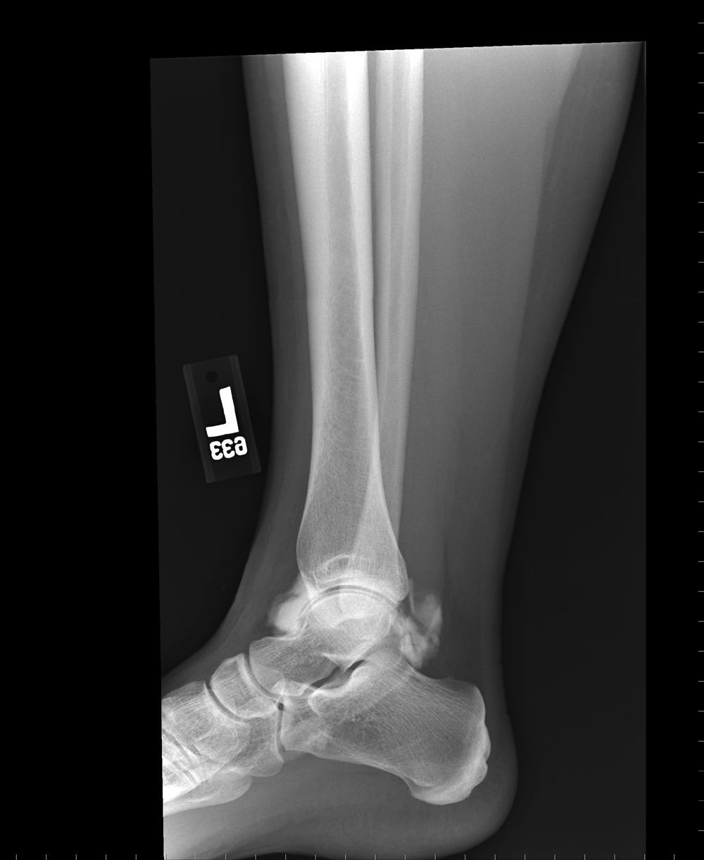
You are shown a closeup view of the hand. Identify the parts of the joint indicated by the links. What is the mystery study?
Further Explanation:
