

Case 2-arthritis
This case will compare and contrast various types of arthritis on imaging, particularly osteoarthritis and rheumatoid arthritis
Question 1:
That are some imaging features that can be helpful in distinguishing rheumatoid arthritis (RA) from osteroarthritis (OA) on radiography?
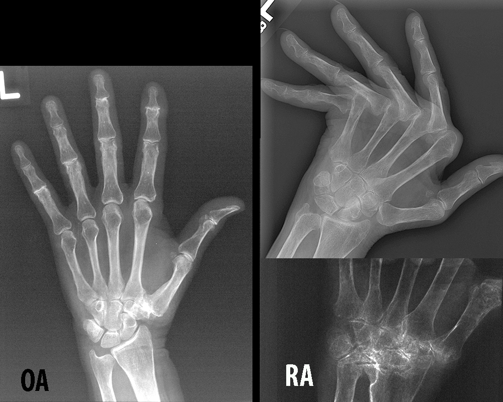
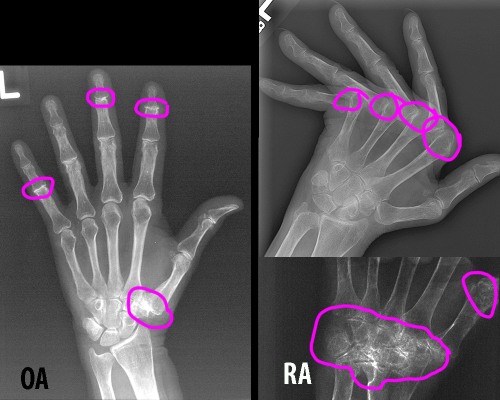
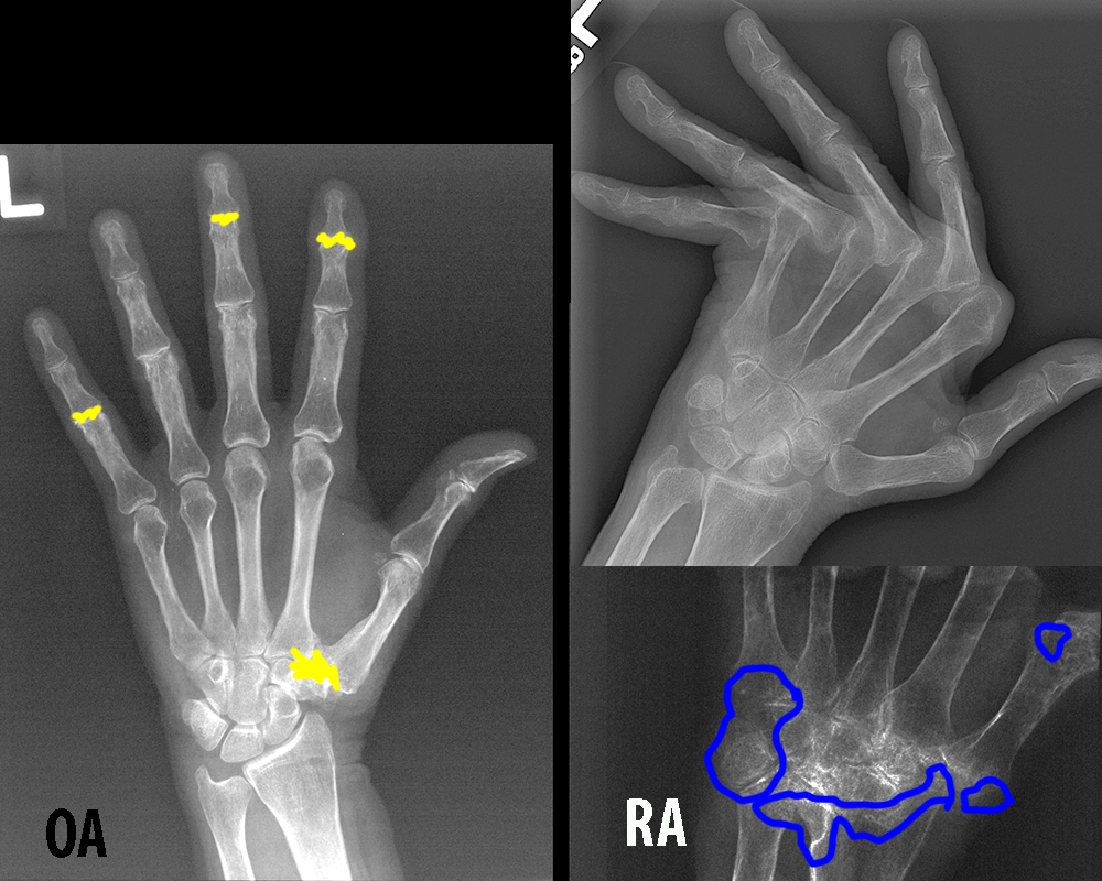
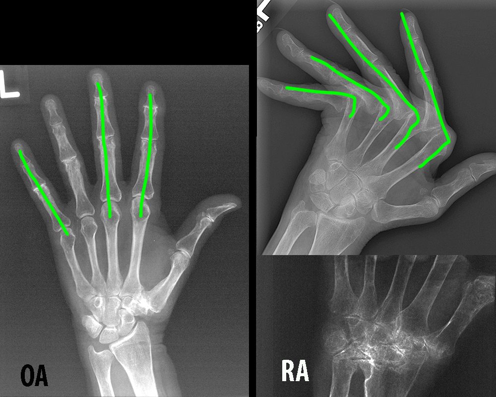
Some features that are most helpful include 1) distribution: RA tends to involve more proximal joints in the hand (MCP and carpal), while OA most often involved more distal joints (DIP); 2) bone mineralization: RA often produces bone loss (erosions) particularly in the periarticular regions, while OA most often produces increased bone and new bone formation as spurs; 3) alignment: RA often produces malalignment, particularly with ulnar deviation at the MCP joints, while OA less often produces severe malalignment. You can click the links below for illustrations of these findings.
Case 2-arthritis
It is important to understand that there can be considerable overlap in different types of arthritis in terms of imaging features.
Question 2:
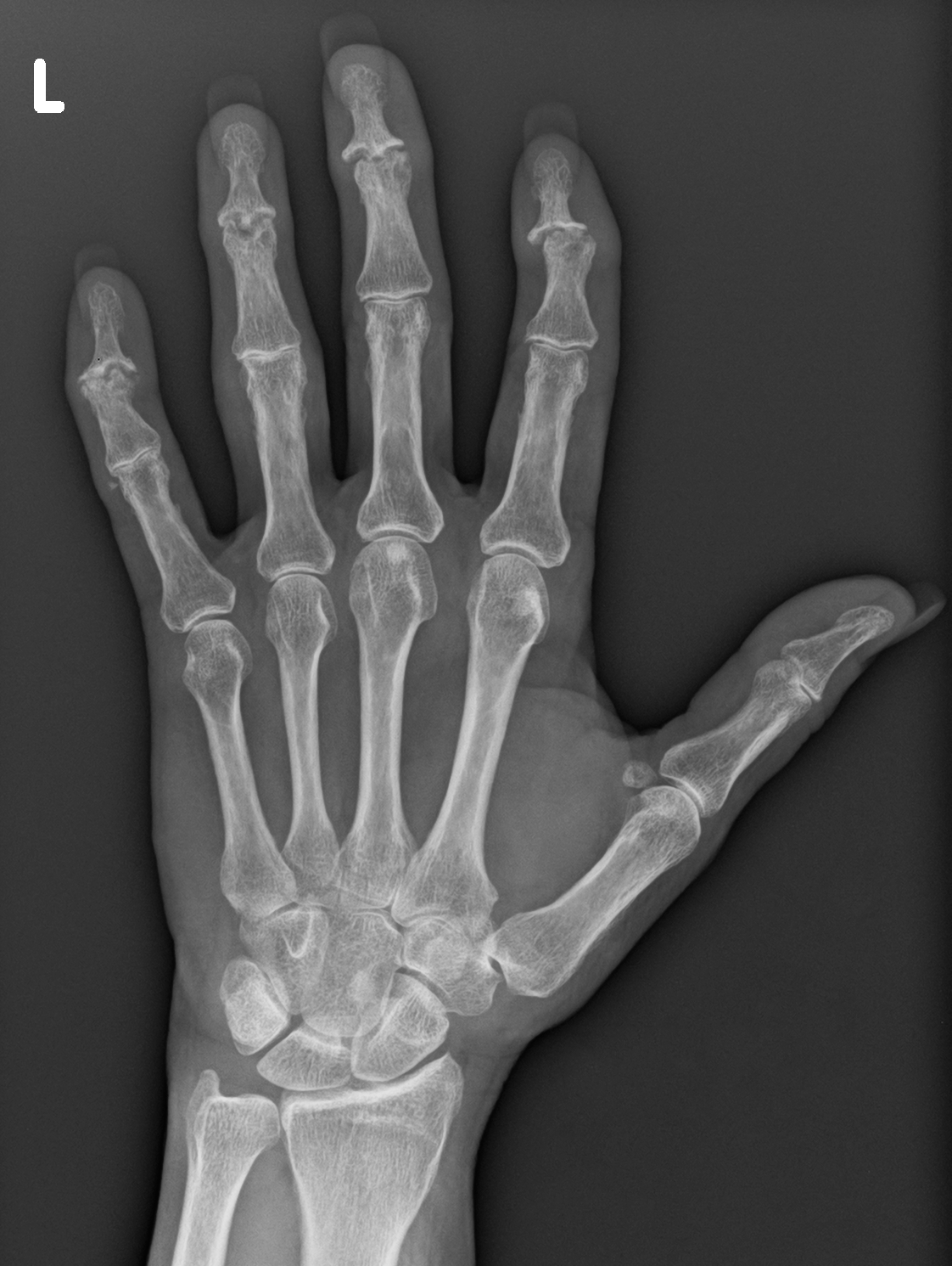
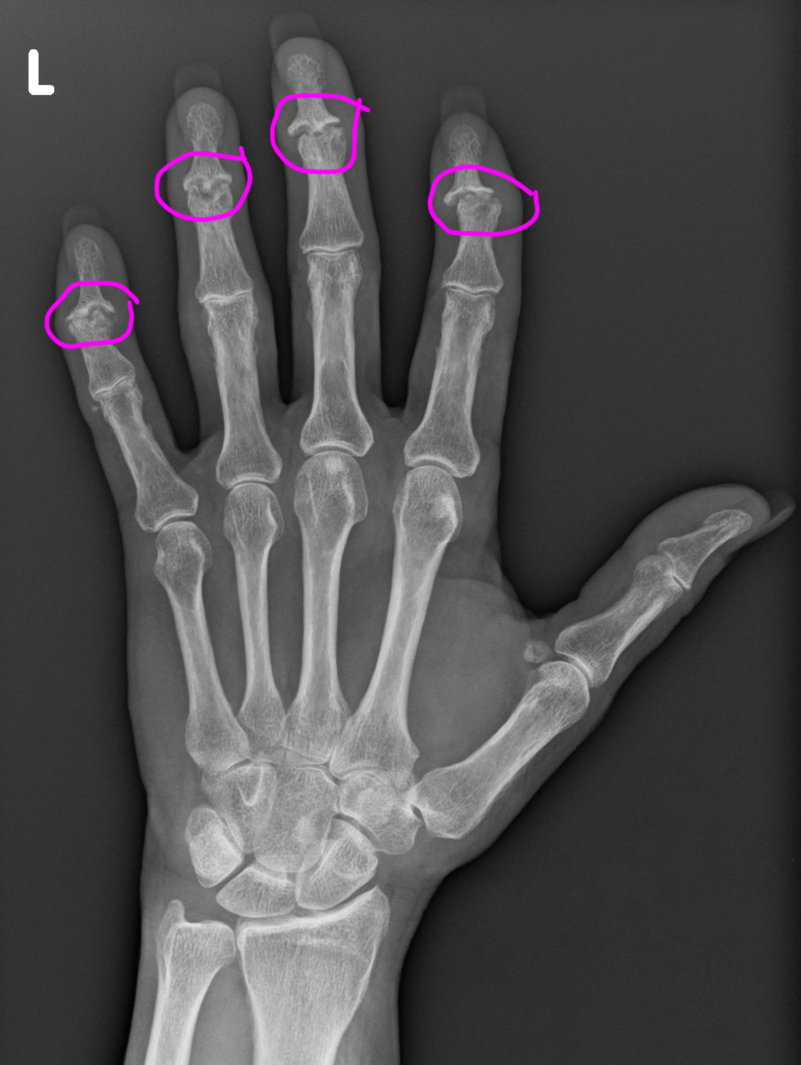
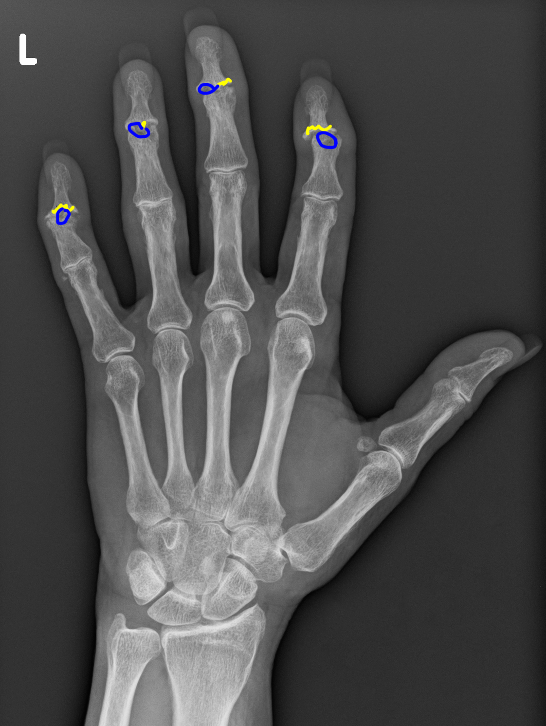
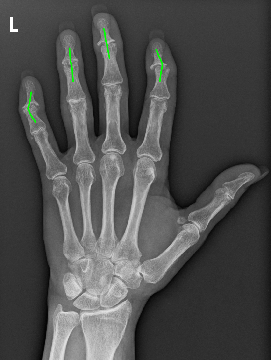
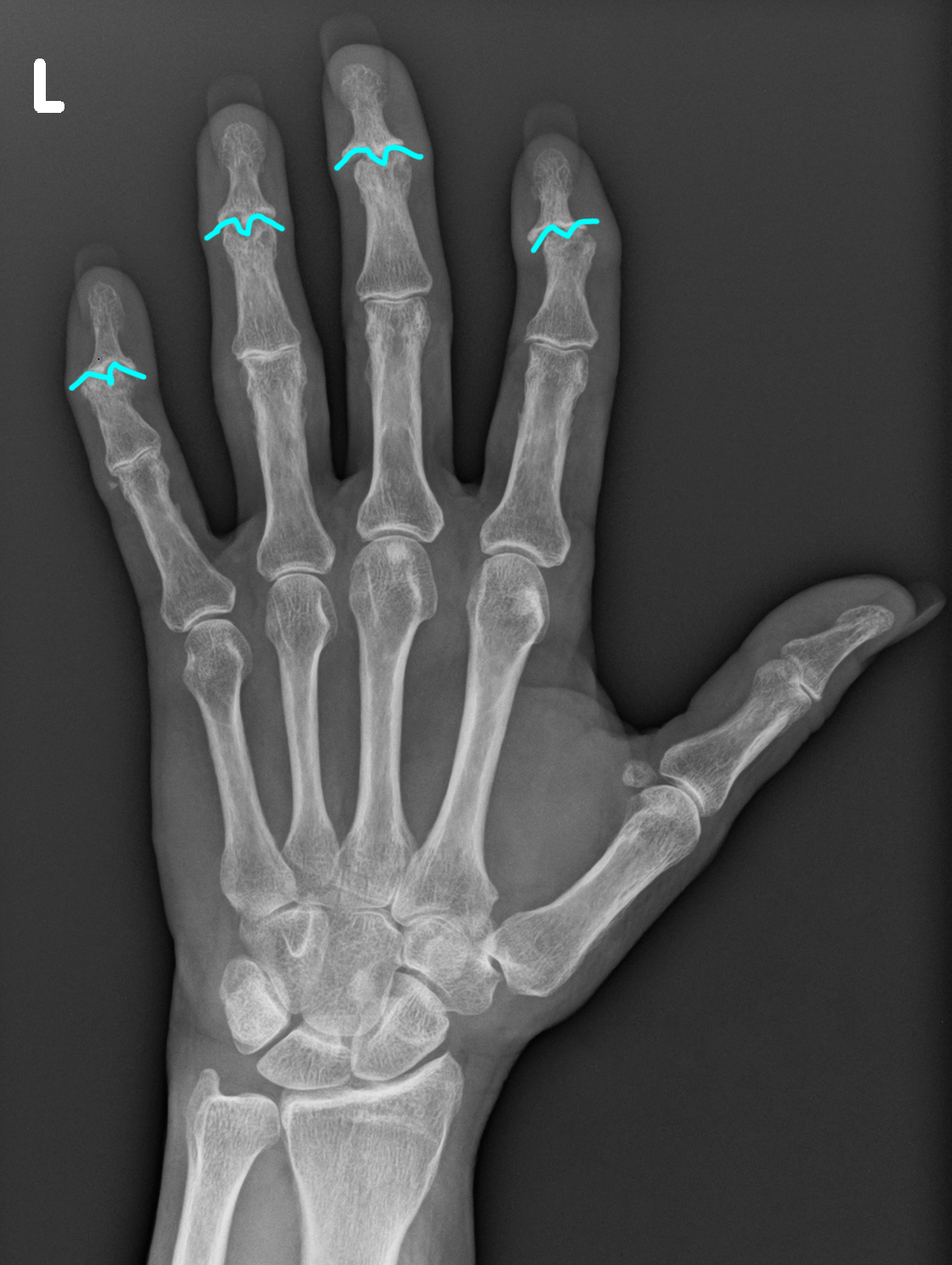
a) How would you analyze the distribution, mineralization and alignment of this case of a patient with hand pain?
In this case, the distribution is DIP joints, consistent with OA. However the mineralization at the involved joints includes both new bone and erosions (loss of bone). And the alignment at some joints is not disturbed but shows some ulnar deviation at other joints. This is an example of a variant of OA called 'erosive OA', with more inflammatory features leading to bone loss. The ultimate shape of involved joints has been called a 'gullwing' deformity.
b) What other imaging can be helpful in arthritis besides radiography?
Ultrasound can detect inflammatory changes in accessible joints. MRI can also be very sensitive for early erosions and bony reaction to inflammation, before changes can be seen on radiographs.
Case 2-arthritis
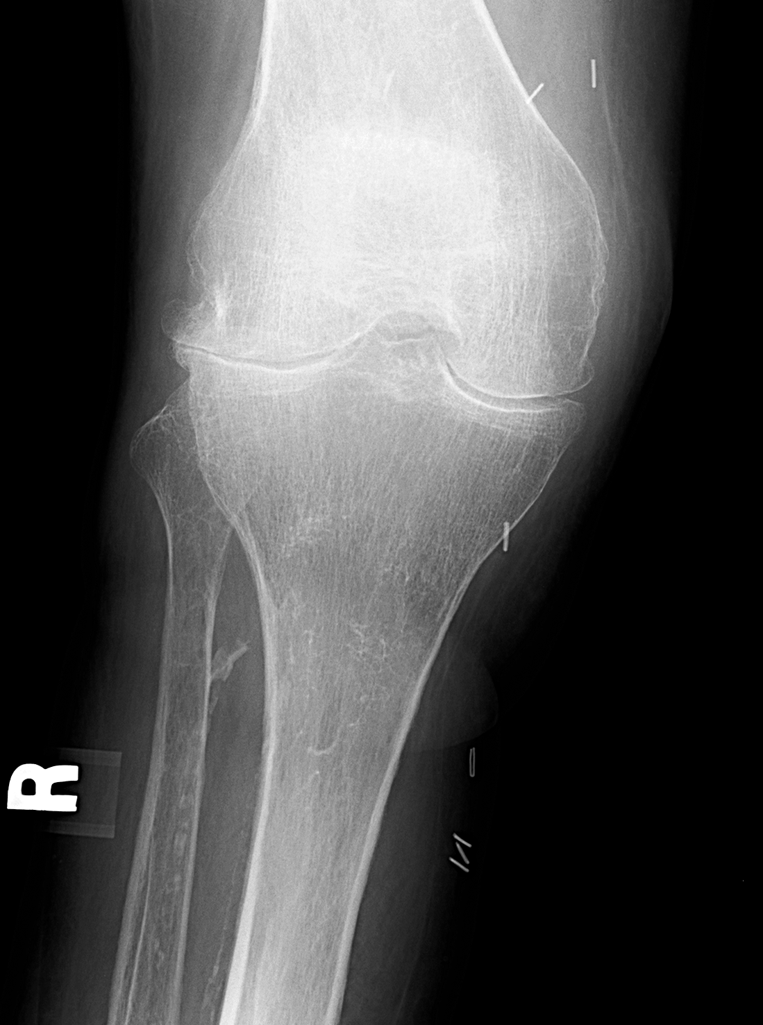
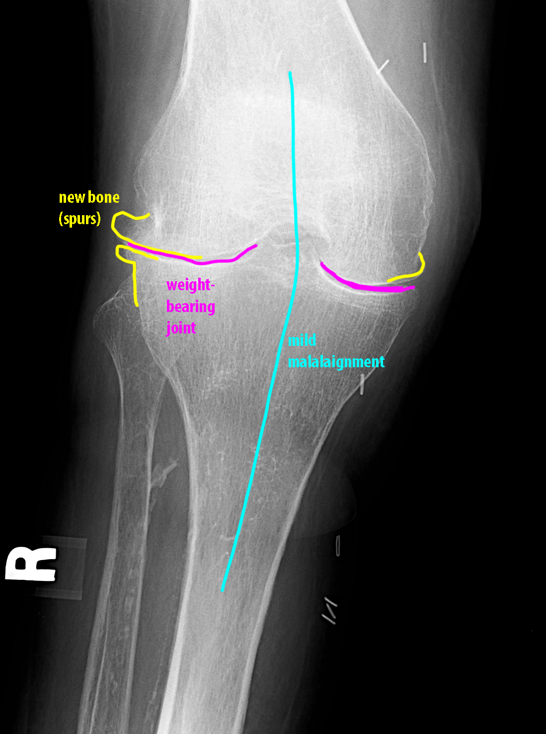
The hands are often involved in many types of arthritis, and offer a view of many joints at once. Large joints can also be involved, depending on the specific type of arthritis. Weight-bearing joints (knee, hip, spine) are often involved with OA, as shown in this example.
Question 3:
What features of this knee radiograph are most consistent with OA?
The knee is a weight-bearing joint, and both the medial and lateral joint compartments are narrowed, but the lateral is slightly more involved than the medial compartment. There is new bone formation with spurs, larger laterally than medially, and there are no erosions (bone loss) seen. There is mild valgus malalignment.
