

Case 3-systemic diseases
This case will demonstrate imaging findings in the chest and in the MSK system for diseases with widespread findings, particularly systemic lupus erythematosis (SLE) and scleroderma
Question 1:
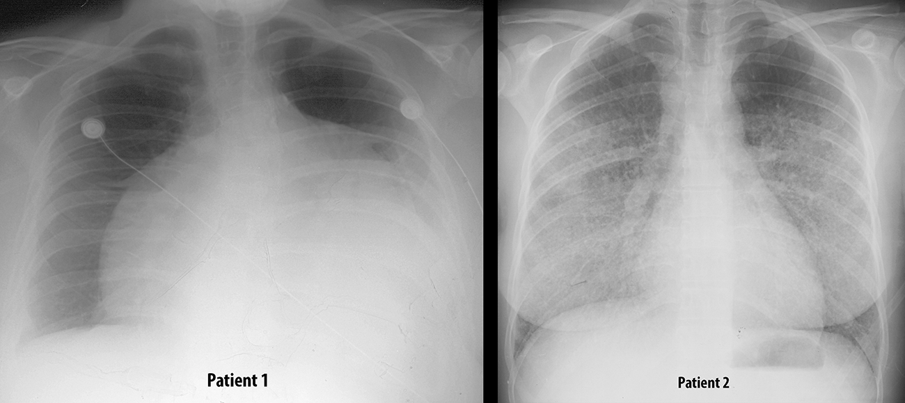
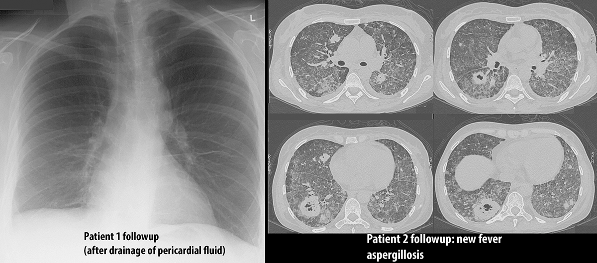
You are shown chest radiographs in two different patients with SLE. What diagnoses would you consider for the findings that are related to this underlying disease?
Patient 1 has a very abnormal cardiac contour, much too large and rather shapeless. This appearance is sometimes called a 'water-bottle heart' and suggests pericardial effusion, which is often seen in SLE along with pleural effusions. Patient 2 has diffuse hazy lung opacities bilaterally with a normal appearing heart, which could represent a number of different abnormalities, but one thing to consider in SLE is pulmonary hemorrhage. She did have hemoptysis as well.
Case 3-systemic diseases
Scleroderma is another systemic disease with many imaging manifestations in many organ systems.
Question 2:
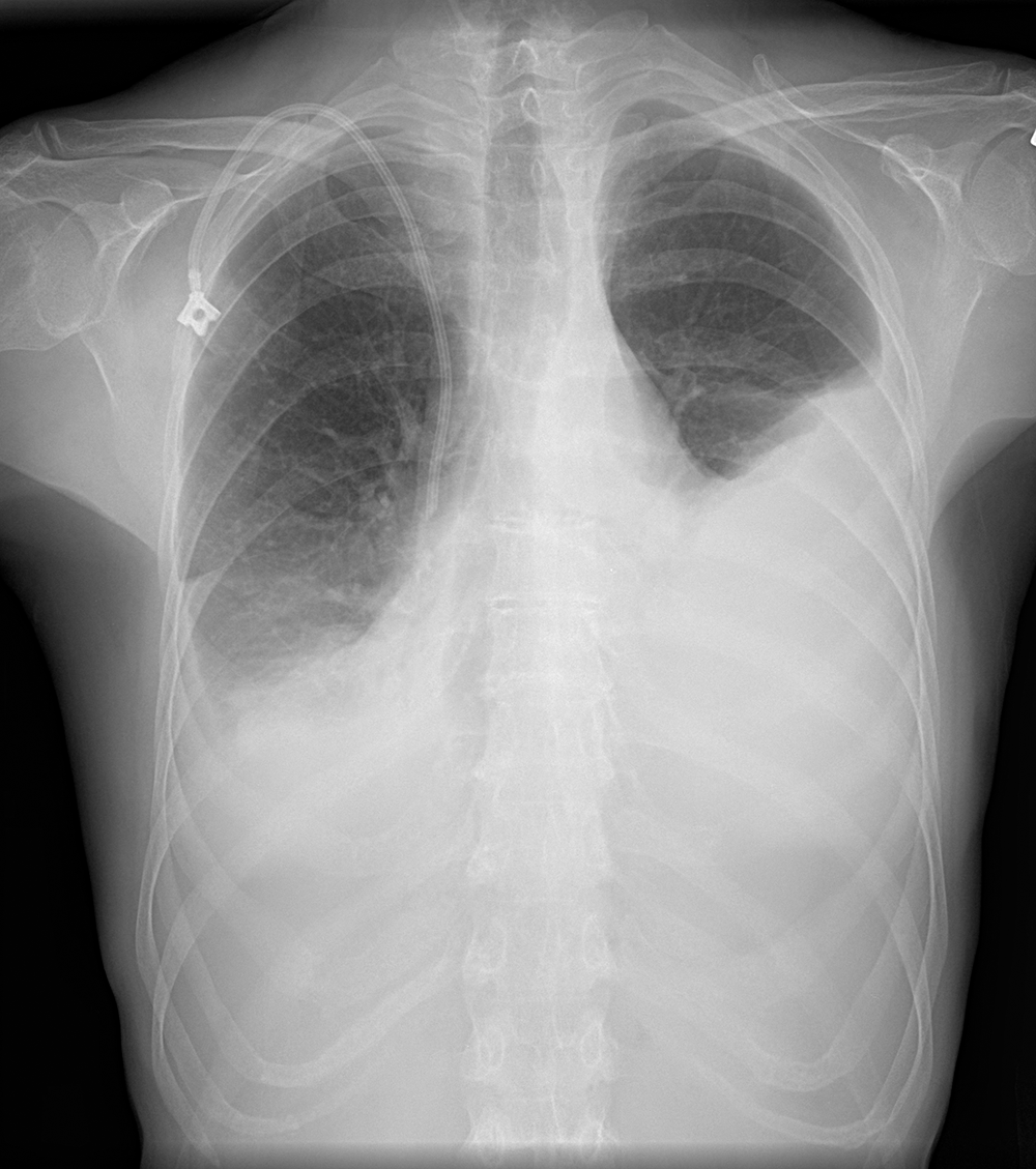
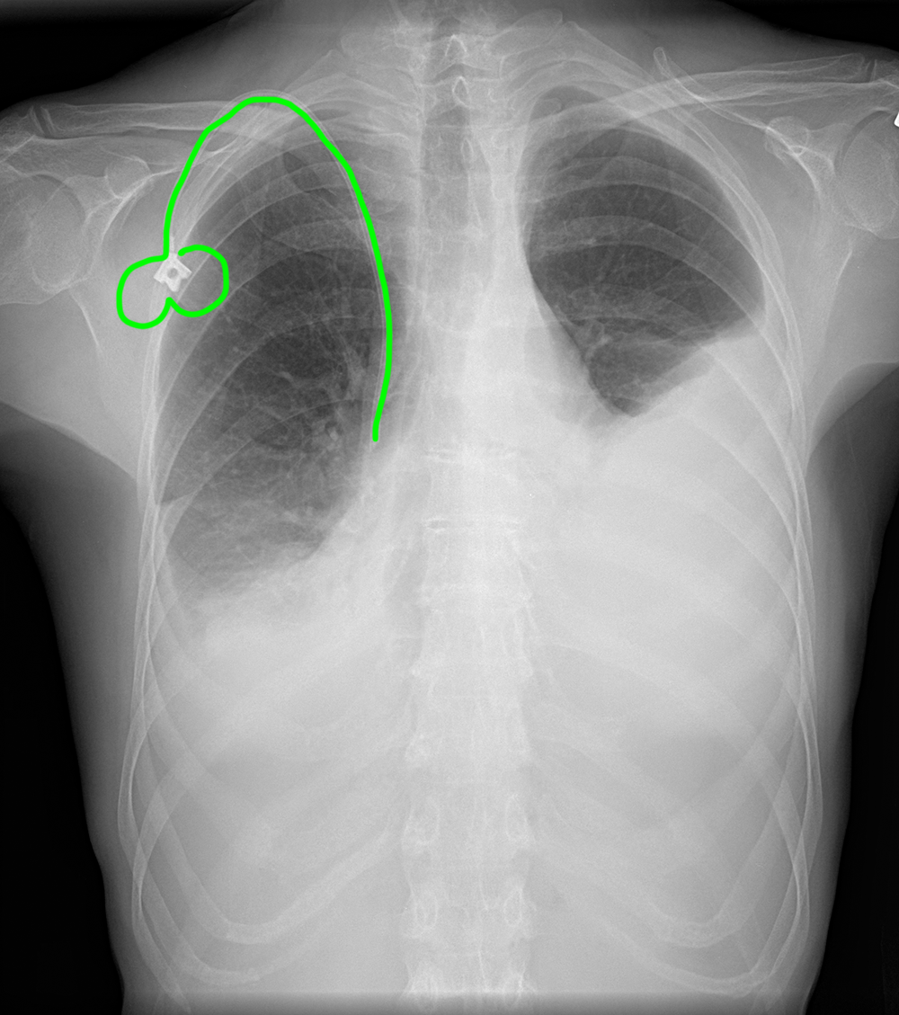
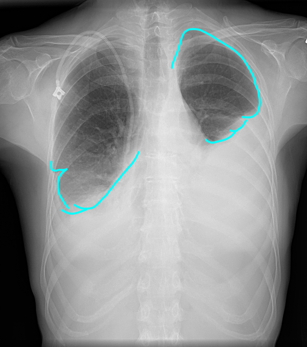
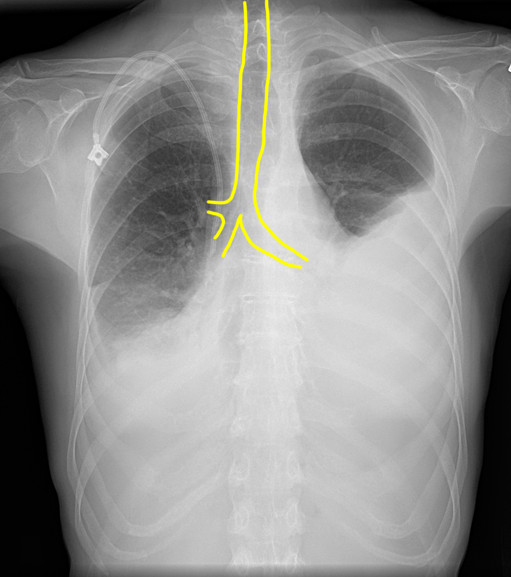
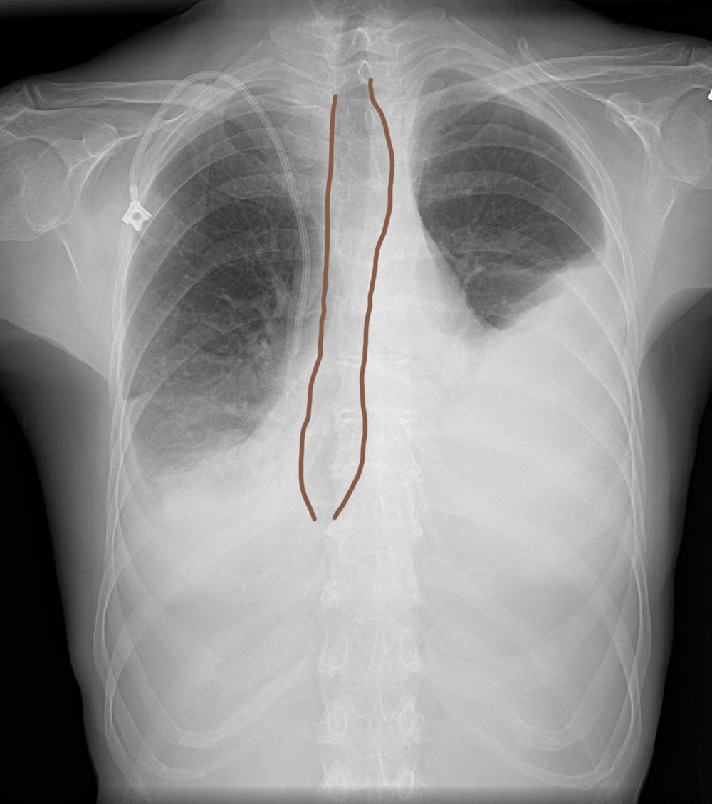
What do you think of the chest radiograph shown below of a patient with a known diagnosis of scleroderma?
There are obvious bilateral pleural effusions and a portacath in place. The other findings are more subtle. At first glance, the trachea looks a bit dilated, but if you follow it down, you can see that this is actually a dilated esophagus, as it continues down to the level of the diaphragm. Scleroderma can often result in GI disturbances and problems with motility.
Case 3-systemic diseases
The other body systems that are often involved in scleroderma are the skin and subcutaneous tissues and the lungs. You are shown images from two different patients with scleroderma to consider.
Question 3:
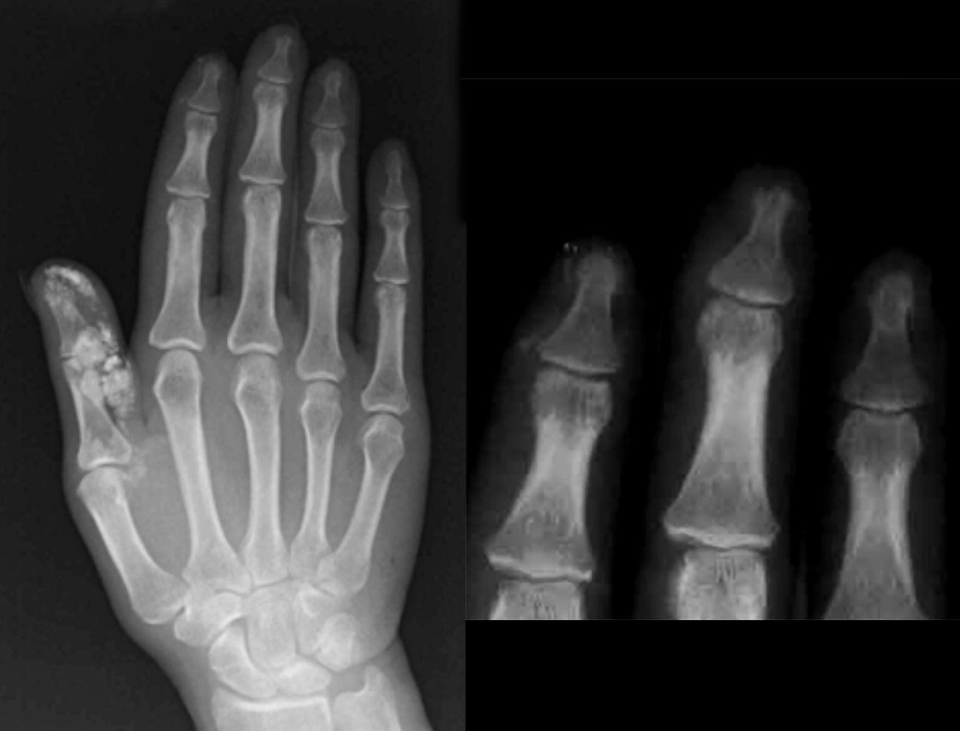
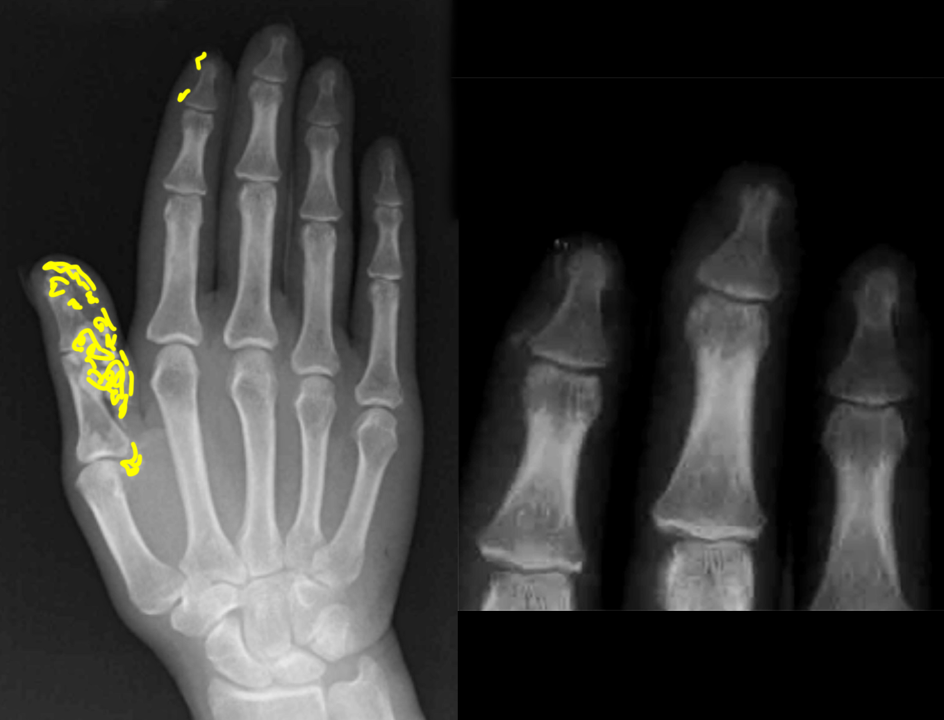
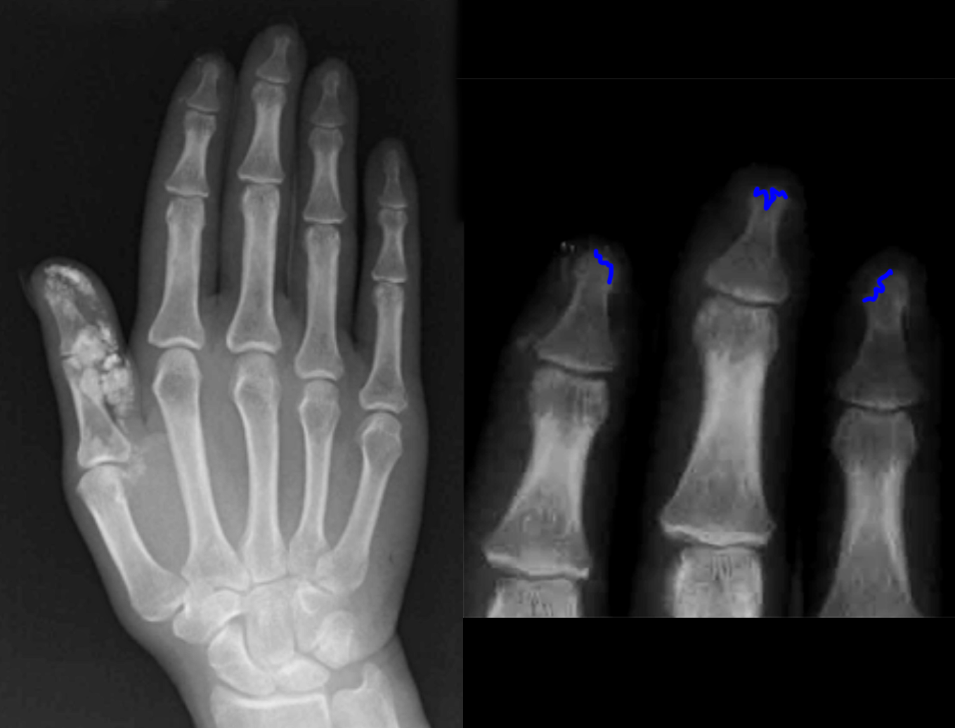
a) How would you describe the hand findings on this radiograph?
There is calcification in skin and/or subcutaneous tissues, mostly in the palmar surface of the thumb. There is also resorption of the distal tufts of several of the phalanges, called acro-osteolysis, which is a finding seen in scleroderma.
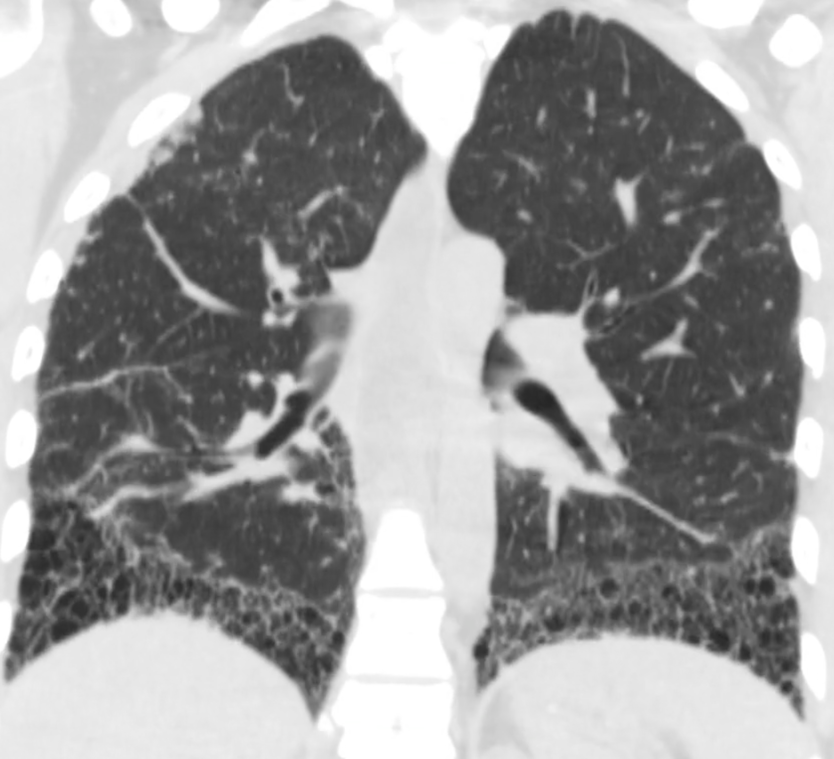
b) What is most striking about the distribution of the lung disease in Patient 2 below?
Scleroderma can cause interstitial lung disease often with a marked distribution limited to the lowermost portions of the lungs. The coronal reconstructed image you are shown makes this distribution more evident than might be noted on axial images.
