

Question 1:
a) To what does the term "ultrasound" refer?
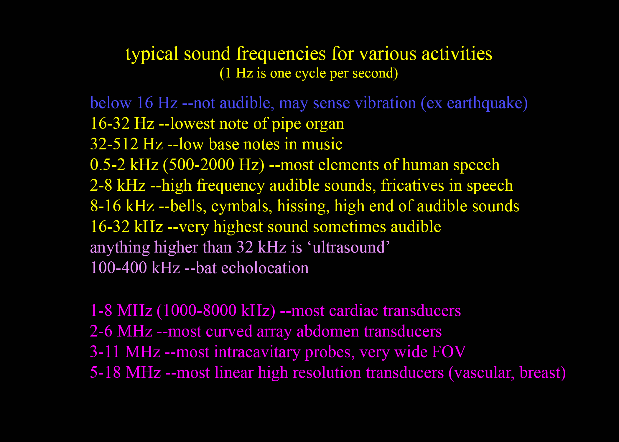
"Ultrasound" refers to sound waves with higher frequency than can be heard by the human ear. Most auditory sound is in the KHz range in terms of frequency, while medical US is in the MHz range.
b) Why do we use gel when performing an ultrasound exam?
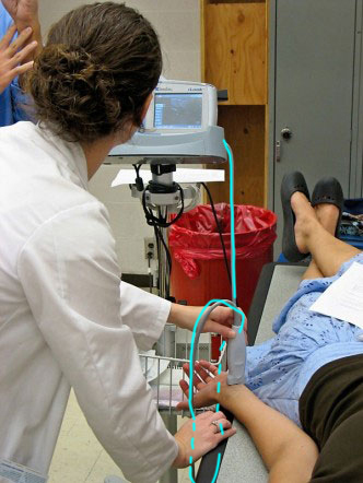
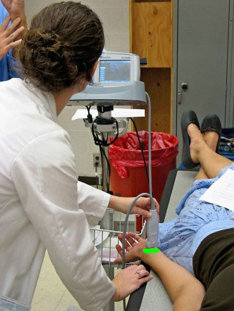
Air blocks high frequency sound waves, so the gel allows us to exclude any air that may be between the surface of the transducer (the piece of equipment you hold up to the patient to do the study, also called a probe) and the patient's skin.
c) How do we display the right and left parts of the body on an ultrasound image?
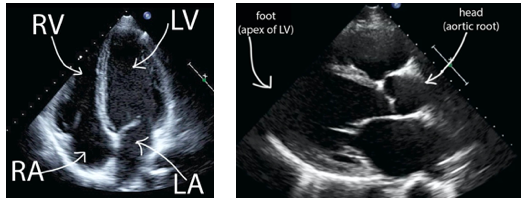
We use the same convention for displaying axial slices (or nearly axial slices) obtained with ultrasound as for other cross-sectional imaging (CT, MR). That means that the RIGHT side of the patient will be on the LEFT side of the image, as if you are approaching a supine patient from the feet (which is how you actually interact with patients). For the coronal plane, the same is true. The RIGHT side of the patient will be on the LEFT side of the image, as if you are approaching a seated or standing patient from the front (as you actually do when interacting with patients). For the sagittal plane, the usual radiological convention is to put the HEAD of the patient on the LEFT side of the image (and FEET to the right), but for echocardiography, the cardiologists display the images in the opposite orientation (Head to the Right). If you can recognize the anatomy, this flipped image should not cause you a problem.
Question 2:
a) What are advantages of using higher frequency for ultrasound examinations, and what are disadvantages?
The higher the frequency, the more fine detail that can be displayed. But the higher the frequency, the less penetration that is possible in tissues. So for very high resolution transducers, you can only visualize a few cm deep into the body, so they are mostly used to examine very superficial structures.
b) What does the term 'footprint' mean in ultrasound?
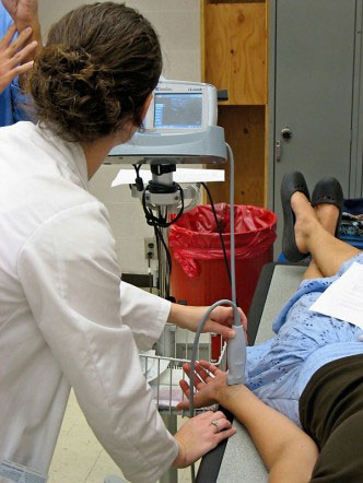
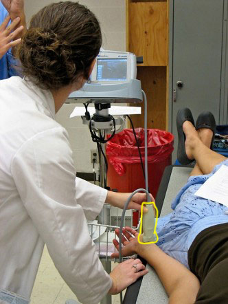
The 'footprint' of a transducer describes the shape and size of the part that touches the patient's body. For examining large structures, like the liver, a large footprint is helpful so that the images show a broad field of view. But for examining the heart, a small footprint is needed, since the probe must send sound through a very small area (between ribs or alongside the sternum).
Technology
Images show orientation for axial and sagittal cardiac ultrasound. This study is also called 'cardiac echo', or 'sonography'.
Further Explanation:
Image on the left is a four-chamber view of the heart in an angled axial plane. The RIGHT side of the heart is on the LEFT side of the image. The image on the right is a para-sternal long-axis view of the left part of the heart, in an angled sagittal plane. The INFERIOR part of the patient's body is to the LEFT (which is the opposite of the orientation for all other ultrasound examinations, but this is the standard view used by cardiologists).
