

Question 1:
What tissues or materials appear WHITE on ultrasound images?
×
Answer:
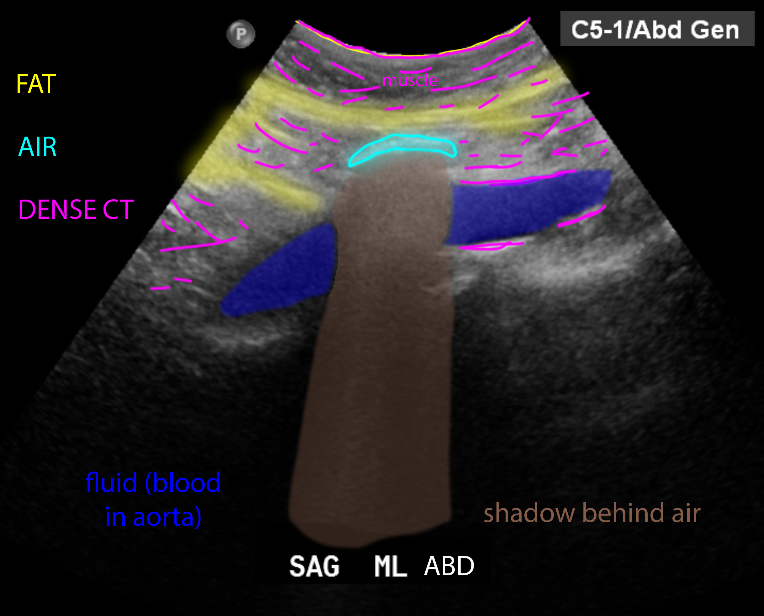
White areas on ultrasound images occur because a large amount of the sound wave energy is reflected back to the transducer. Tissues that do this include BONE (or calcifications), AIR, and DENSE CONNECTIVE TISSUE. For BONE and AIR, virtually all of the sound is reflected, so a white line is seen at the surface of the material that is closest to the transducer. White areas can be called or 'echogenic', 'hyper-echoic'.
White areas on ultrasound images occur because a large amount of the sound wave energy is reflected back to the transducer. Tissues that do this include BONE (or calcifications), AIR, and DENSE CONNECTIVE TISSUE. For BONE and AIR, virtually all of the sound is reflected, so a white line is seen at the surface of the material that is closest to the transducer. White areas can be called or 'echogenic', 'hyper-echoic'.
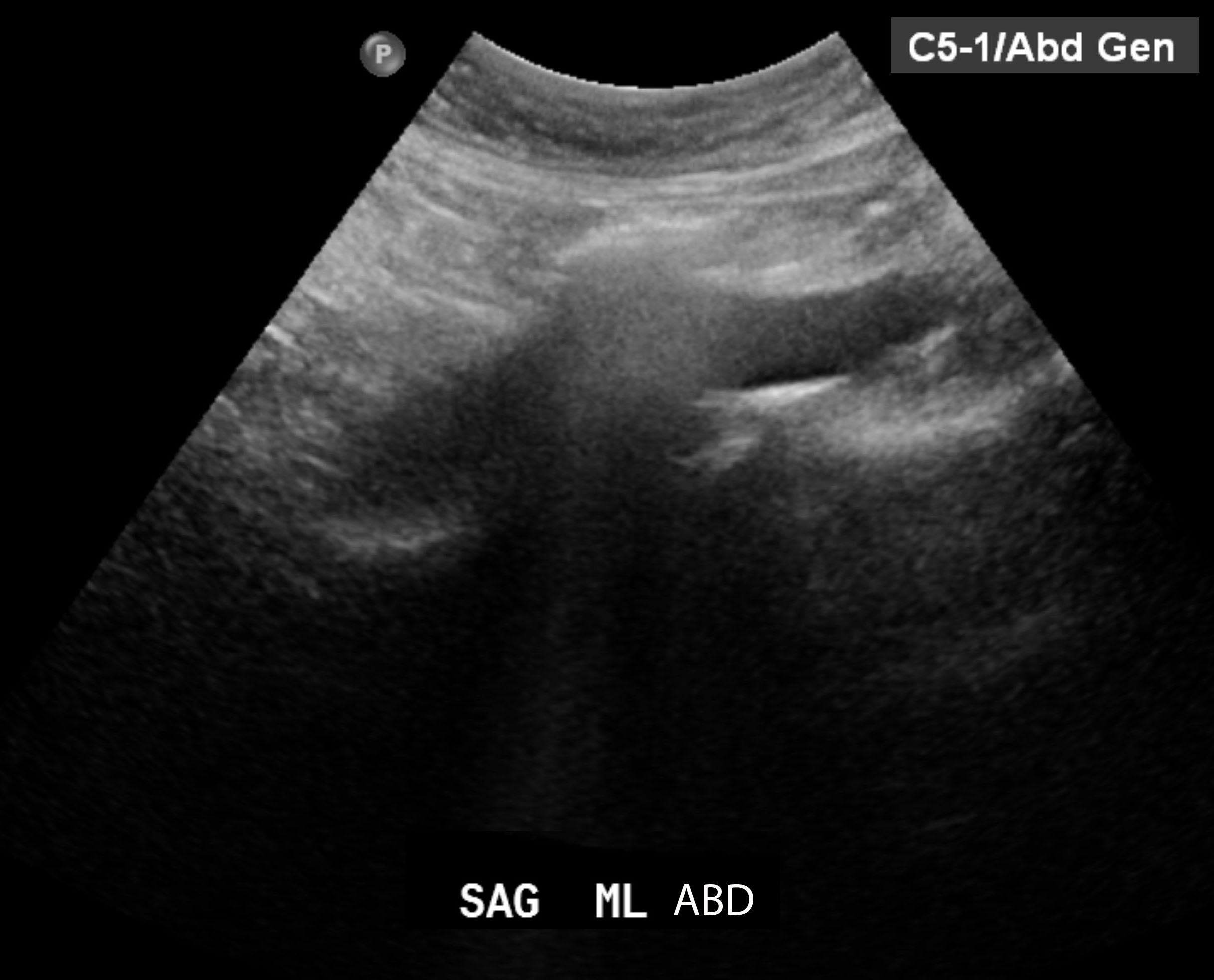
The image shown is an sagittal view of the abdomen in the midline.


Question 2:
What tissues or materials appear DARK or BLACK on ultrasound?
×
Answer:
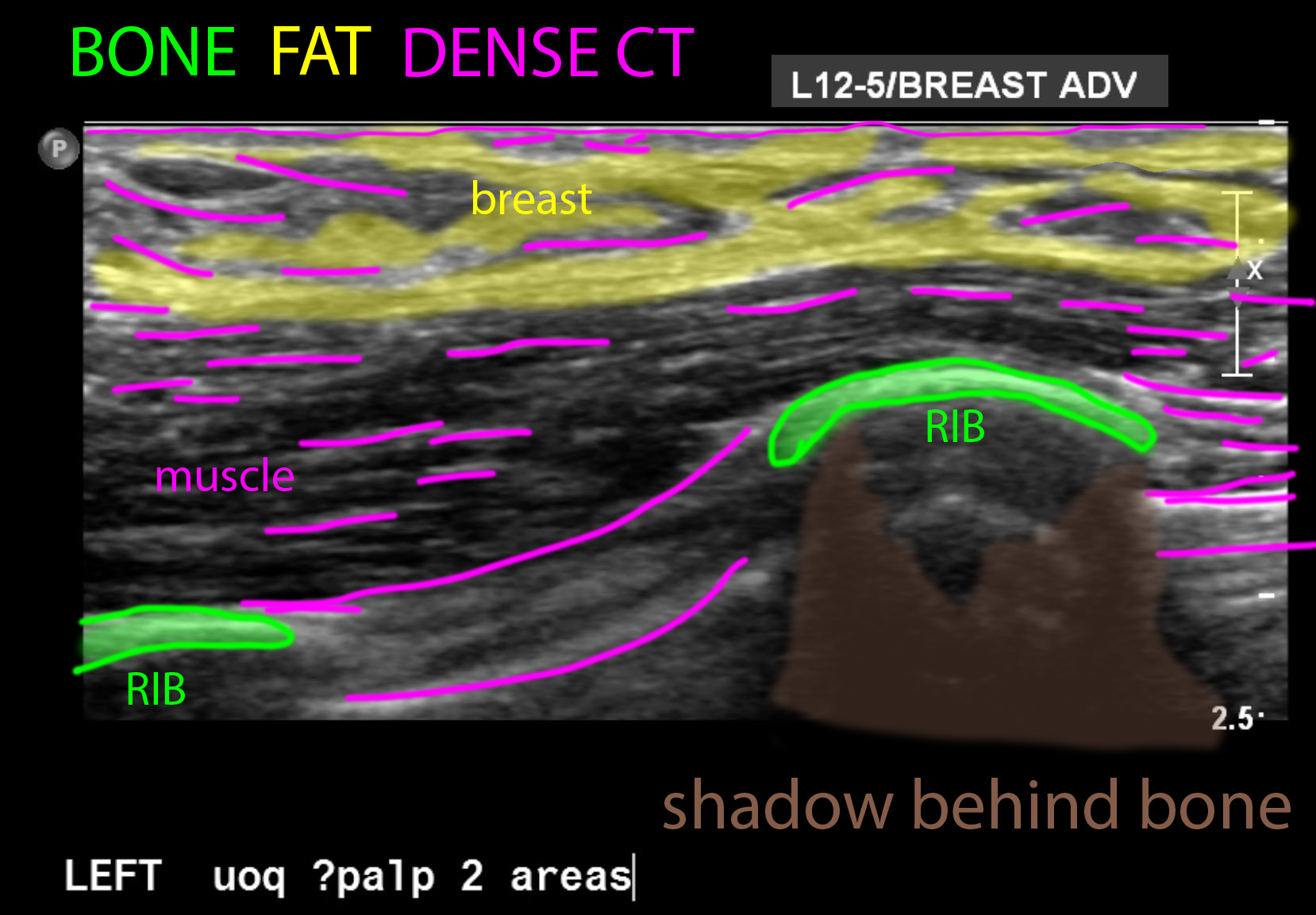
The main material that appears BLACK on ultrasound is fluid. This can be blood, urine, bile, cerebrospinal fluid, or the fluid within the eyeball. Anything that is liquid will transmit the sound wave energy rather than reflecting it, producing a dark area of the image. Other tissues (like muscle) appear various shades of grey. Another cause of BLACK areas on an ultrasound image is an artifact called SHADOWING. When a very bright reflector (like bone or air) is encountered, all of the sound is reflected back, leaving no sound deep to the reflecting structure to generate image data. So a band of dark is seen distal to the structure. The darkness of shadowing varies with the type of material that is producing it. Shadowing from bone or calcification tends to be very dark, while shadowing from air is often not as dark. Dark areas on on US image can be called 'anechoic' (if completely BLACK), or 'hypo-echoic' (if they are dark but still have a few echos). Another term that can be used is 'echo-lucent'.
The main material that appears BLACK on ultrasound is fluid. This can be blood, urine, bile, cerebrospinal fluid, or the fluid within the eyeball. Anything that is liquid will transmit the sound wave energy rather than reflecting it, producing a dark area of the image. Other tissues (like muscle) appear various shades of grey. Another cause of BLACK areas on an ultrasound image is an artifact called SHADOWING. When a very bright reflector (like bone or air) is encountered, all of the sound is reflected back, leaving no sound deep to the reflecting structure to generate image data. So a band of dark is seen distal to the structure. The darkness of shadowing varies with the type of material that is producing it. Shadowing from bone or calcification tends to be very dark, while shadowing from air is often not as dark. Dark areas on on US image can be called 'anechoic' (if completely BLACK), or 'hypo-echoic' (if they are dark but still have a few echos). Another term that can be used is 'echo-lucent'.
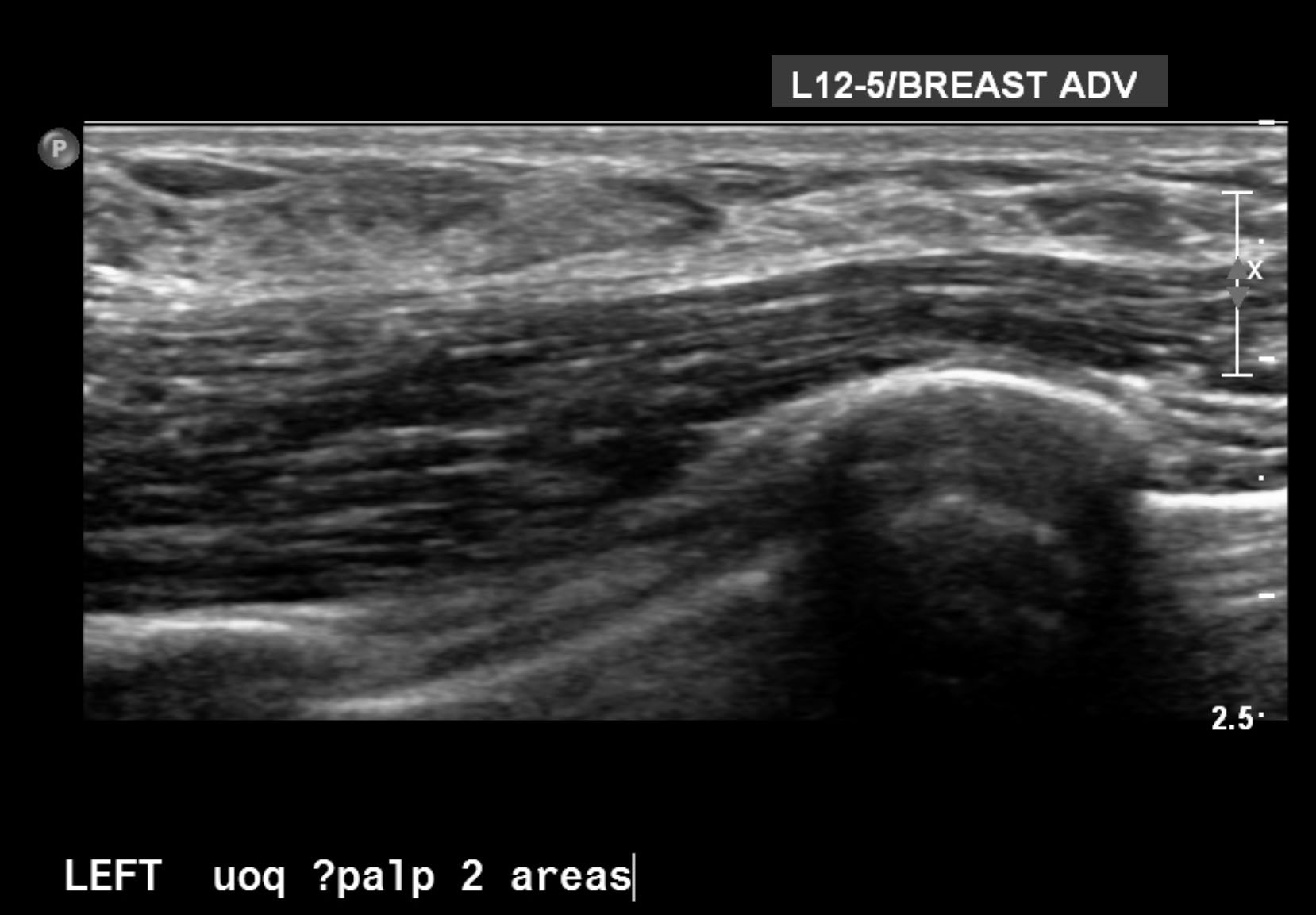
The image shown is an axial view of the chest wall in the region of the breast.


