

Case 2
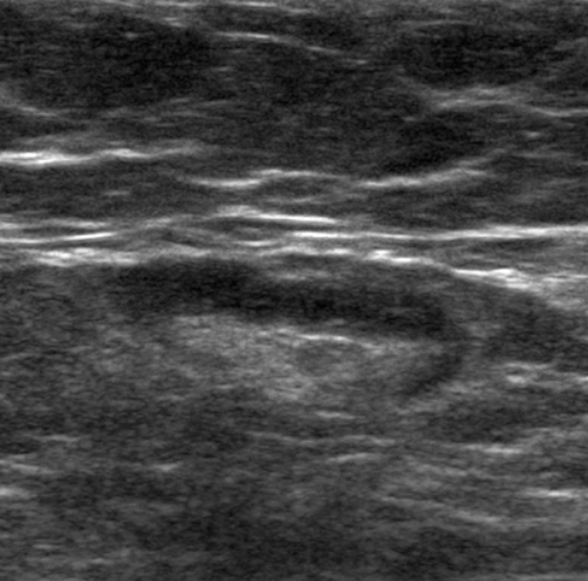
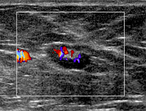
This is an ultrasound study of a structure that could be found in the neck.
Question 1:
What is this normal structure?
×
Answer:
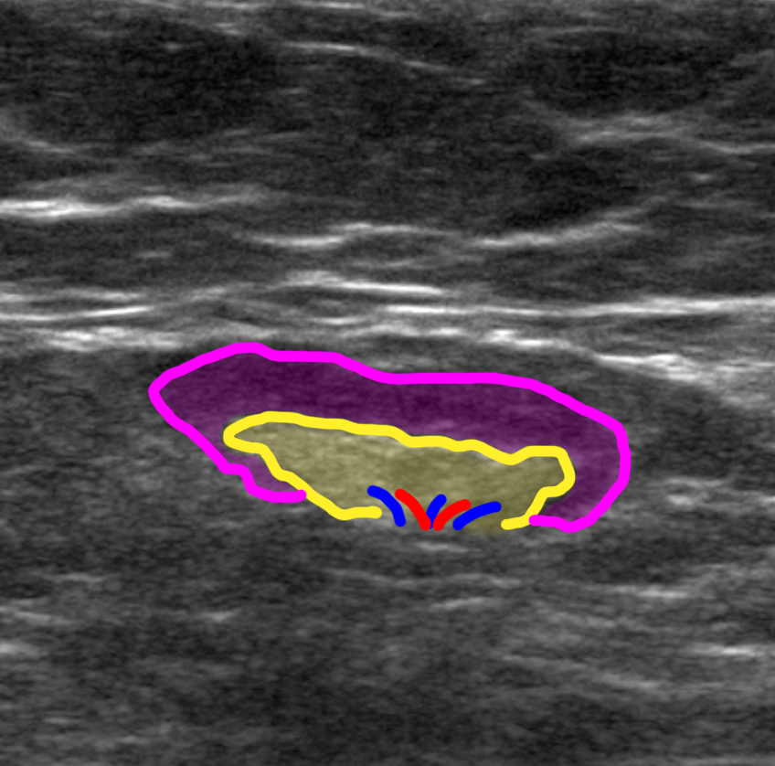
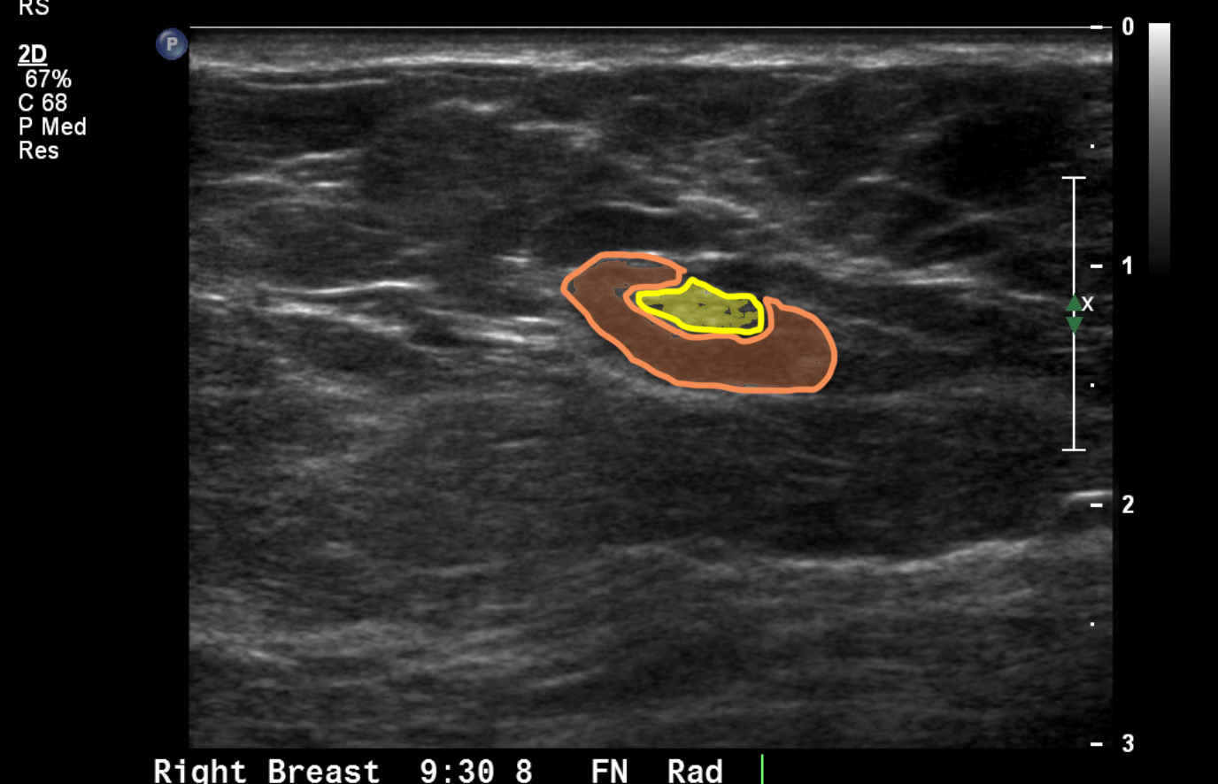
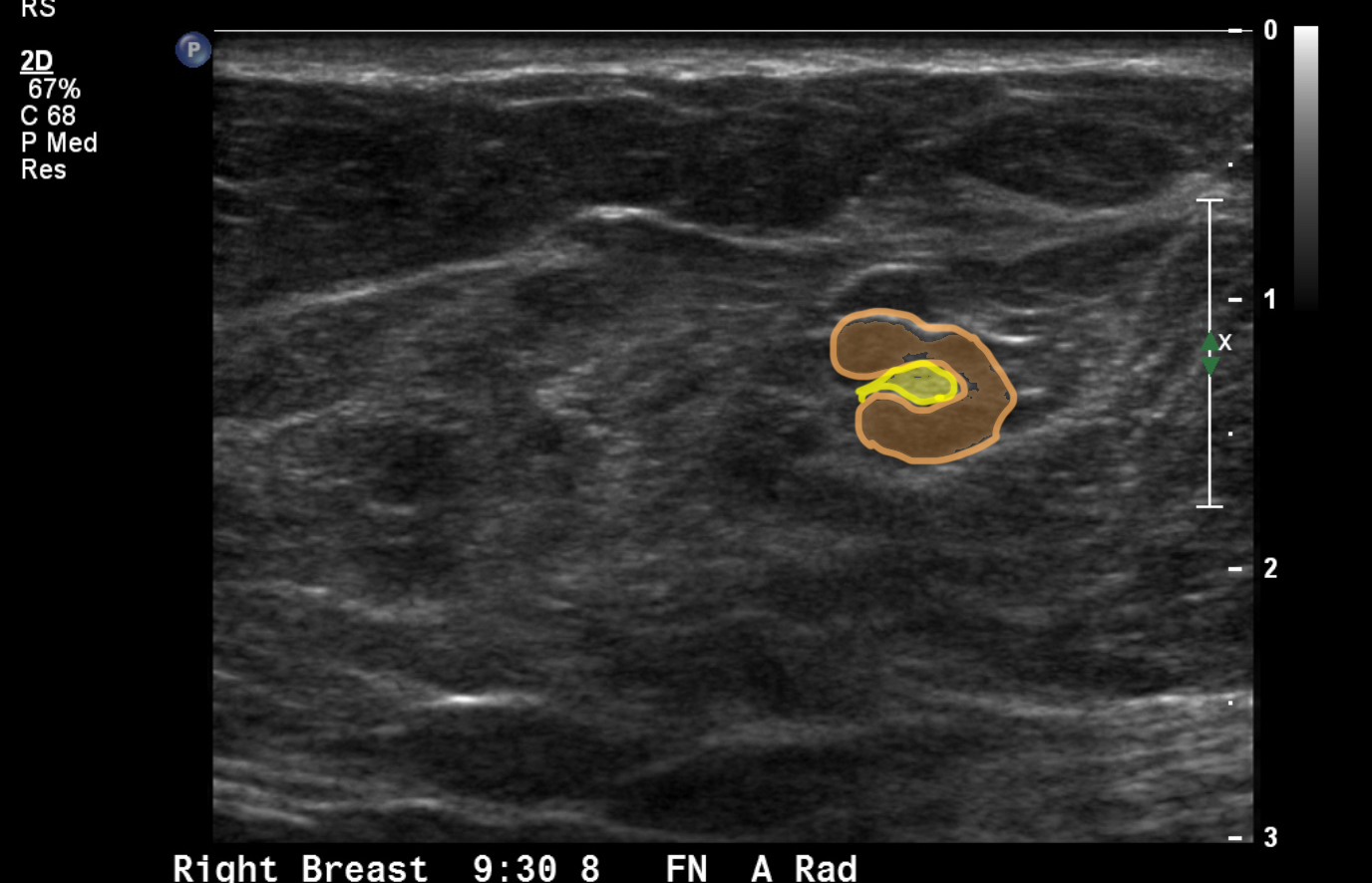
This is the typical appearance of a lymph node on ultrasound. Just as for other organs, like the thyroid or the kidney, we typically try to get perpendicular views of the node, which are typically labeled as 'longitudinal' and 'transverse', since they refer to the structure itself and may not correspond to classic 'sagittal', 'coronal' or 'axial' planes. The labels below show the appearance of the cortex and medulla of the node. The medulla is typically echogenic because it contains fat. The cortex is typically hypoechoic due to the uniform sheets of lymphocytes present.
This is the typical appearance of a lymph node on ultrasound. Just as for other organs, like the thyroid or the kidney, we typically try to get perpendicular views of the node, which are typically labeled as 'longitudinal' and 'transverse', since they refer to the structure itself and may not correspond to classic 'sagittal', 'coronal' or 'axial' planes. The labels below show the appearance of the cortex and medulla of the node. The medulla is typically echogenic because it contains fat. The cortex is typically hypoechoic due to the uniform sheets of lymphocytes present.
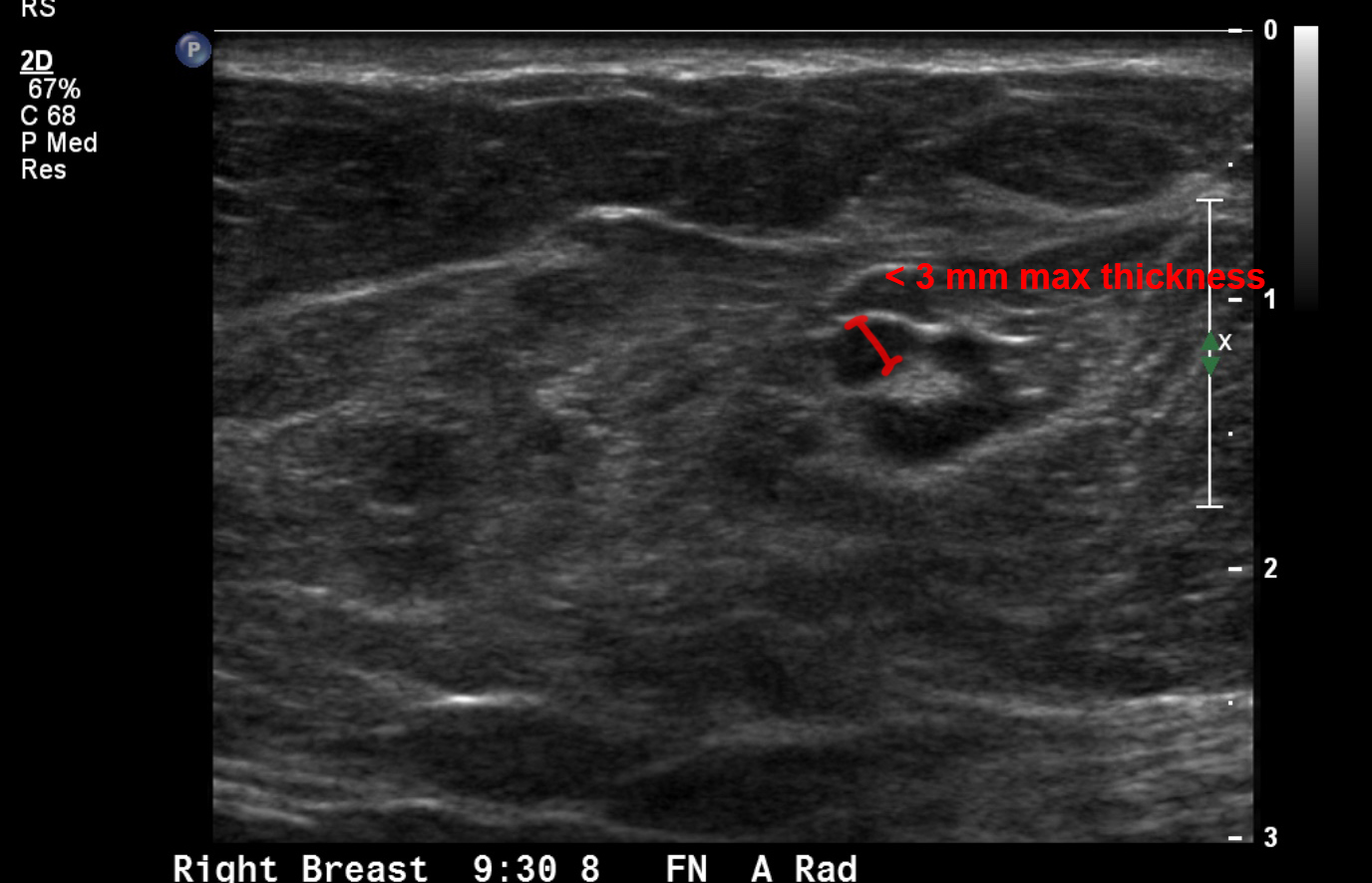
Case 2
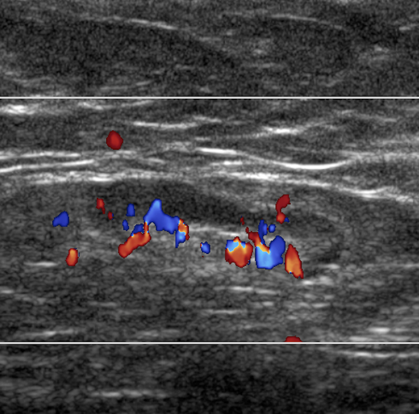
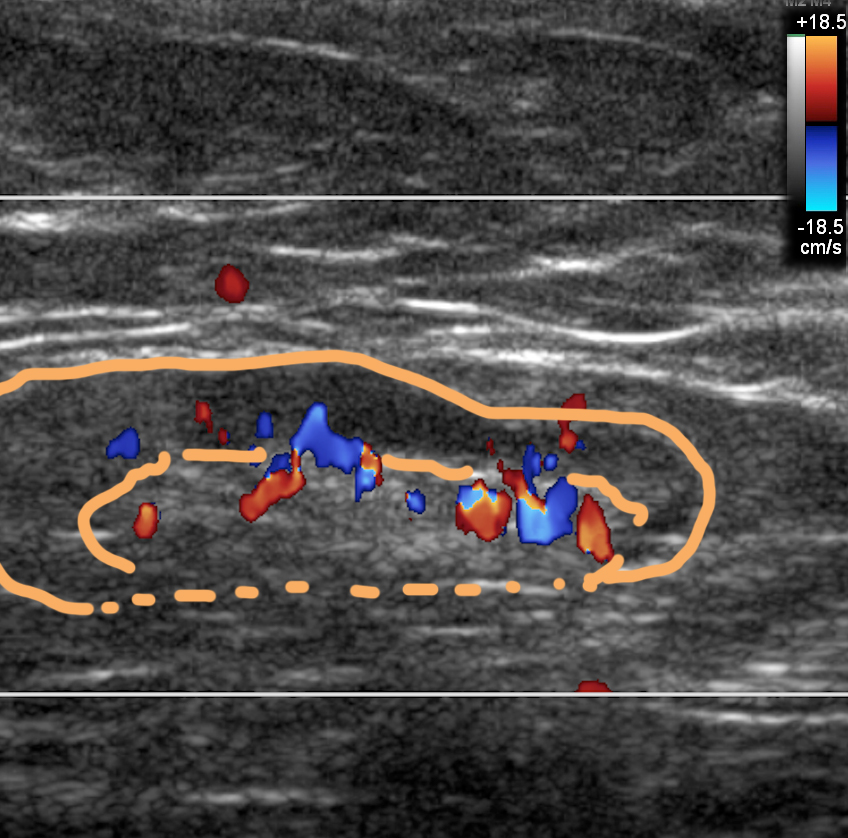
This is another ultrasound of a lymph node, located in the axilla. No matter where nodes are located, they typically look similar in structure.
Question 2:
What is the study shown?
×
Answer:
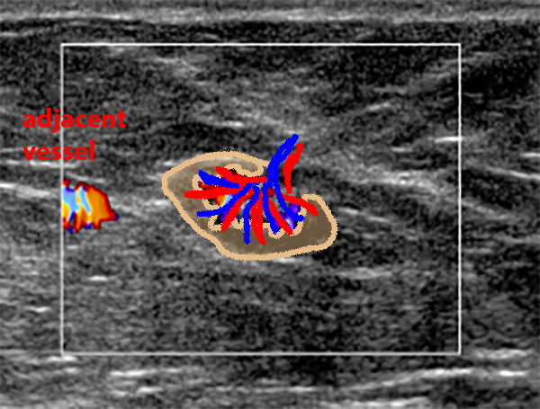
This is a doppler study of the node. This will typically show a radiating array of vessels passing in an orderly fashion from the medulla out into the cortex of the node. This is helpful in confirming that this is actually a node and not some other type of mass. The cortex should be uniform and thin in a normal node. An inflamed node or a node containing tumor will often show thickened cortex, and in the case of tumor, the cortex may appear lumpy and may extend inward to obliterate the echogenic medulla.
This is a doppler study of the node. This will typically show a radiating array of vessels passing in an orderly fashion from the medulla out into the cortex of the node. This is helpful in confirming that this is actually a node and not some other type of mass. The cortex should be uniform and thin in a normal node. An inflamed node or a node containing tumor will often show thickened cortex, and in the case of tumor, the cortex may appear lumpy and may extend inward to obliterate the echogenic medulla.
