

Case 3
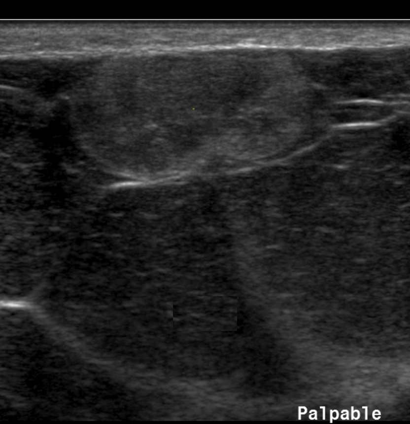
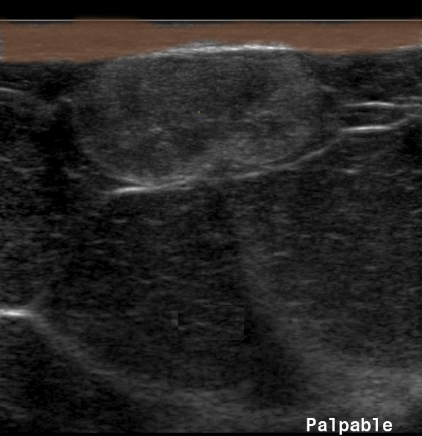
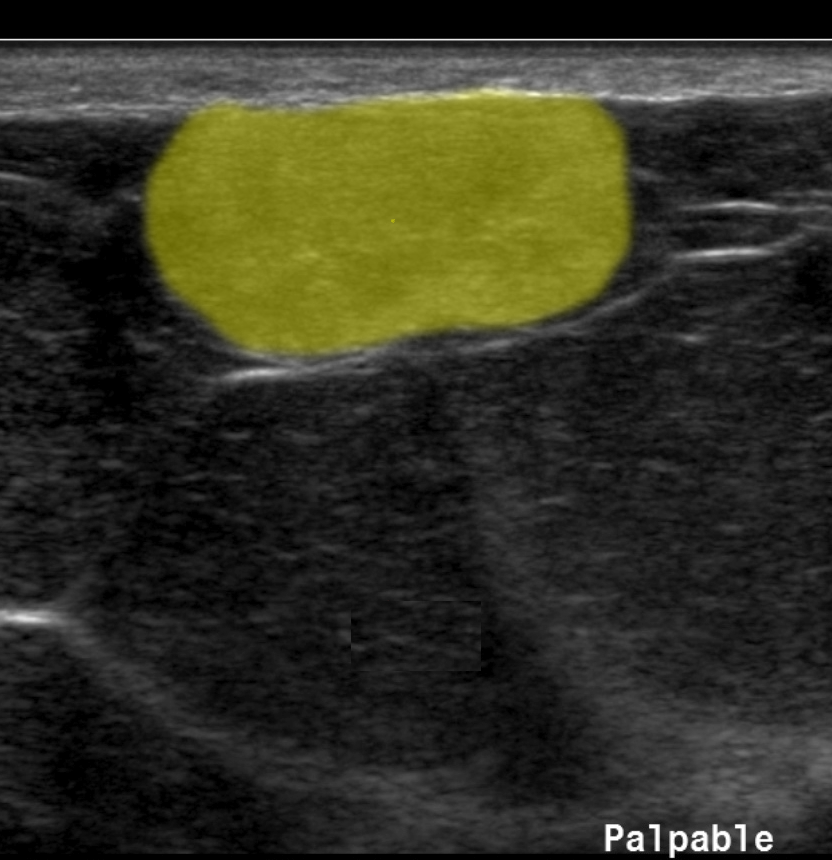
This is a superficial ultrasound of a lesion that can appear anywhere in the body, including the neck. It was palpable.
Question 1:
What does the echogenicity of this mass suggest about its likely etiology?
×
Answer:
It is fairly uniformly echogenic, but with no shadowing. This suggests the presence of fat, which is typically echogenic (white) but does not shadow. An echogenic area with shadowing could represent calcification. The mass is very smoothly marginated and oval, and its location just below the dermis explains why it is palpable. It is just under the skin. It was soft and squishy, which also goes along with a fatty lesion. This appearance is consistent with a lipoma, a benign growth of fat cells that is quite common and benign.
It is fairly uniformly echogenic, but with no shadowing. This suggests the presence of fat, which is typically echogenic (white) but does not shadow. An echogenic area with shadowing could represent calcification. The mass is very smoothly marginated and oval, and its location just below the dermis explains why it is palpable. It is just under the skin. It was soft and squishy, which also goes along with a fatty lesion. This appearance is consistent with a lipoma, a benign growth of fat cells that is quite common and benign.
Case 3
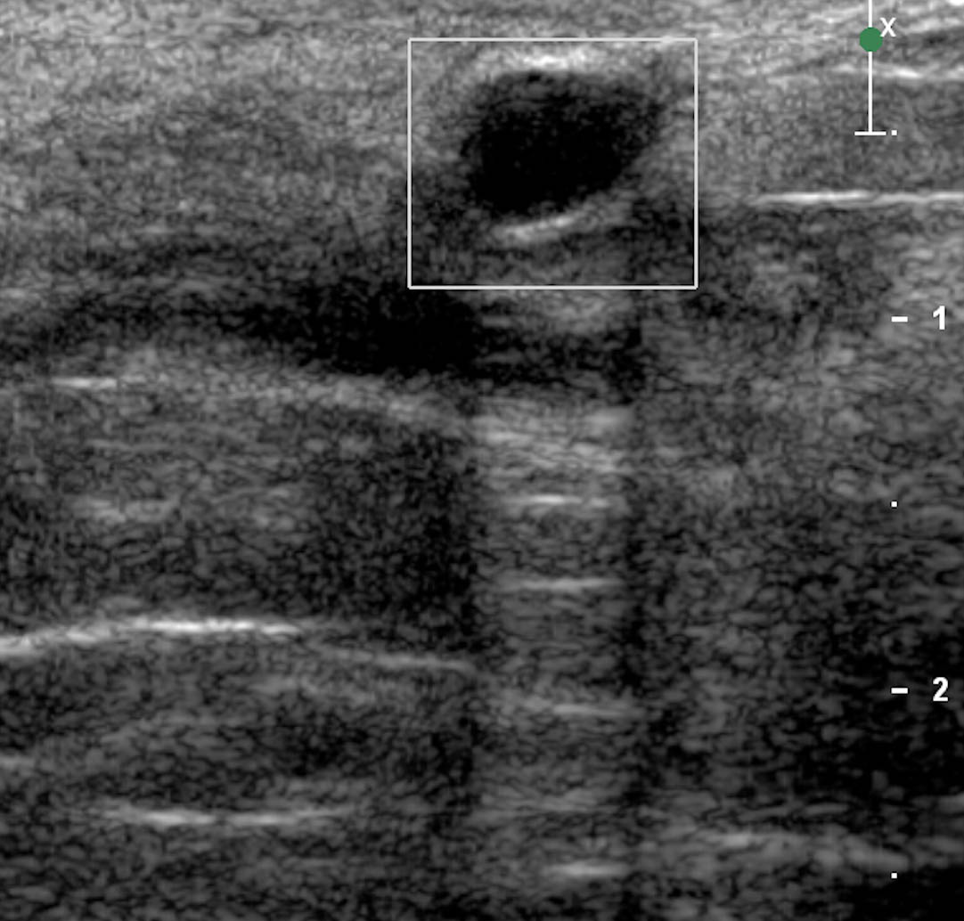
This is another common ultrasound lesion that can occur in many parts of the body, including the thyroid. This example is in the breast.
Question 2:
How would you describe the ultrasound appearance of this lesion?
×
Answer:
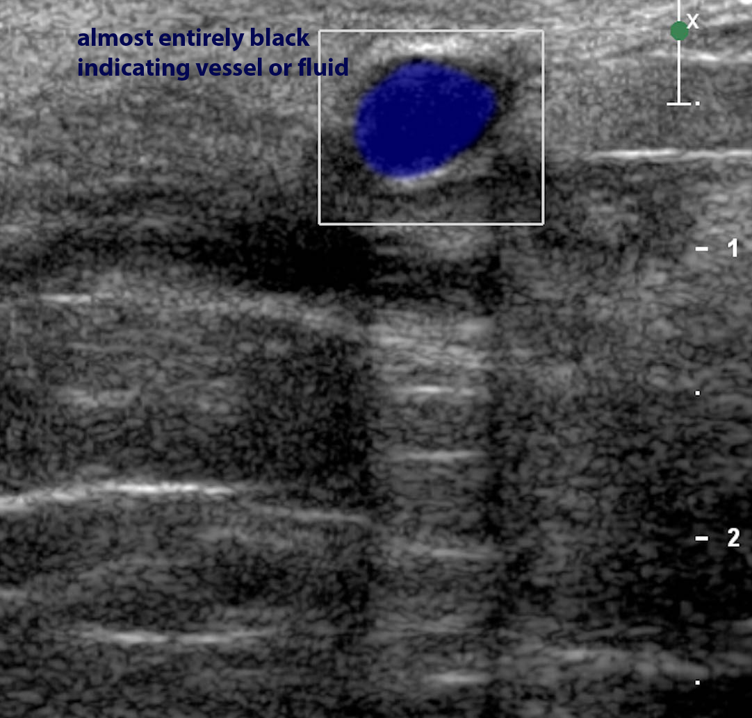
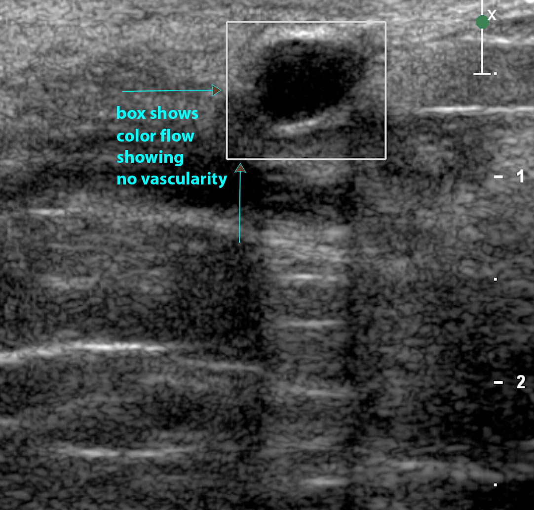
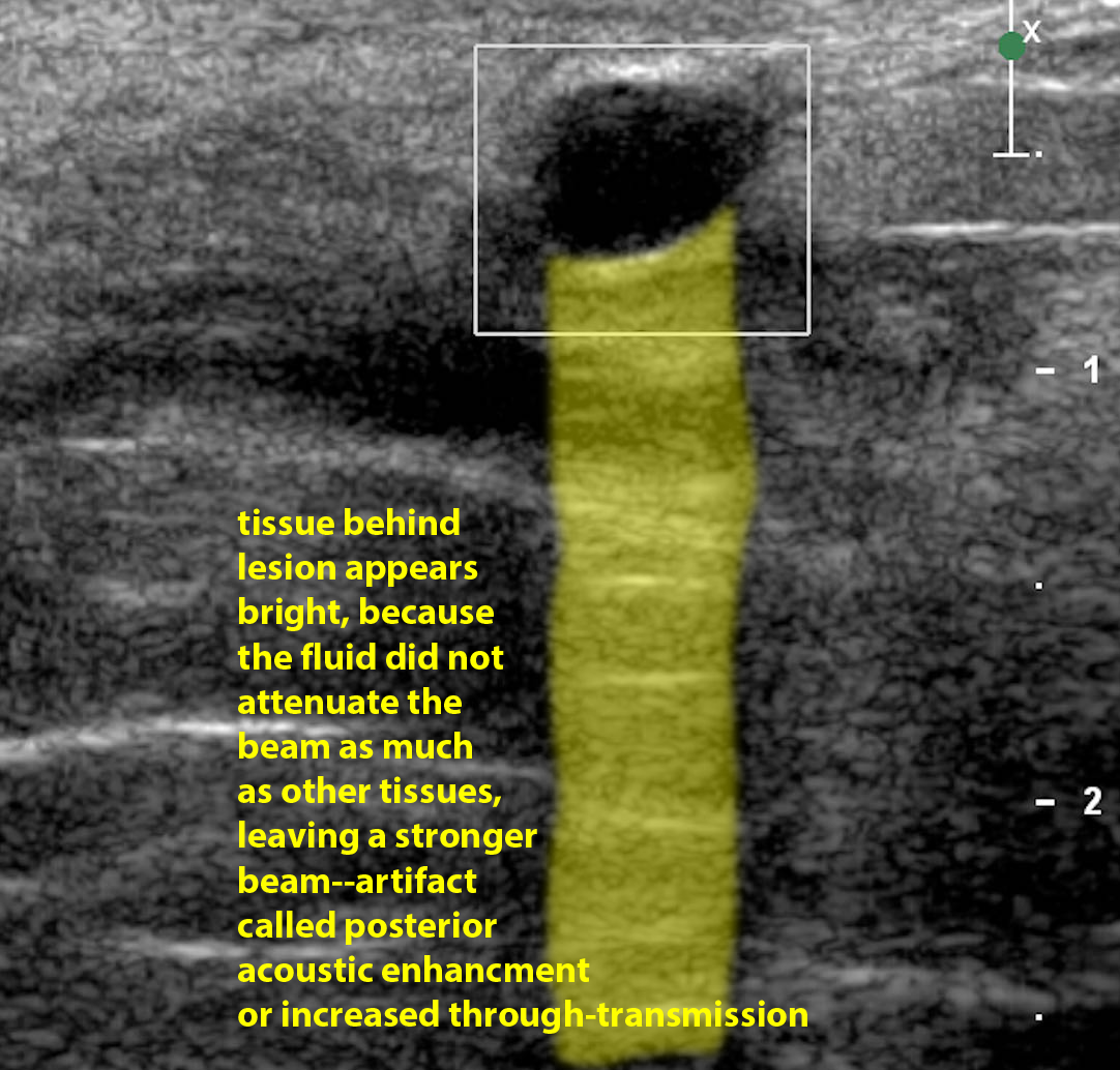
It is virtually black and homogeneous, so it can be called 'anechoic'. This usually means either a vascular structure or something filled with fluid. In this case, the lesion was round in every imaging plane, so it is not tubular but spherical in shape, consistent with a cyst. In most parts of the body, cysts that are smooth and round like this are benign. Note the artifact distal to the lesion--a bright band indicating 'posterior acoustic enhancement' also called 'increased through-transmission'. This artifact confirms that the lesion consists of fluid, which is not attenuating the ultrasound beam as much as normal tissue, resulting in a brighter appearance of the tissue just beyond the fluid collection.
It is virtually black and homogeneous, so it can be called 'anechoic'. This usually means either a vascular structure or something filled with fluid. In this case, the lesion was round in every imaging plane, so it is not tubular but spherical in shape, consistent with a cyst. In most parts of the body, cysts that are smooth and round like this are benign. Note the artifact distal to the lesion--a bright band indicating 'posterior acoustic enhancement' also called 'increased through-transmission'. This artifact confirms that the lesion consists of fluid, which is not attenuating the ultrasound beam as much as normal tissue, resulting in a brighter appearance of the tissue just beyond the fluid collection.
