

Case 2
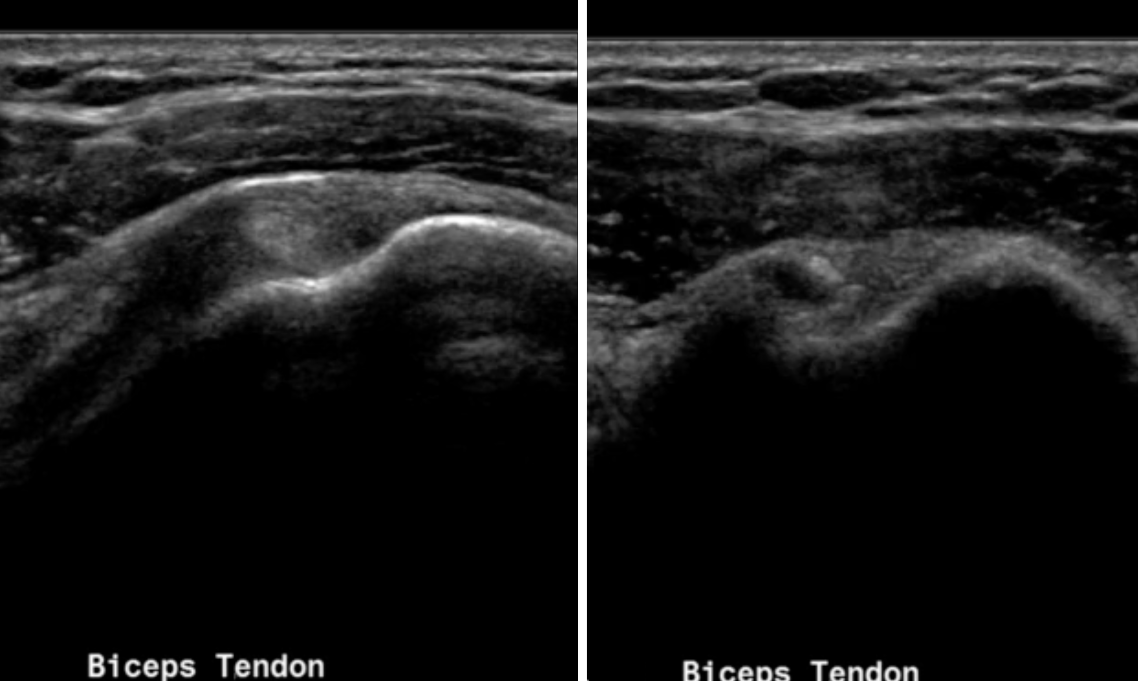
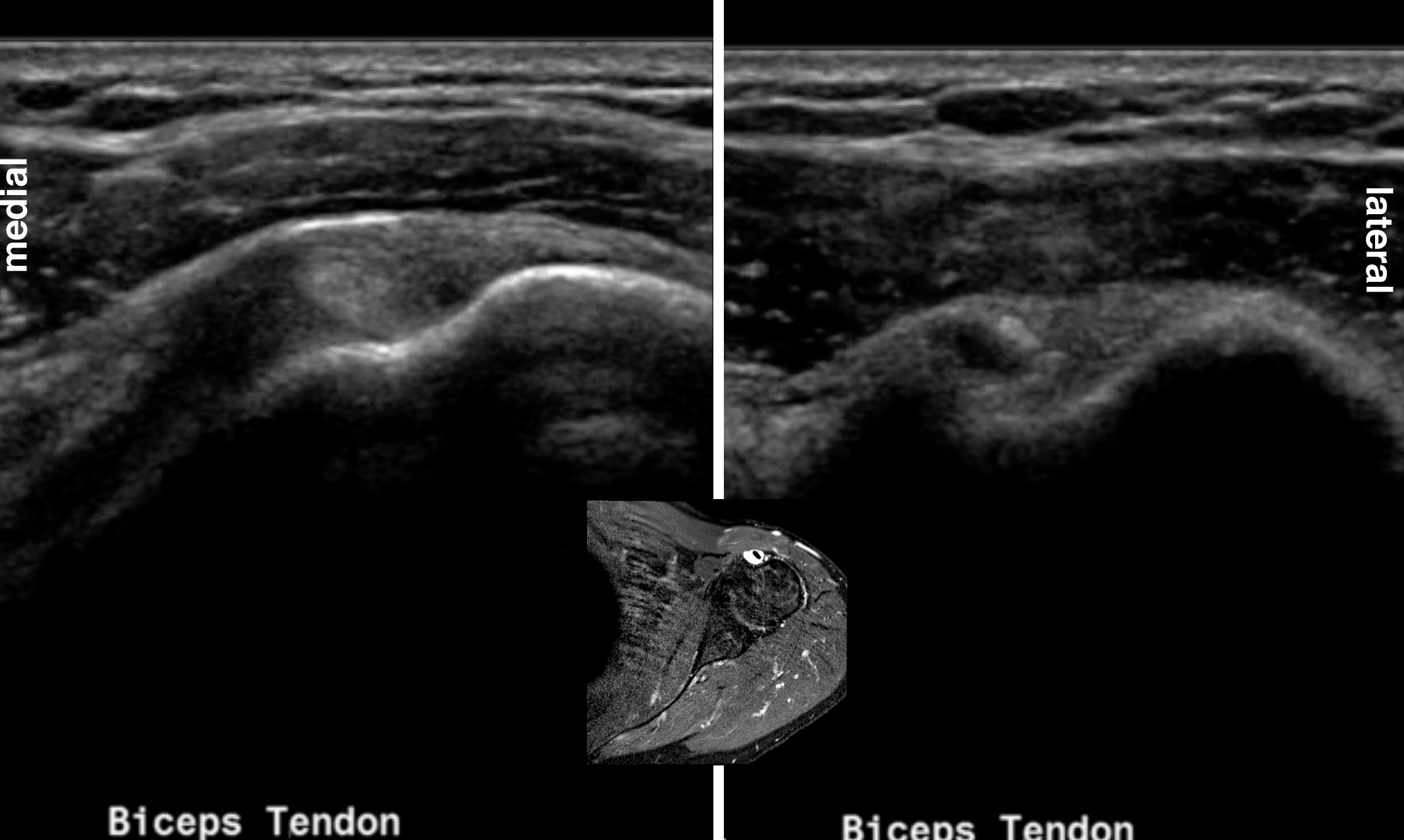
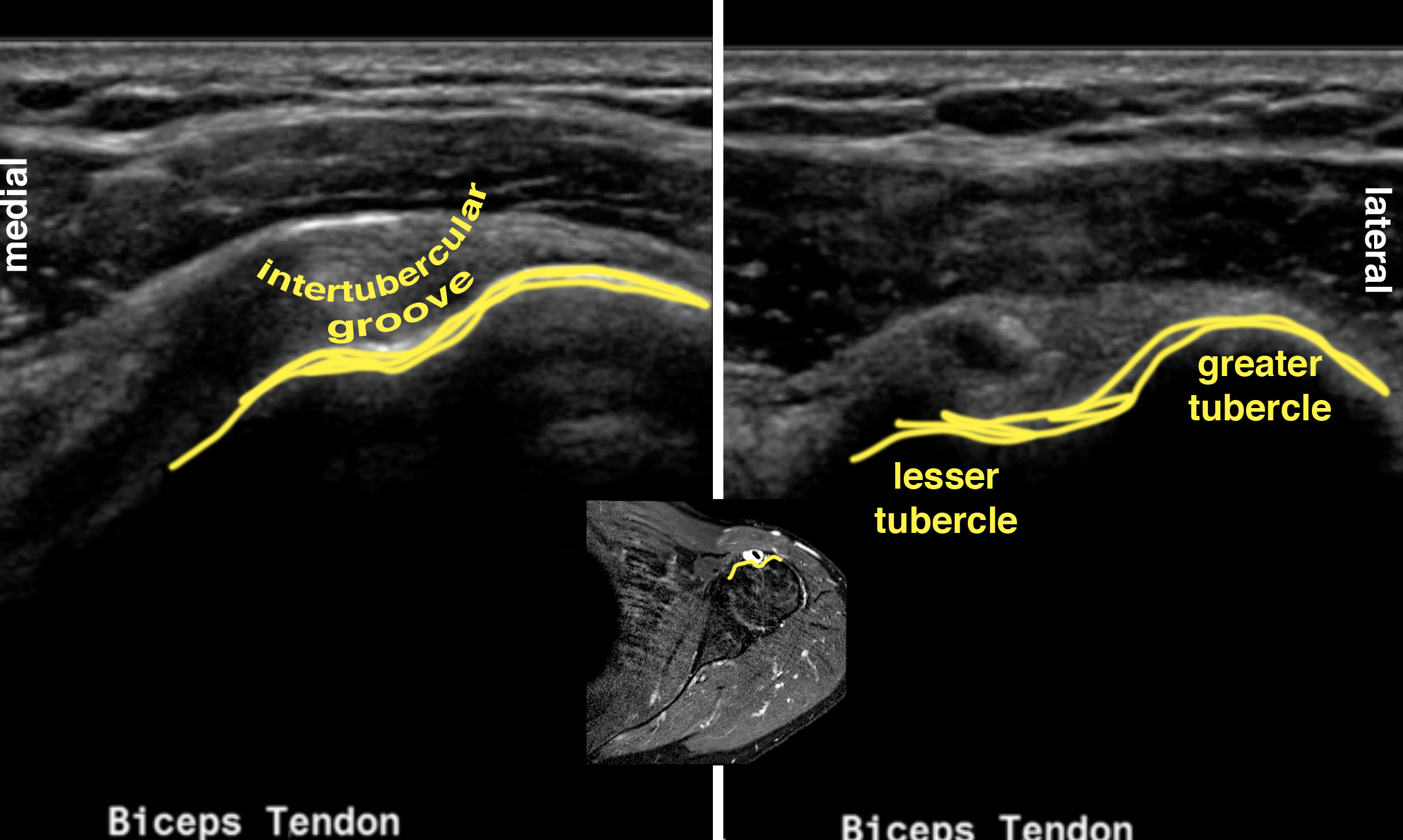
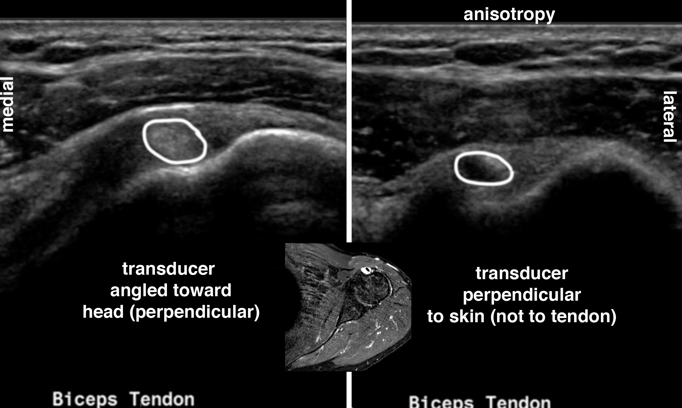
Nerves and tendons exhibit an artifact on ultrasound called anisotropy. This can be used to help identify these structures. Anisotropy occurs when ultrasound beams are directed toward a structure with highly orderly linear organization histologically. The angle of the beam relative to the linear structures of a nerve or tendon can produce either a dark appearance or a light appearance, depending on the angle at which it reaches the structure. If the beam is perpendicular to the nerve or tendon, it will appear echogenic. If the beam is not perpendicular, the structure will appear hypoechoic or anechoic. Examples of this appearance are shown below for the biceps tendon.
Further Explanation:
Case 2
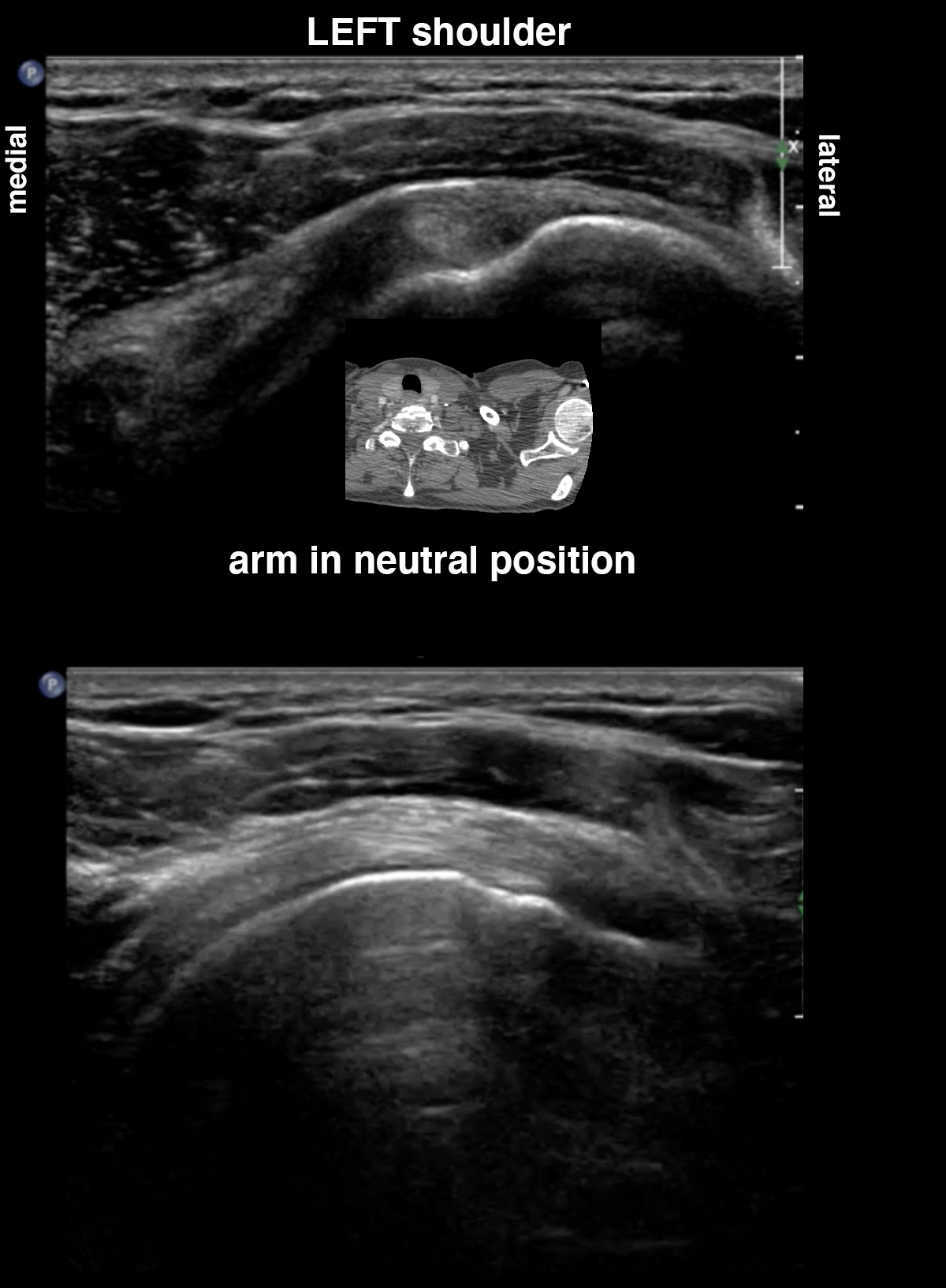
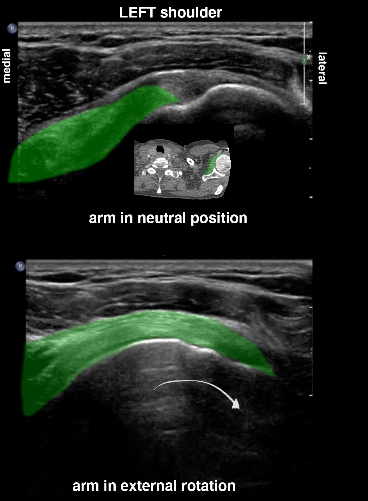
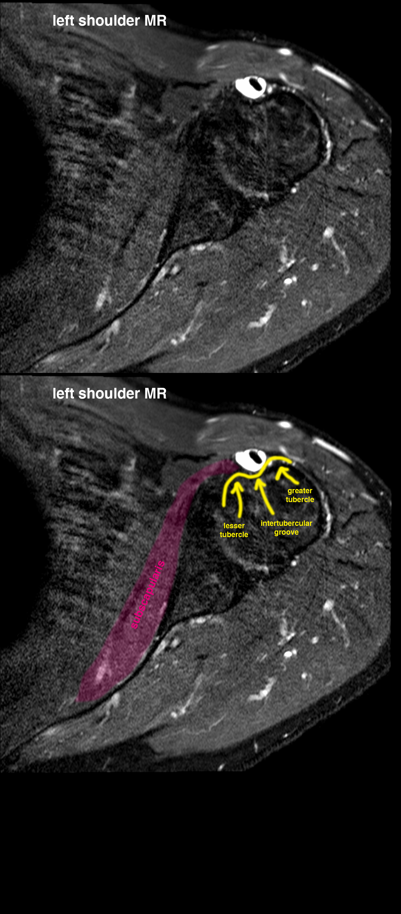
This is an example of anisotropy in the subscapularis tendon on US. Moving the humerus into external rotation brings the subscapularis tendon into a position where the ultrasound beam is perpendicular to the tendon, allowing it to appear normally echogenic and fibrillar.
Further Explanation:
