

CT
This is a CT scan of the lower chest, abdomen and pelvis.
Question 1:
How is CT similar to radiography and how does it differ?
CT is similar to radiography in that it also uses x-rays to image the body. But instead of sending the beam of ionizing radiation through the whole body at once (which produces a projection image with lots of overlapping structures), for CT the beam is shaped into a fan, and rotated around the body to produce a series of slices. This takes a lot more radiation to do, but it eliminates overlap, which is a challenge when interpreting radiographs. The same basic tissue density spectrum is used when looking at CT images as when looking at radiographs, but since we no longer are summing up all of the tissue, even subtle differences in density (like between fat and muscle) are easier to see. Try to identify fat, muscle, bone and air as you look through the stack of CT images.
CT
These are another set of CT images to consider on a different patient.
Question 2:
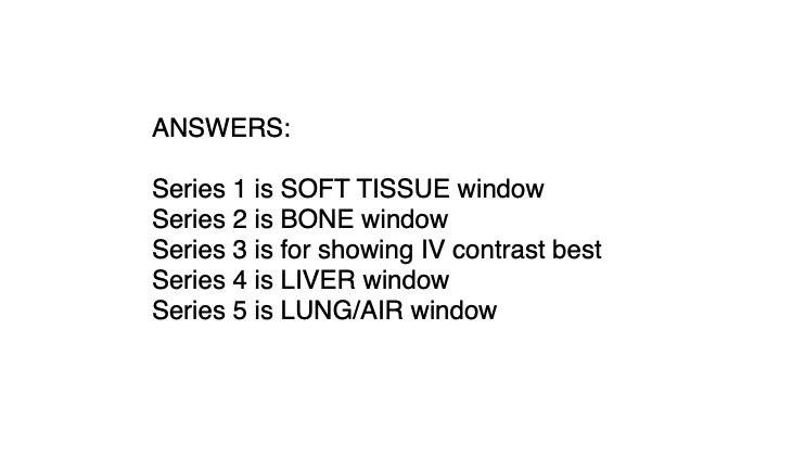
What is the imaging plane shown, and what tissue is best seen on each of the set of images below?
In the early days, CT scans were called 'CAT scans', meaning Computed Axial Tomography. The data is always obtained initially in the AXIAL plane, and that was all that was available on early machines. With improved technology, it is now routine to digitally manipulate that axial data to produce slices in other planes, such as these coronal planes you are shown. The other thing that can be done with CT is to adjust the brightness and contrast of the image to make certain parts of the density spectrum easier to see. So you can focus on the LOW density end and get an image that shows air and air-filled structures well. Or you can focus on the HIGH density end and get images that show lots of bony detail. There are several special brightness and contrast settings (called a 'window' in CT terminology) that are specific for looking at the liver or at soft tissues like muscle and fat.

CT
This is a diagram of how a CT scan is performed
Question 3:
Why do we display the image with the RIGHT side of the patient on the LEFT side of the image?
We actually do the same thing with radiographs. We display the image the same way we would be oriented when we interact with the patient in real life. For a patient lying on an exam table, we would enter from the direction of their feet, facing up from below toward their head. For a chest radiograph, we display it as if we are face-to-face with the patient.
Important features of CT to keep in mind:
-much higher radiation dose than radiography
-relatively high spatial resolution, but not as high as radiography
-slices remove overlap, but take longer to acquire than a single radiograph
-can reformat the data in different planes or with different windows
(all of this can be done after the patient is off the table)


