

US
This image is from a normal ultrasound study of the thyroid gland.
Question 1:
How does this image differ from a radiograph?
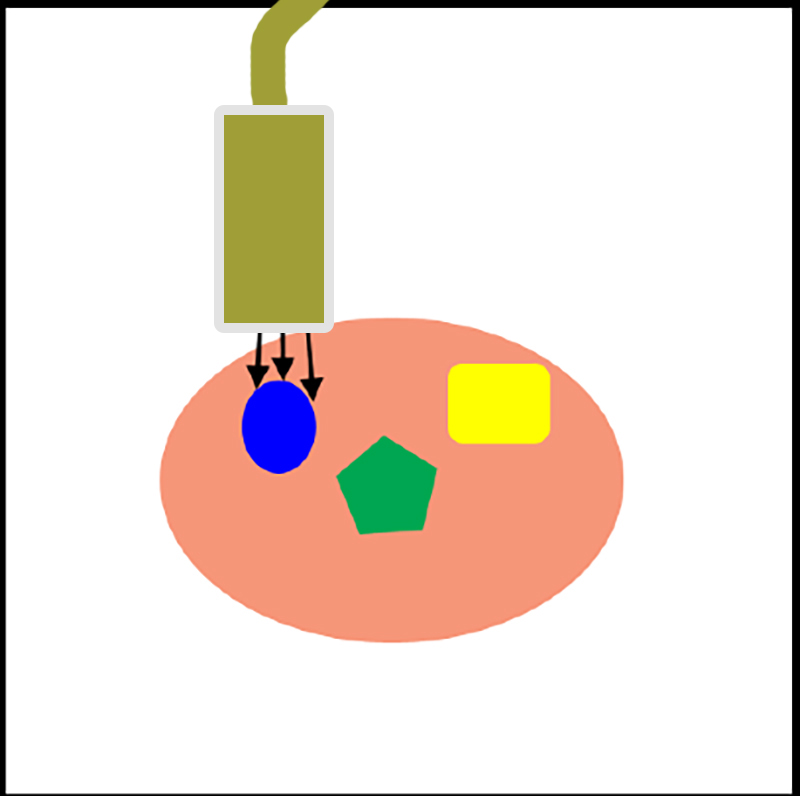
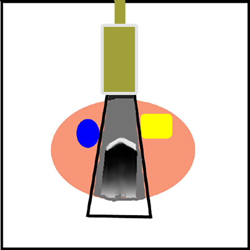
Ultrasound uses sound waves that are sent into the patient and bounce off of various tissues, returning to the transducer (the object that sends and receives sound, and is placed against the patient's body) to produce an image. This imaging modality does not use any ionizing radiation. The spatial resolution of US is much lower and the image much grainier than CT or radiography, and the sound beam is weak so it cannot penetrate very far into the body, so ultrasound is best used for superficial structures. The whiteness and blackness of tissues has nothing to do with density, and is much more complex than for radiography or CT. Certain tissues BLOCK transmission of the high frequency sound used in US: air, bone and metal.
US
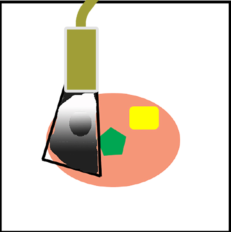
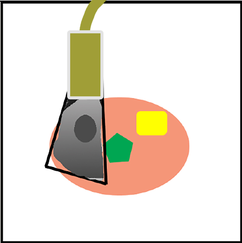
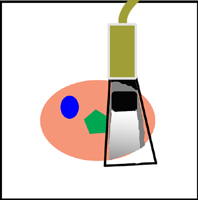
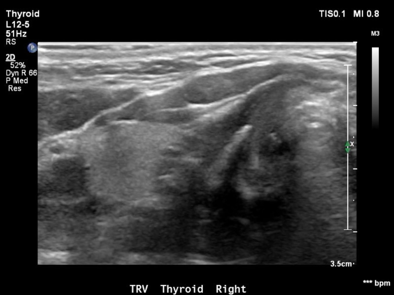
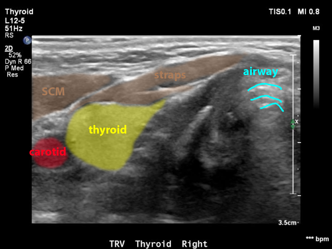
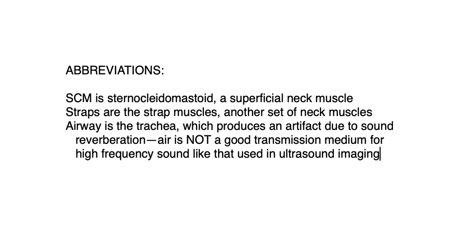
This is a sweep through the thyroid gland in the neck (in the axial plane) with ultrasound.
Question 2:
What is different about the way these images are displayed compared to CT?
Because the process of sending and receiving US waves is so quick, you can use US to obtain 'real time' imaging, meaning you can see motion pictures of things in the body, which is not true for most CT scans. This can be particularly useful for interventional procedures, such as placing a needle into a structure. You can watch the needle move and see when it penetrates the target using US. Or you can show the sweep of the transducer (probe) through an organ like the thyroid, to get an appreciation of the 3D shape as you scan through. The US images are slices (like CT) but at variable planes in the body, depending on how the transducer is oriented. Just like for CT, if we are in the axial plane, we show the RIGHT side of the patient on the LEFT side of the image.
US
This is a diagram of how ultrasound is performed
Further Explanation:
Things to remember about US:
-images are slices, and the plane of imaging varies with how the transducer is positioned
-no radiation, low spatial resolution, can show real-time images
-small field of view (limited penetration of sound in tissue), so best for superficial things
-NOT based on tissue density, difficult to get oriented and to predict what will be dark or light
