

MR
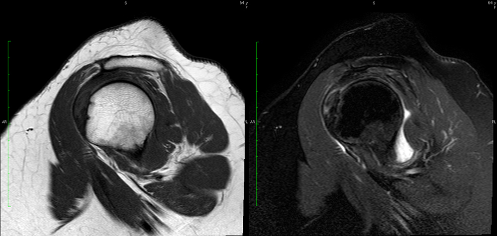
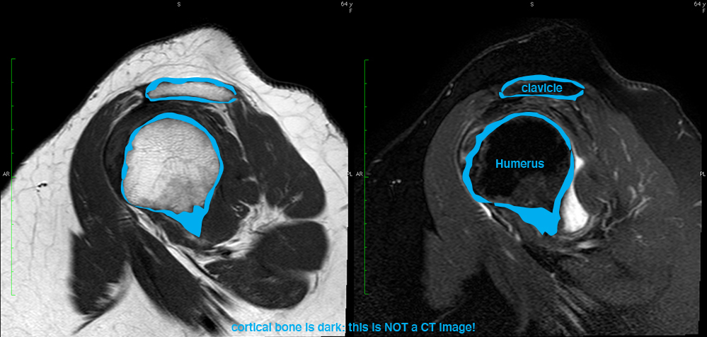
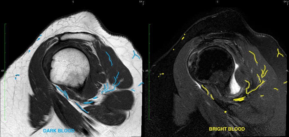
These two images are from the same normal MR study of the shoulder.
Question 1:
How does MR differ from CT?
×
Answer:
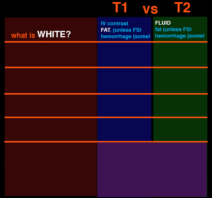
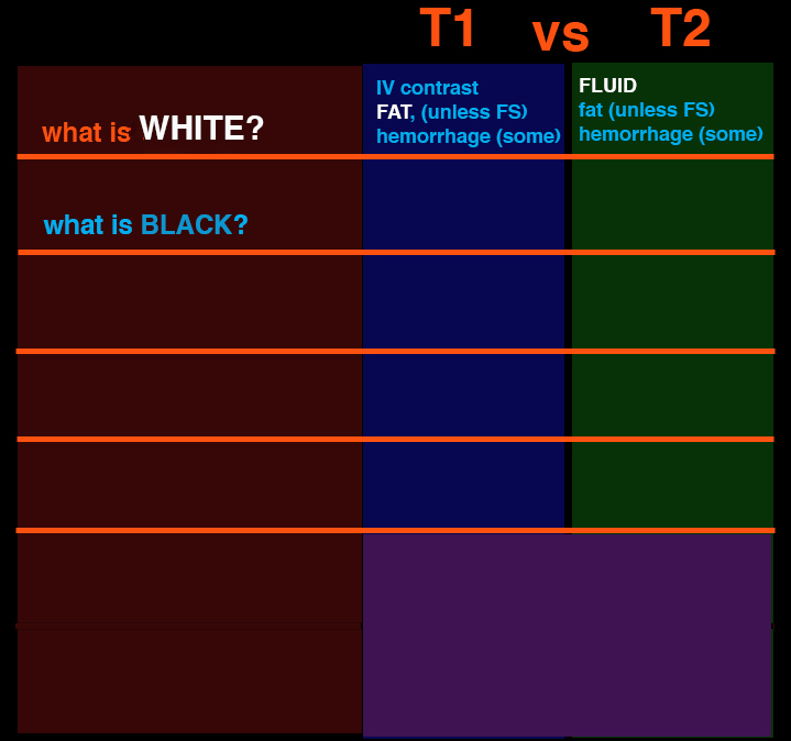
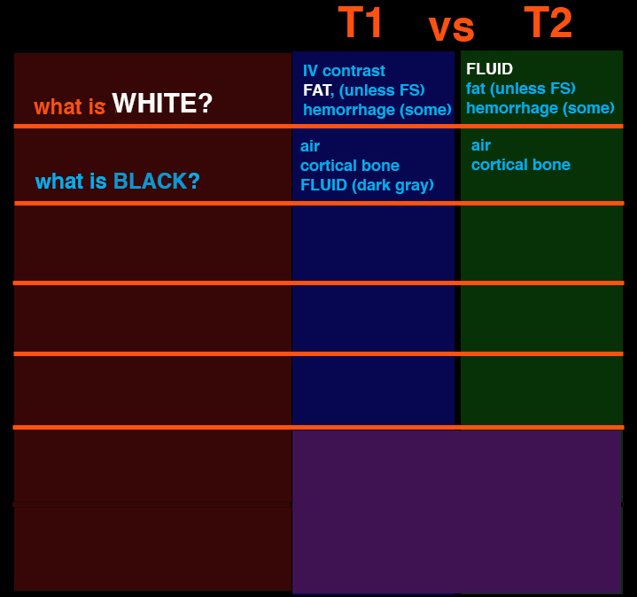
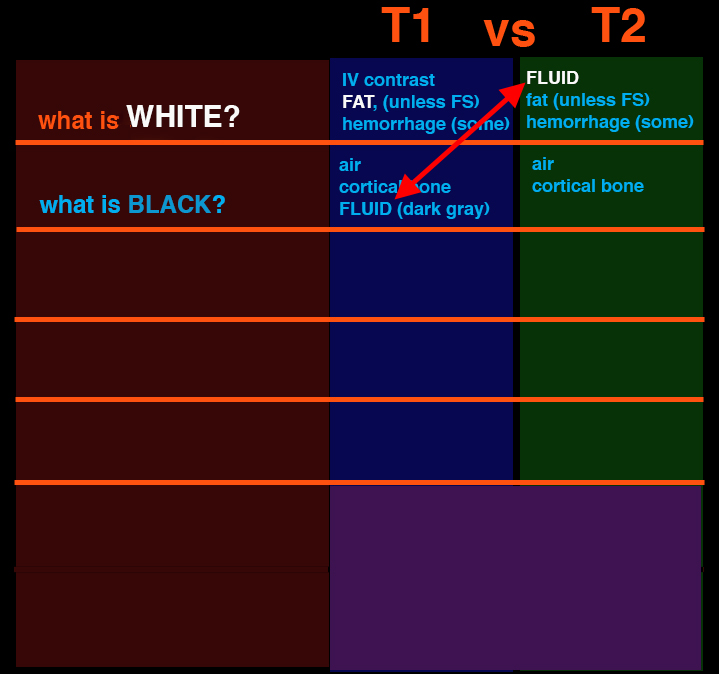
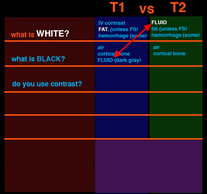
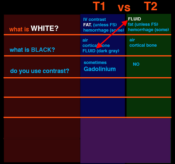
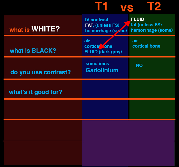
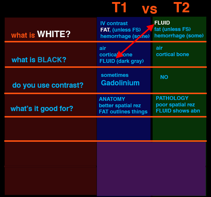
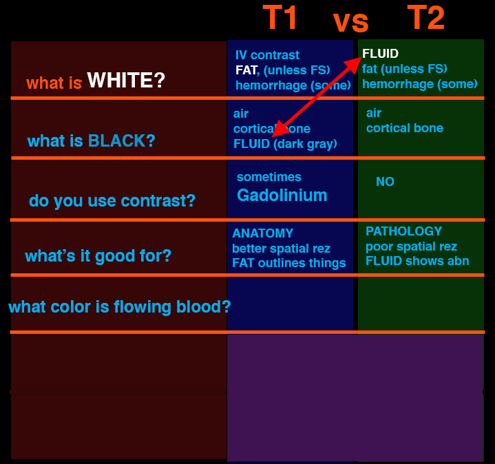
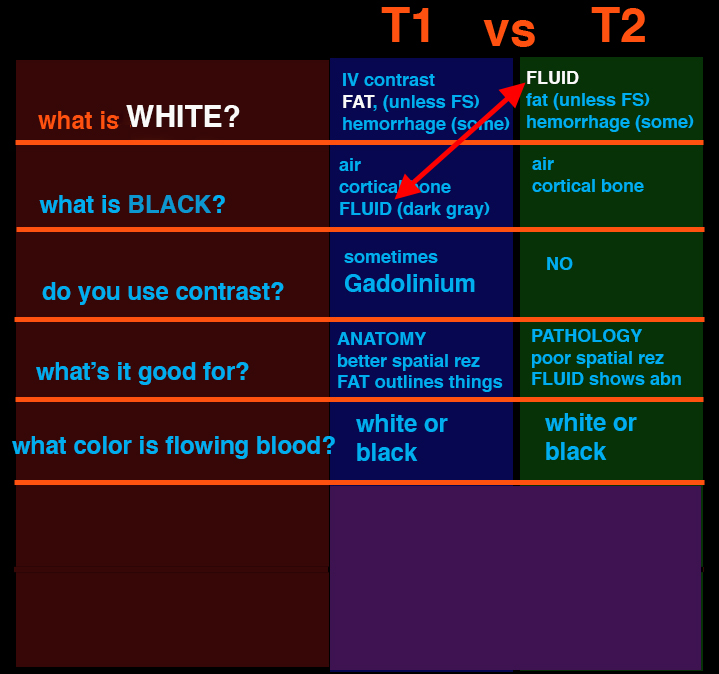
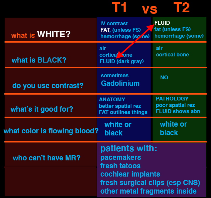
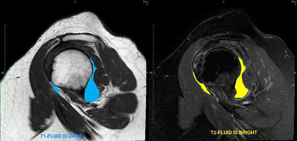
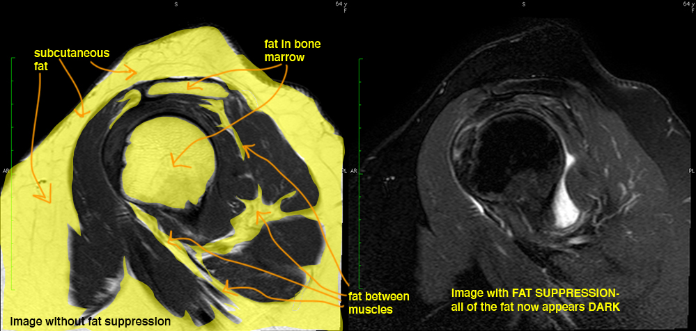
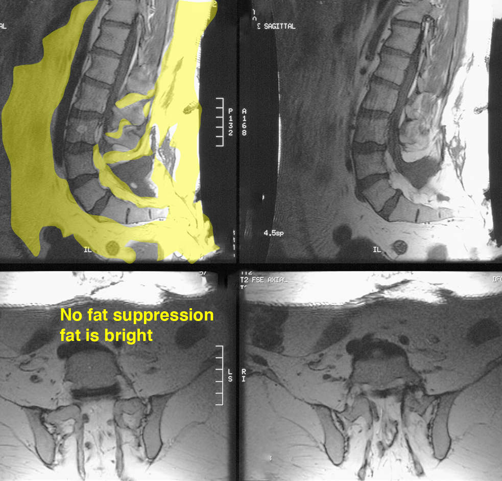
MR uses the same principles as NMR spectroscopy to image body tissues based on the magnetic properties of particular biochemical compounds within the tissues. The physics of this is quite complicated, but a few basic principles can help in looking at images. Imaging is based on presence/concentration of hydrogen, which will produce different signal based on the type of chemical compound it is in. Most of the signal that makes the images comes from water and fat, two of the most common chemicals in the body, both containing hydrogen. The way that the images are obtained can vary a lot and many different parameters can be changed to get very different appearances, as shown in these two images. Some common things to vary are--the appearance of flowing blood (can appear dark or light, depending on how the scan is done), the appearance of fat (is generally bright, but special sequences can make it appear dark, called 'fat suppression'), and the appearance of watery fluid (can be dark--called T1-weighted imaging, or can be bright--called T2-weighted imaging). There are many other ways that the scan can be modified besides these, but these are used most commonly. So MR does not use ionizing radiation, but it does display the body in slices and can be done in many imaging planes like CT. The lightness or darkness of tissues on MR has nothing to do with density, also unlike CT.
MR uses the same principles as NMR spectroscopy to image body tissues based on the magnetic properties of particular biochemical compounds within the tissues. The physics of this is quite complicated, but a few basic principles can help in looking at images. Imaging is based on presence/concentration of hydrogen, which will produce different signal based on the type of chemical compound it is in. Most of the signal that makes the images comes from water and fat, two of the most common chemicals in the body, both containing hydrogen. The way that the images are obtained can vary a lot and many different parameters can be changed to get very different appearances, as shown in these two images. Some common things to vary are--the appearance of flowing blood (can appear dark or light, depending on how the scan is done), the appearance of fat (is generally bright, but special sequences can make it appear dark, called 'fat suppression'), and the appearance of watery fluid (can be dark--called T1-weighted imaging, or can be bright--called T2-weighted imaging). There are many other ways that the scan can be modified besides these, but these are used most commonly. So MR does not use ionizing radiation, but it does display the body in slices and can be done in many imaging planes like CT. The lightness or darkness of tissues on MR has nothing to do with density, also unlike CT.
MR
MRI is used in many parts of the central nervous system.
Question 2:
What type of spine problems might be best imaged with CT vs MR, based on what you know about these modalities?
×
Answer:
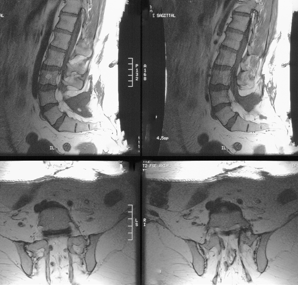
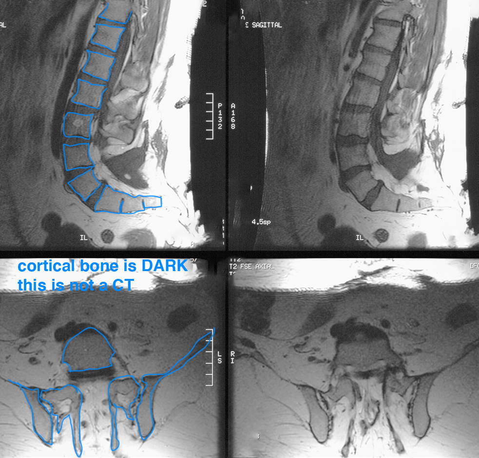
CT is used in spine imaging when a fine detailed view of bony structures is needed, as in complicated fractures or degenerative diseases. MR is used in spine imaging when the focus is on the neural components and soft tissue structures such as discs and ligaments. Many of these structures are the same DENSITY, so are hard to distinguish with CT. But they can have very different appearances on MR based on their biochemical composition.
CT is used in spine imaging when a fine detailed view of bony structures is needed, as in complicated fractures or degenerative diseases. MR is used in spine imaging when the focus is on the neural components and soft tissue structures such as discs and ligaments. Many of these structures are the same DENSITY, so are hard to distinguish with CT. But they can have very different appearances on MR based on their biochemical composition.
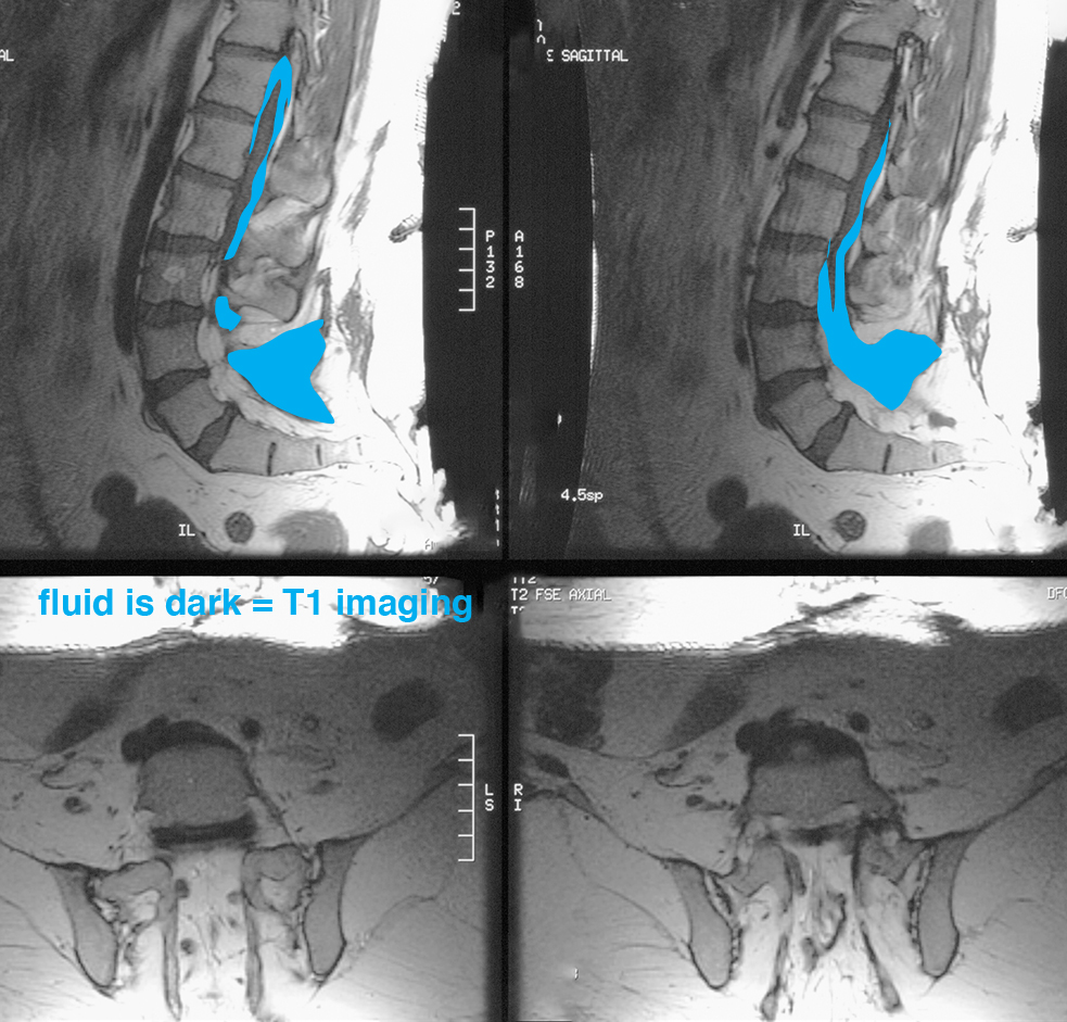
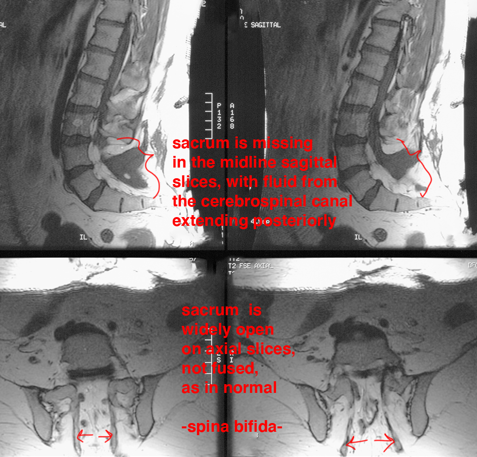
MR
Here are some technical hints for dealing with MRI images
Further Explanation:
