


NM
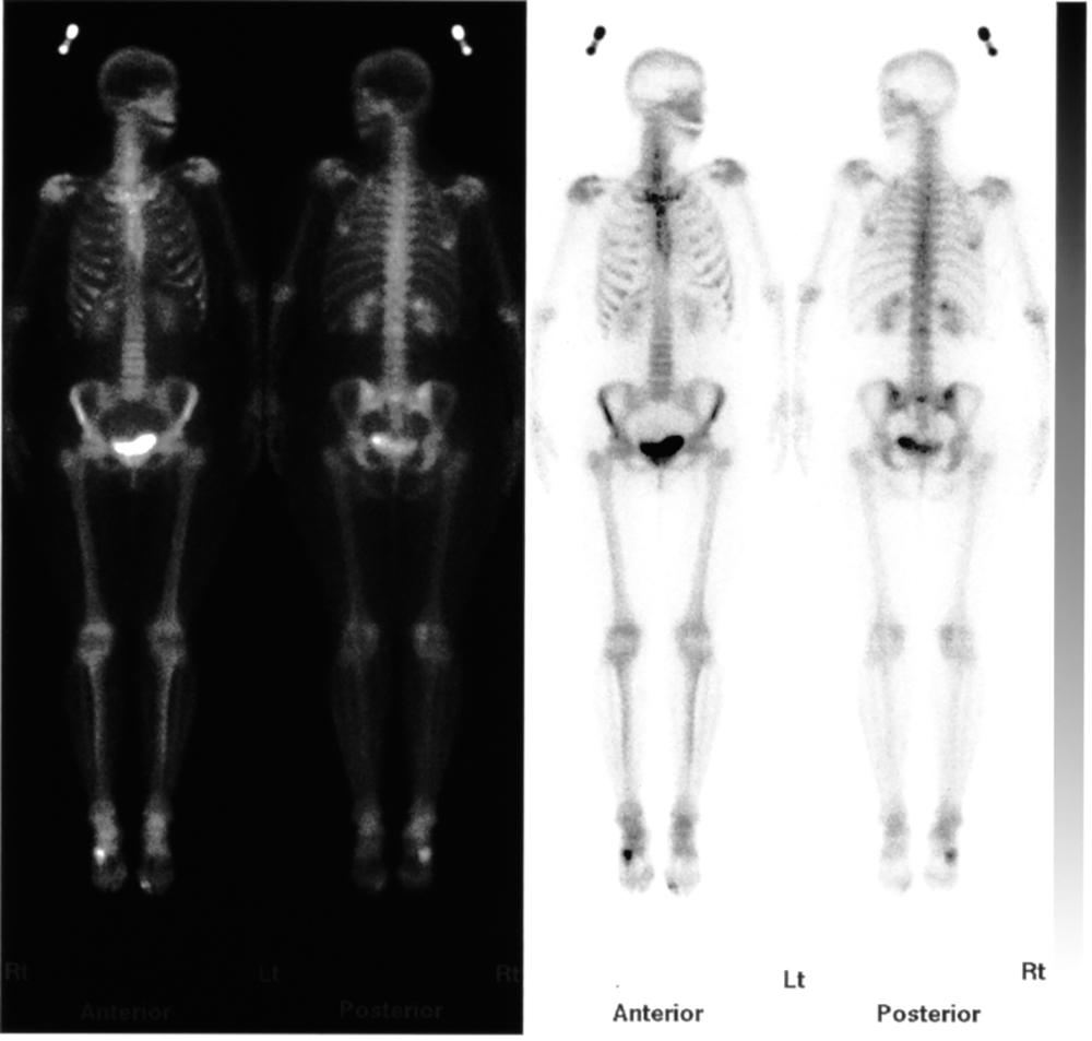
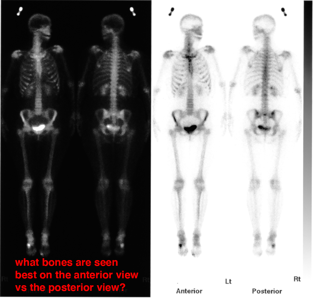
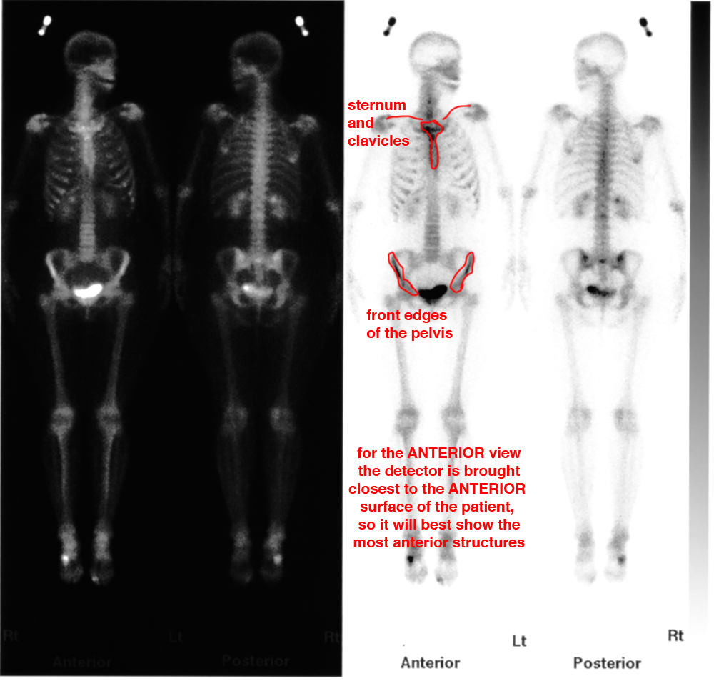
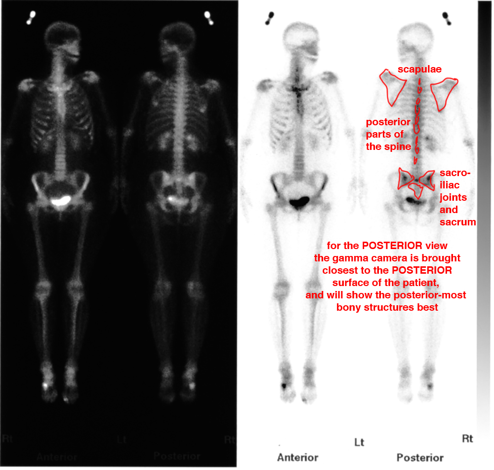
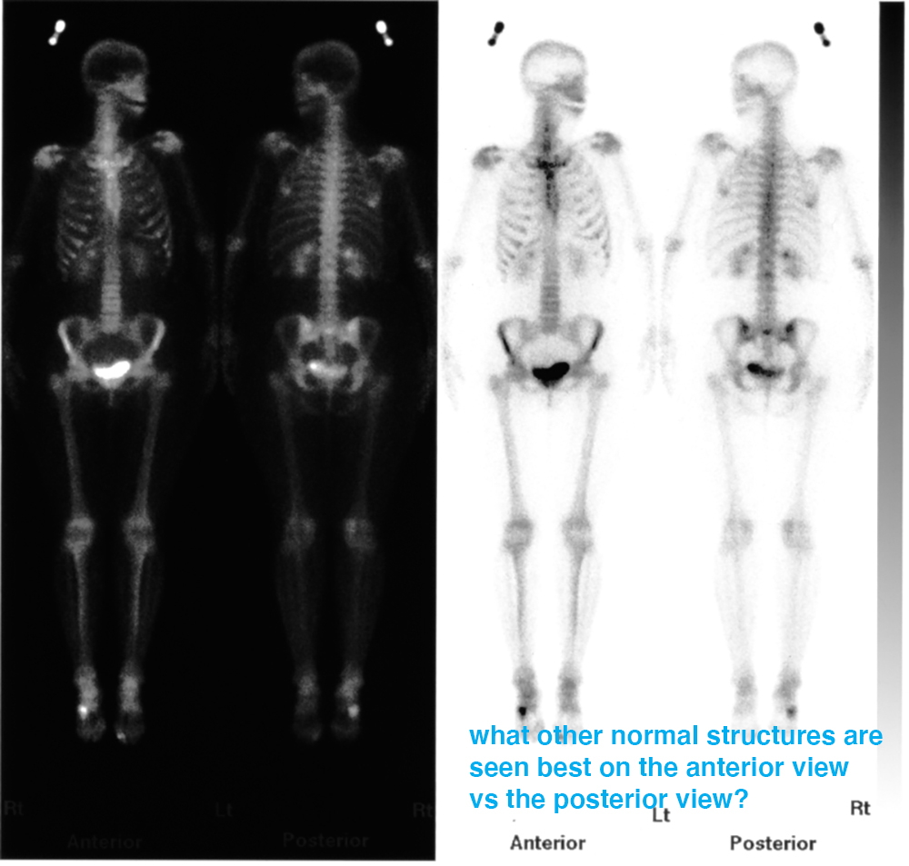
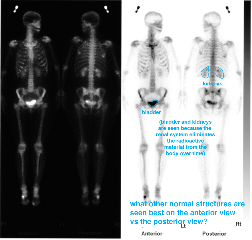
This is an example of a normal bone scan.
Question 1:
How is nuclear medicine imaging different from all the other imaging modalities we have discussed?
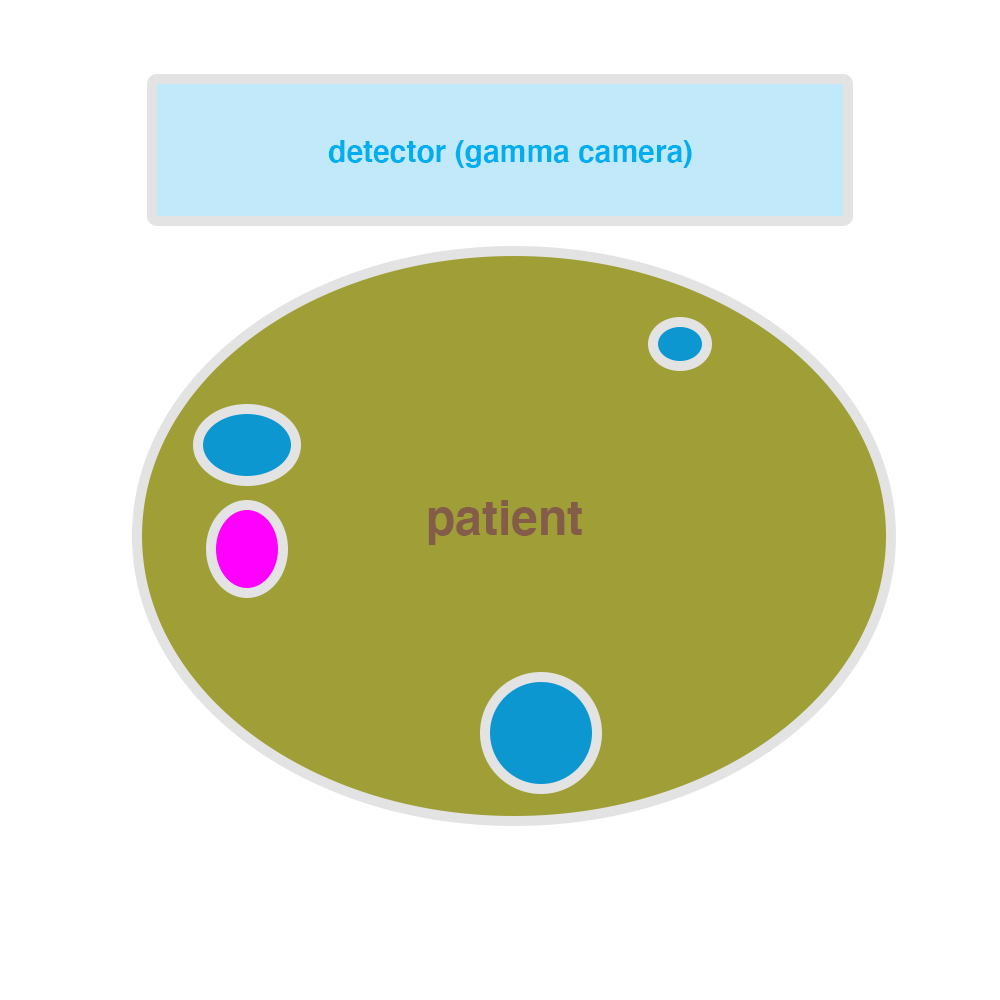
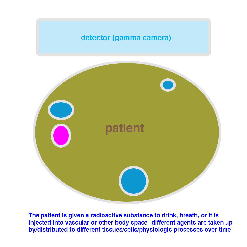
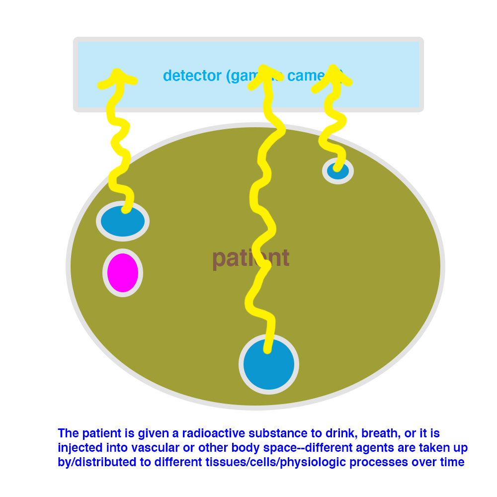
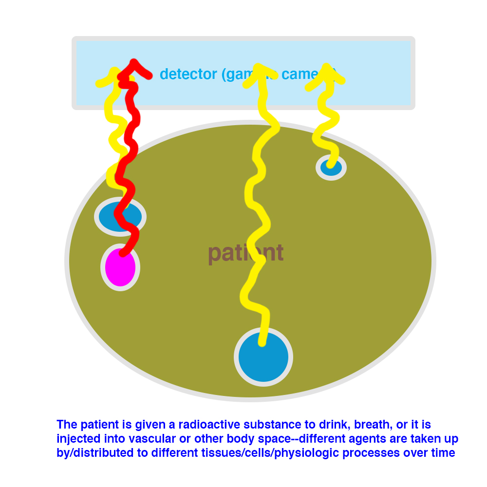
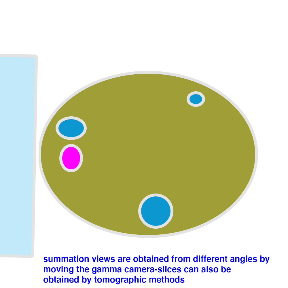
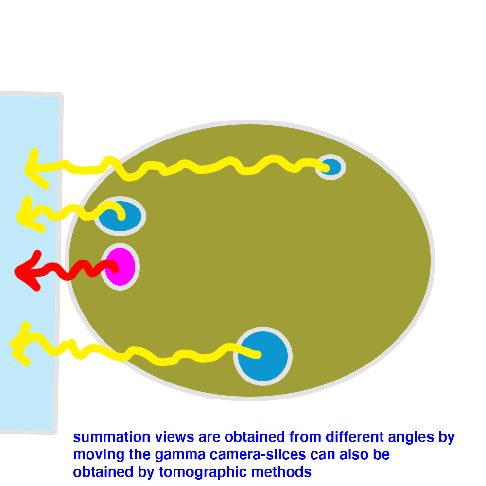
In radiography, CT, US, and MR, we send some sort of beam (x-rays, sound waves, radio frequency) into the patient and record its results to make the image. For nuclear medicine imaging, we put a radioactive substance into the patient, and record where the radiation is accumulated, partly in normal tissues and partly in abnormal tissues or physiological processes. This is the only part of radiology where we use radioactive materials, and this requires a lot of quality control and management of disposal of products. So our images are made with a detector (a gamma camera, which is like a very fancy large Geiger counter) that we bring into proximity with the patient and measure where the radiation is coming from.
NM
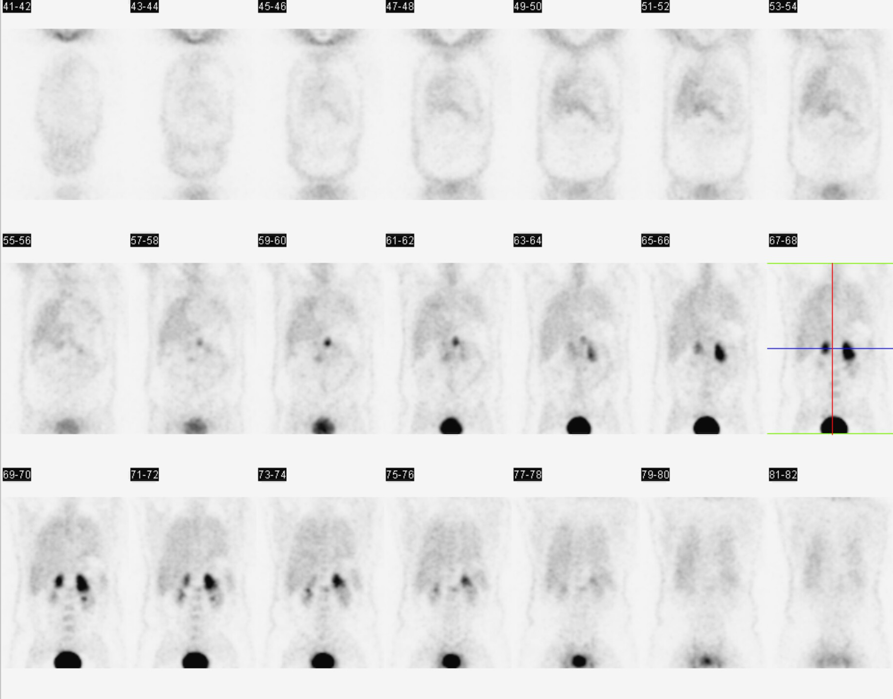
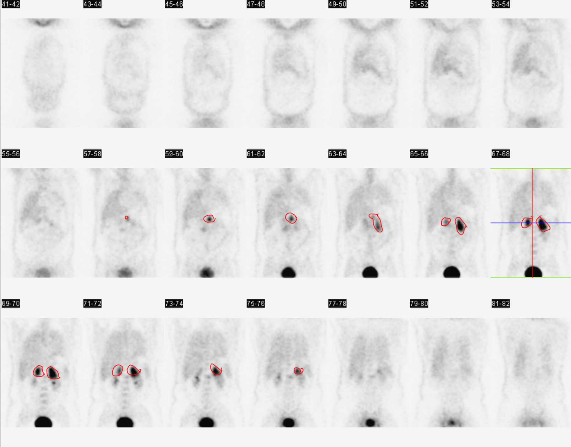
This is an example of a PET scan in a different patient.
Question 2:
How is this set of images different from the prior bone scan images?
These images are coronal slices, not a single image. A nuclear medicine camera can be stationary, collecting radioactive counts from one side of the patient's body (like the previously shown bone scans) or it can be set up to rotate around the patient, similar to a CT scanner. This can produce slices rather than a single image. The agent used in PET scanning is targeted toward any cell that is metabolizing glucose, so many tissues of the body will show up faintly, but areas of rapid growth, particularly by tumors, tend to show much higher radioactivity than normal tissues. There are areas of high activity bilaterally just above the kidneys in this patient, who has tumors involving the adrenal glands.
NM
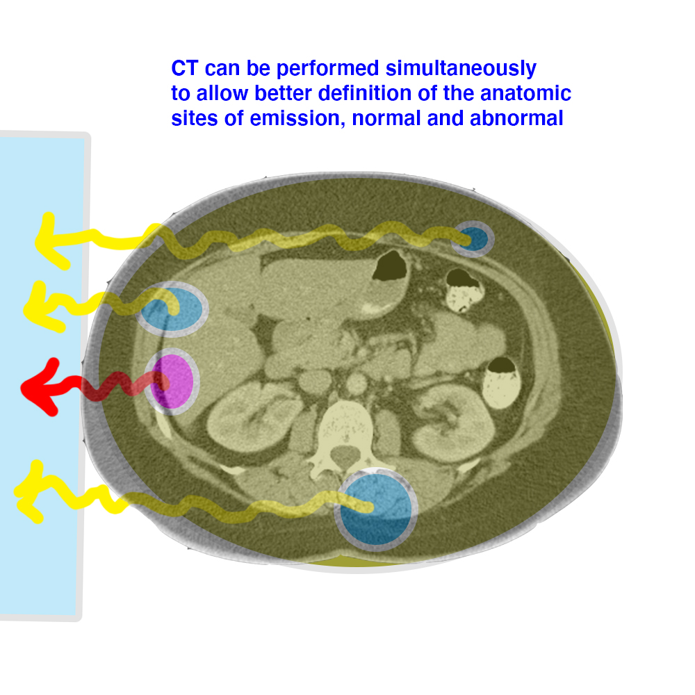
This is a diagram of the basis for nuclear medicine imaging
Further Explanation:
Things to keep in mind about NM studies:
-There are MANY different agents that can be used to show different tissues or physiologic processes
-Radiation dose is generally similar to CT, field of view is very large (often the whole body)
-Images may be acquired as a projection or in slices, and can be done in conjunction with CT (ex. PET-CT)
NM
This completes the review of Imaging Modalities
Further Explanation:
Click this link to return to Imaging Anatomy topic areas for Tufts Medical Students.


