

Case 1
55 year old male with left shoulder pain for many years.
Question 1:
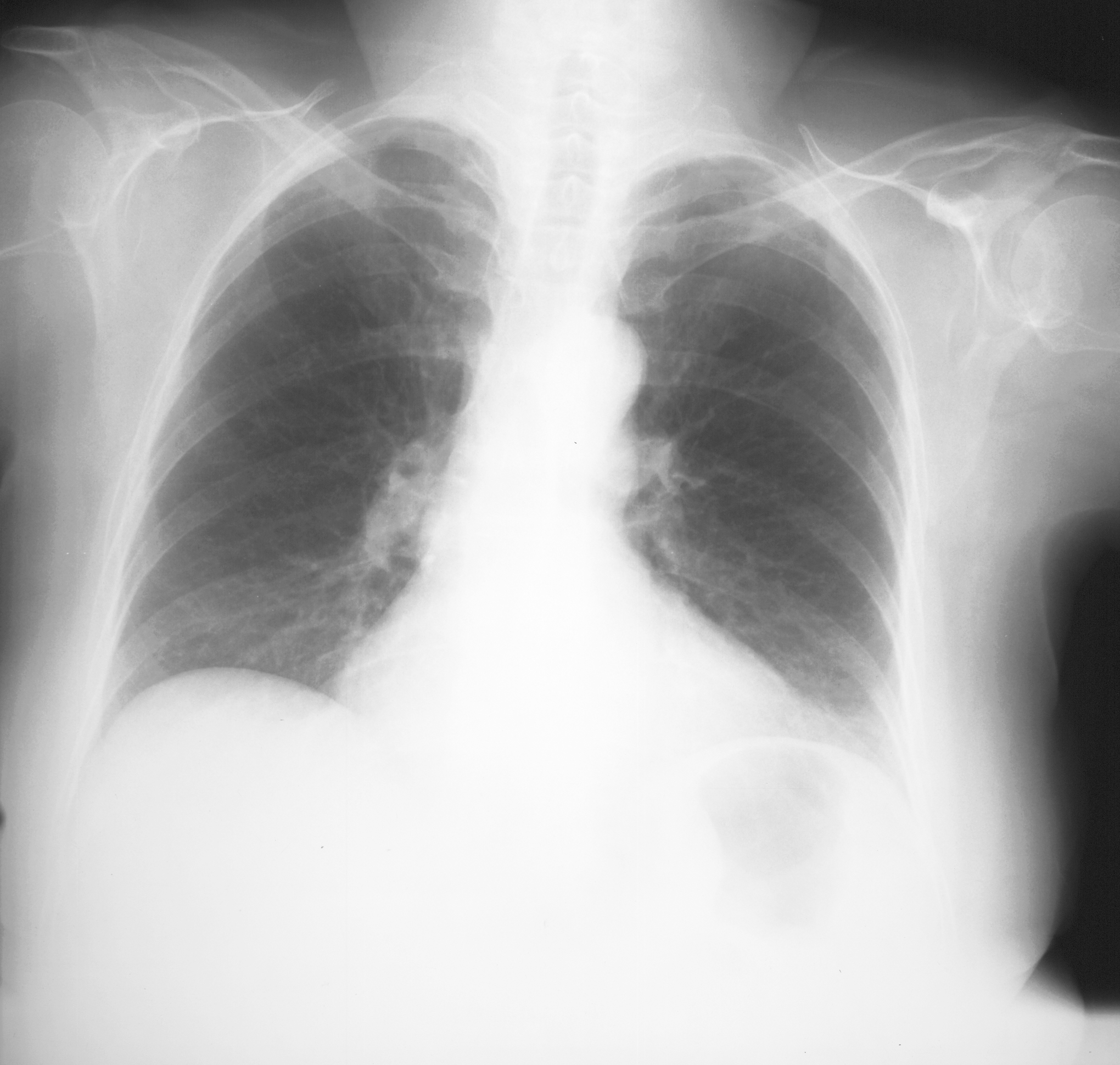
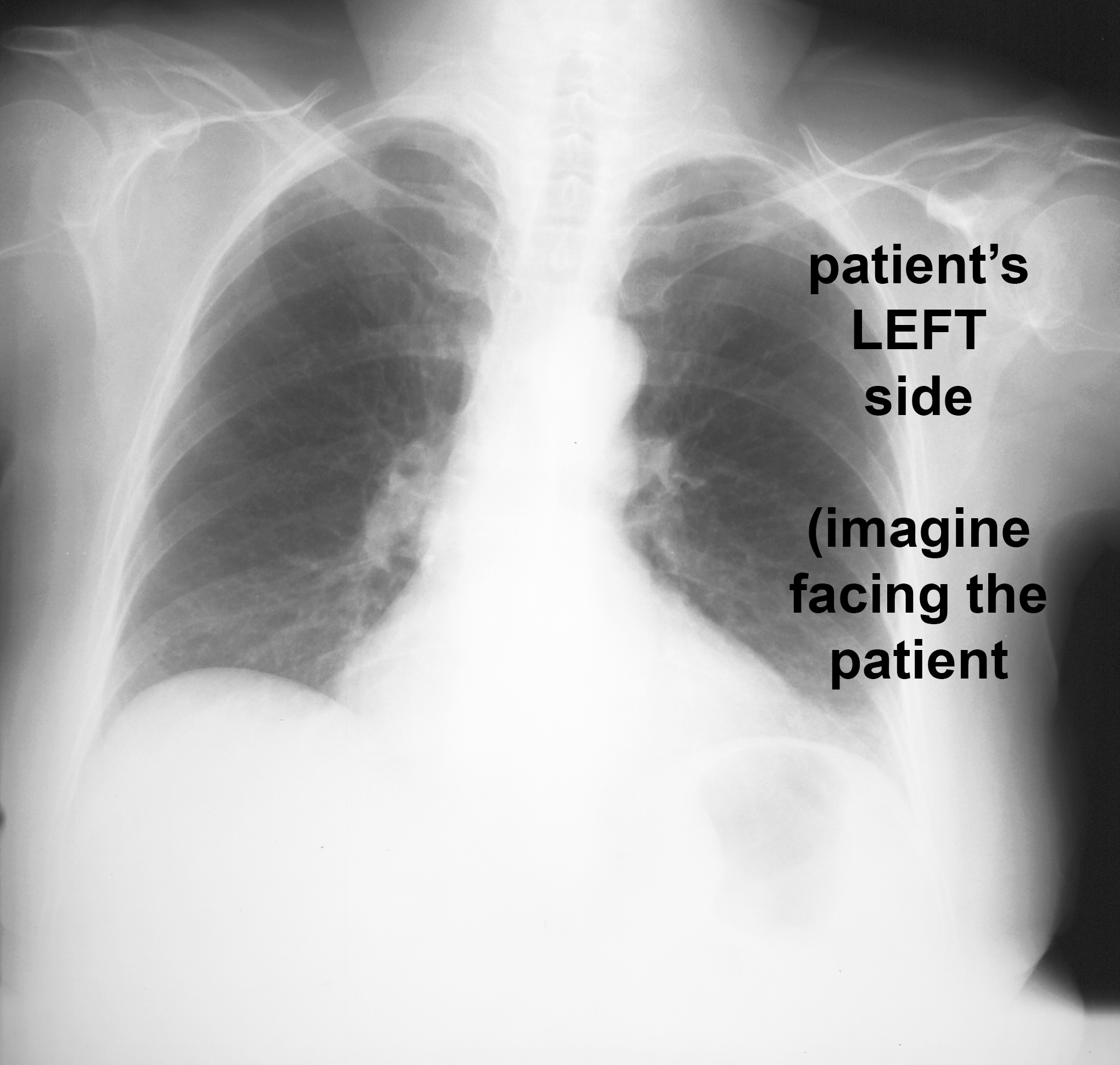
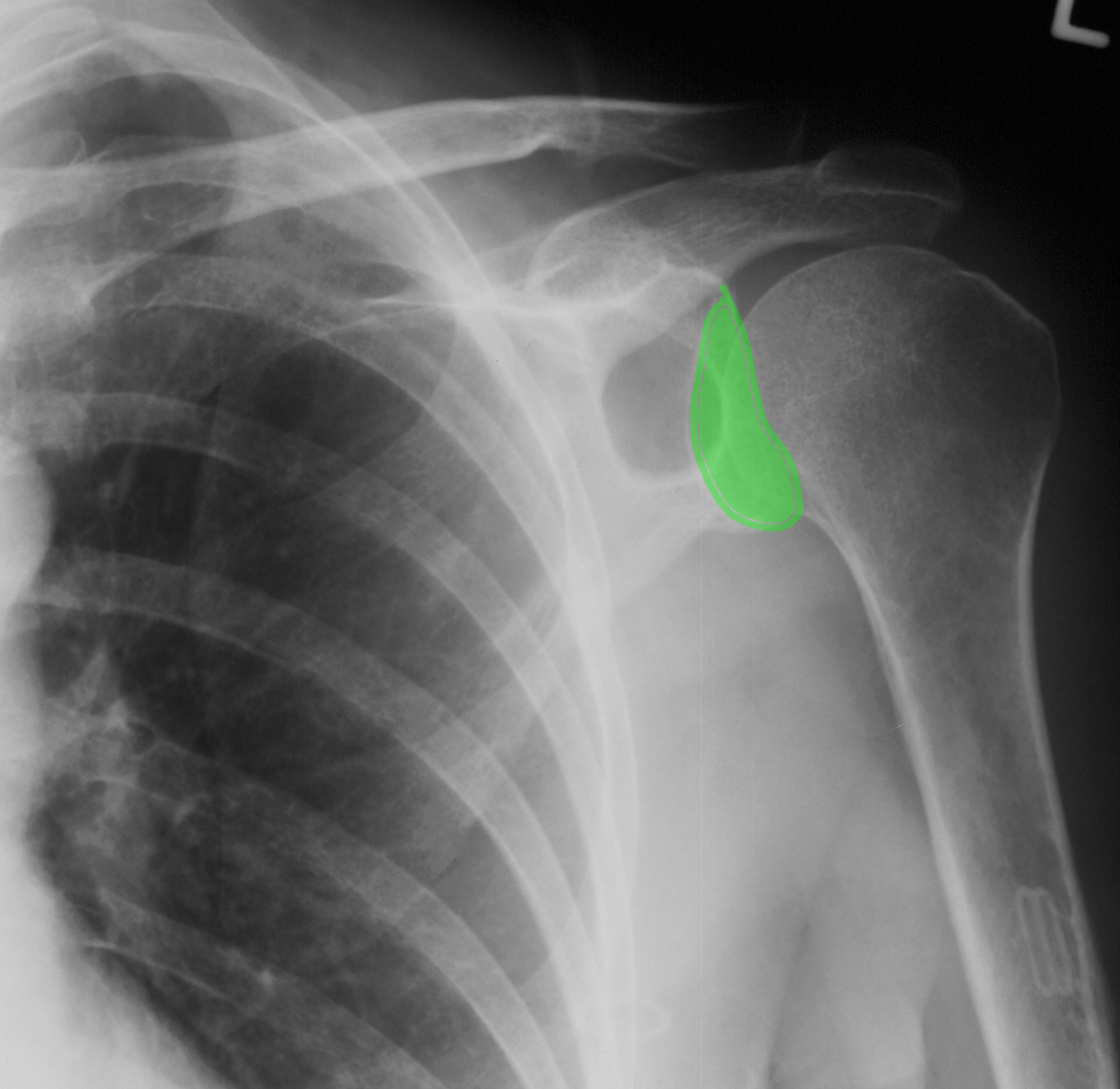
a) What do you think of the view of the shoulders on this image? Which side of the image is the LEFT?
This is a chest radiograph, and it barely includes the shoulders. The angle of the beam relative to the shoulders is far from optimal. Dedicated shoulder radiographs should be done. The image (as for all frontal and coronal images, regardless of modality) is shown as if we are facing the patient, so the left shoulder is on the RIGHT side of the image.
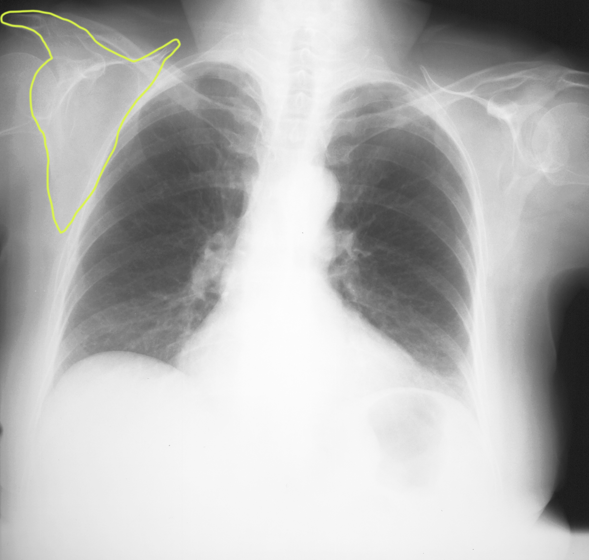
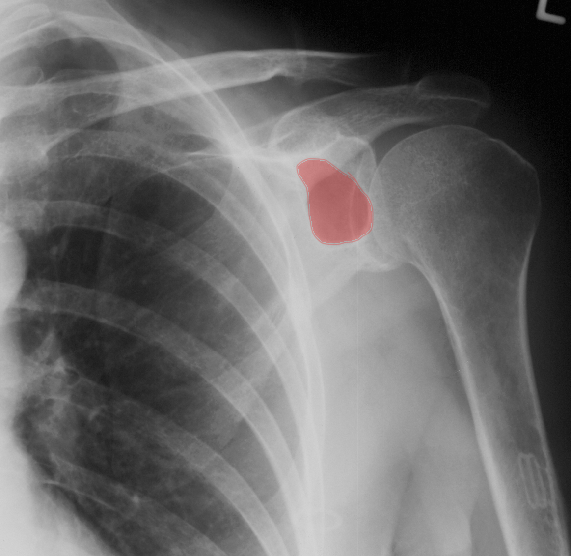
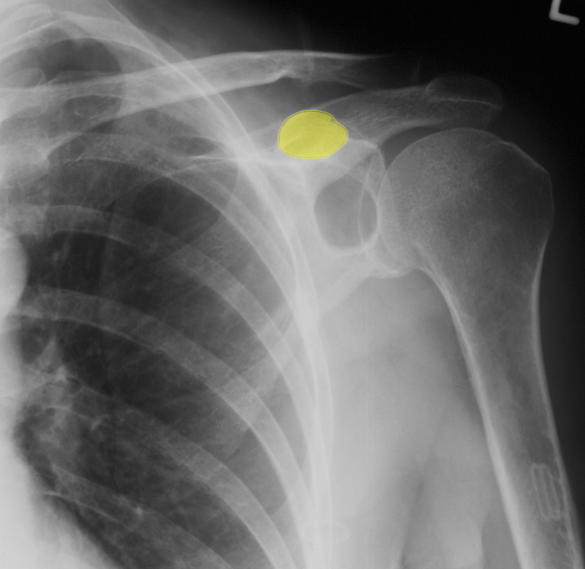
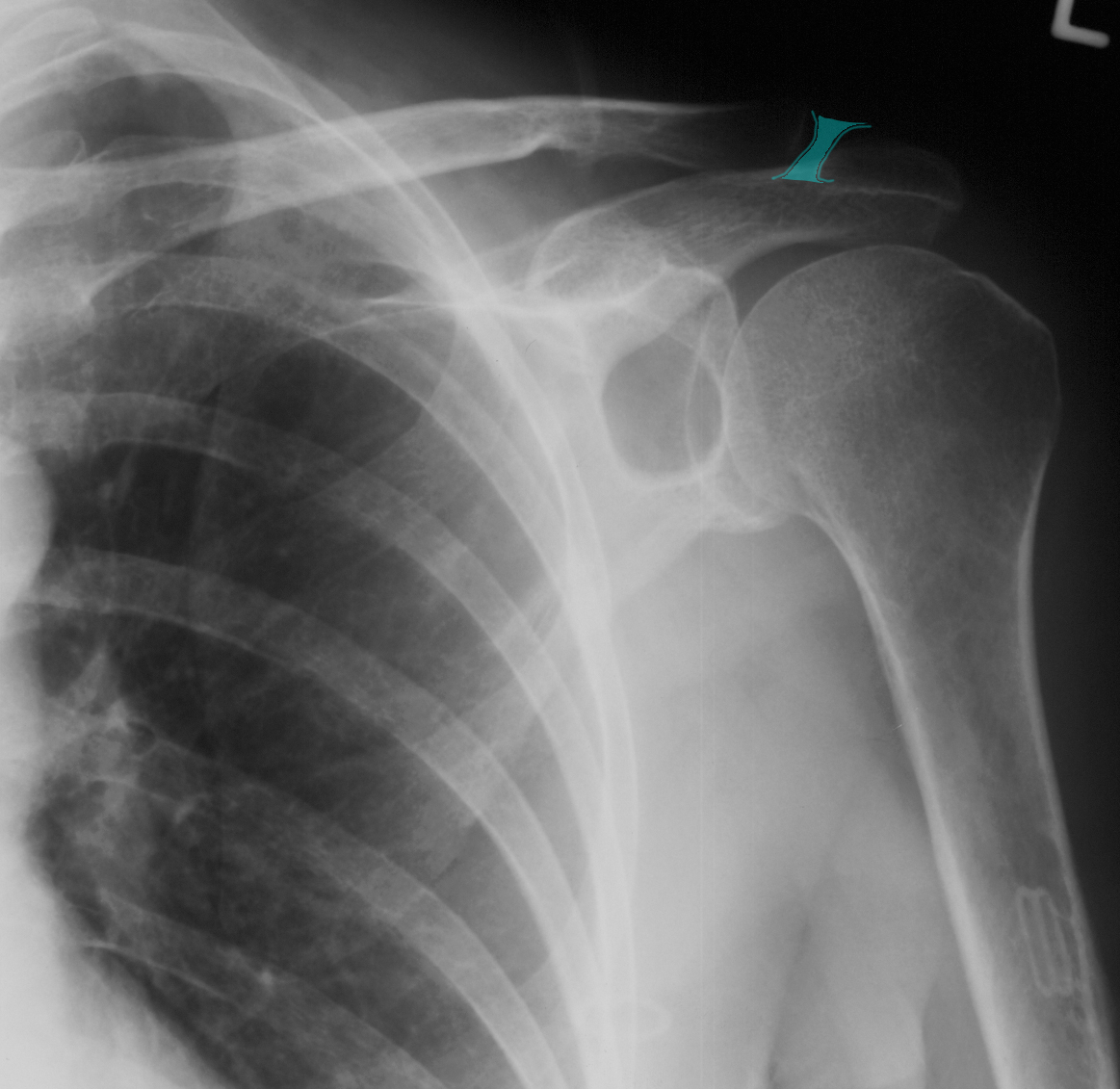
b) Click on the labeled white buttons below to bring up annotation on the image, or additional images. (click the button again to remove the annotation or return to the original image). What bony structure is outlined?
The outline indicates the location of the right scapula, a very complex shaped bony structure that is partly overlapping the humerus, lateral clavicle and right lateral ribs.
Click 'Reveal' to see the answer to each question. Click 'Next' at the bottom right corner of the screen to move to the next page.
Click the pencil icon to enlarge the image and bring up annotation tools to draw on the image, which can be useful if you are working with a group, or to make notes for yourself. Your annotations will not be saved to the website, but you can download your annotated image to your own computer.

Case 1
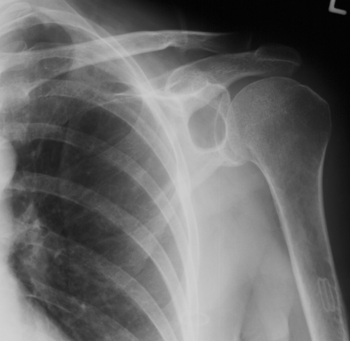
This is the next image that was obtained in this patient.
Question 2:
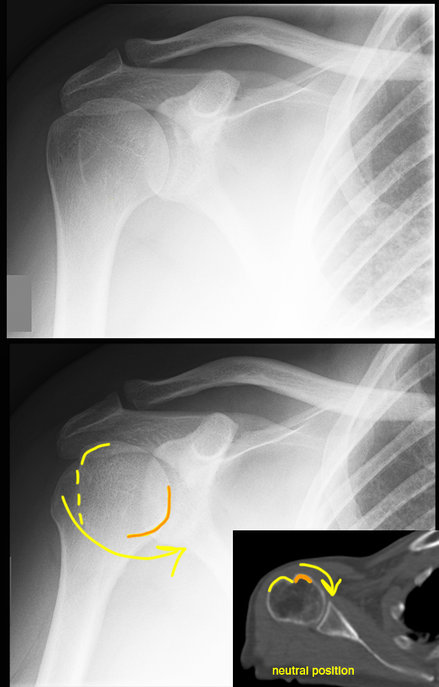
How is this view different from the previous image? Does it look normal?
This is a dedicated radiograph of the shoulder. When describing a radiograph, we name the view based on how the beam passed through the patient. This was done with the beam entering from the front, so it is called an 'AP' view, or 'anterior-posterior', following the courser of the x-ray through the patient. It is centered to show the shoulder well. There is a smoothly rounded low density area in the scapula that is not normal.

Case 1
This is the next study that was done in this patient.
Question 3:
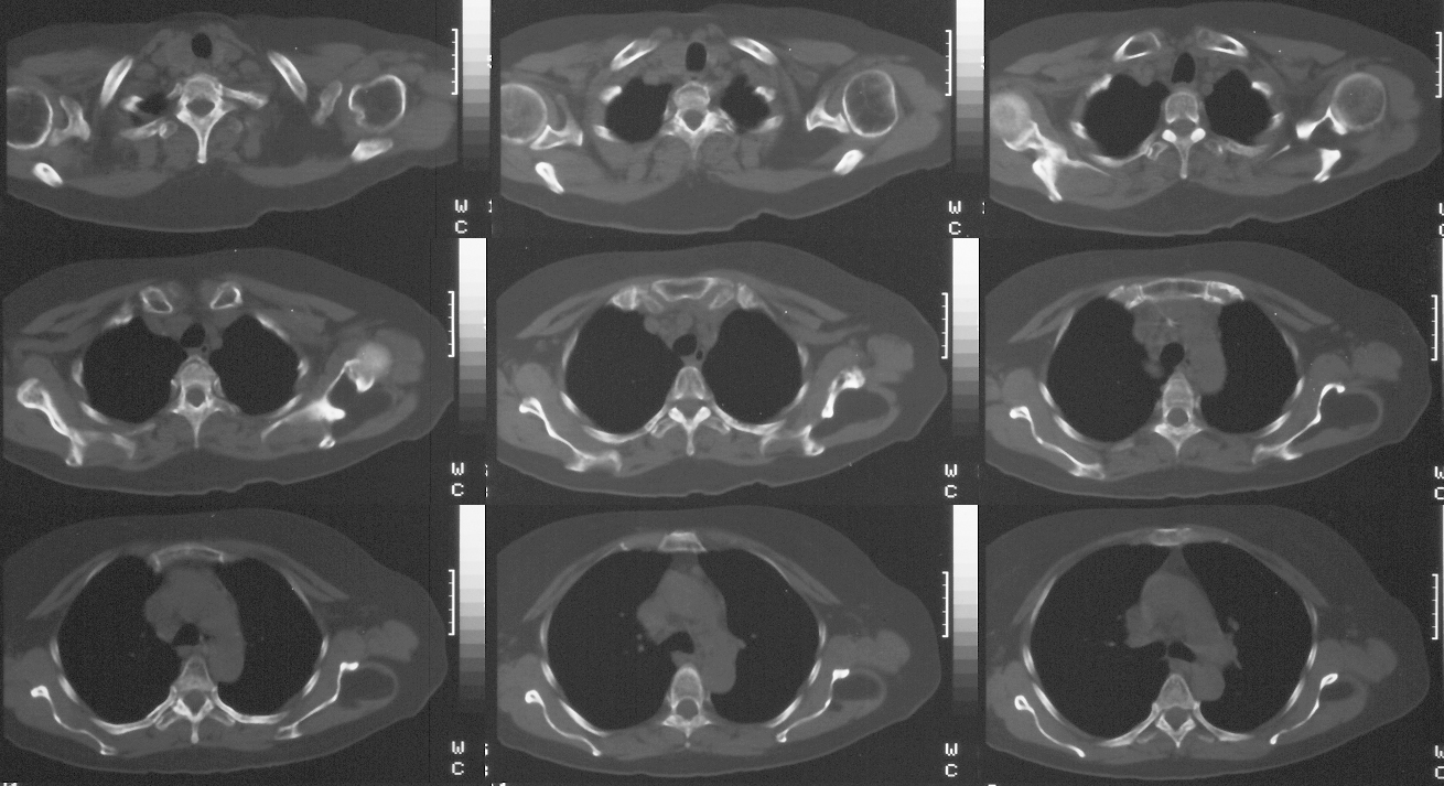
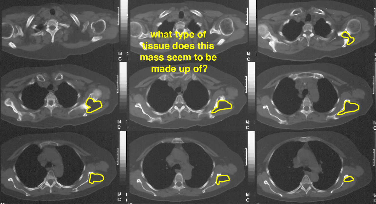
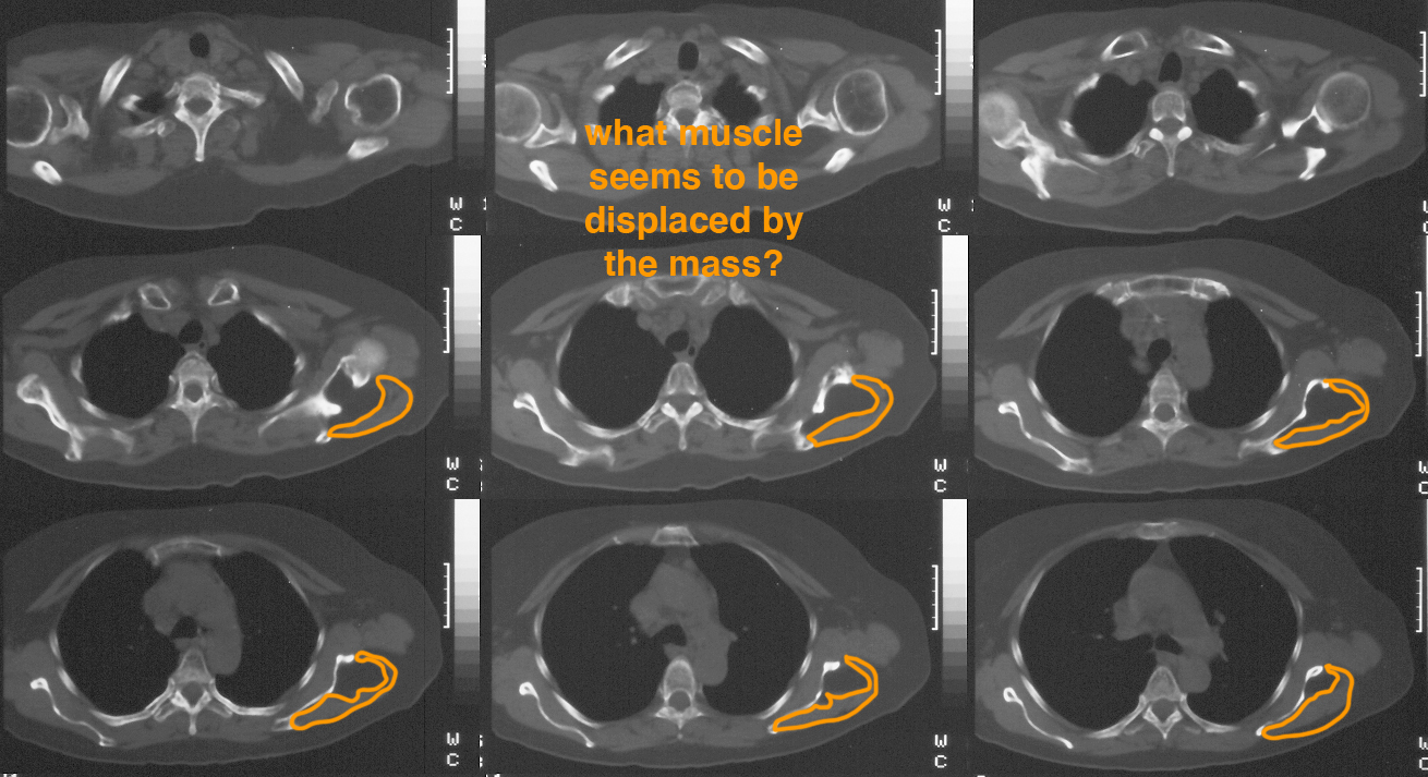
What type of study is this and how do you know?
This is a CT scan of the upper chest and both shoulders. You can tell it is a CT because cortical bone is white. On MR, cortical bone is ALWAYS dark. Other tissues may change depending on the particular type of MR that it is, but cortical bone never changes. It is not an US because it is too sharp and clear and the field of view is much too large. It is not a NM study, again because the anatomy is too sharp and clear.
Case 1
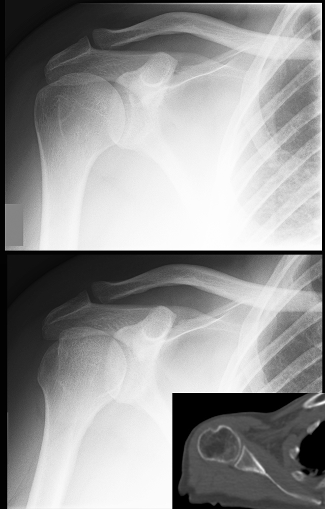
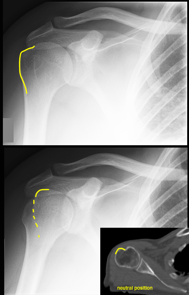
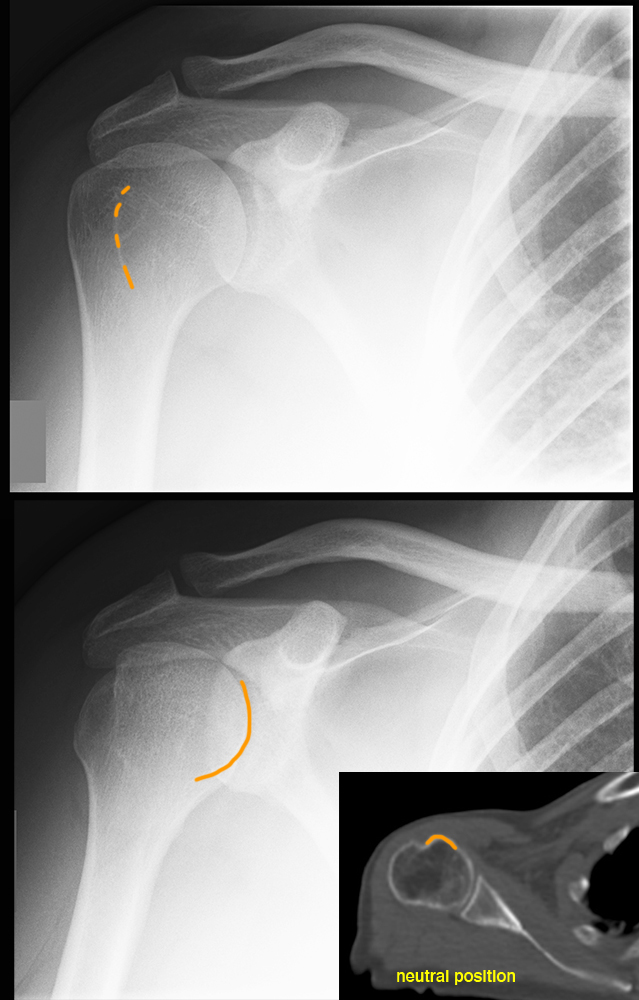
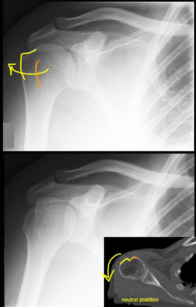
These are normal comparison images for this case.
Question 4:
How do the top and bottom images differ, in terms of patient positioning?
Both are most likely AP views, since is usually more cumbersome to do a PA view and there is no advantage over the AP view for the shoulder. The top view is with the humerus in EXTERNAL rotation and the bottom view is in INTERNAL rotation. The CT image is shown to help you visualize the greater and lesser trochanters and how they would move in these two positions. The CT image is in neutral (anatomic) position. There are many other views that can be done in patients with shoulder complaints. In general, the more complex a bony area, the more views that may be needed to adequately radiograph the area.
