

Case 2
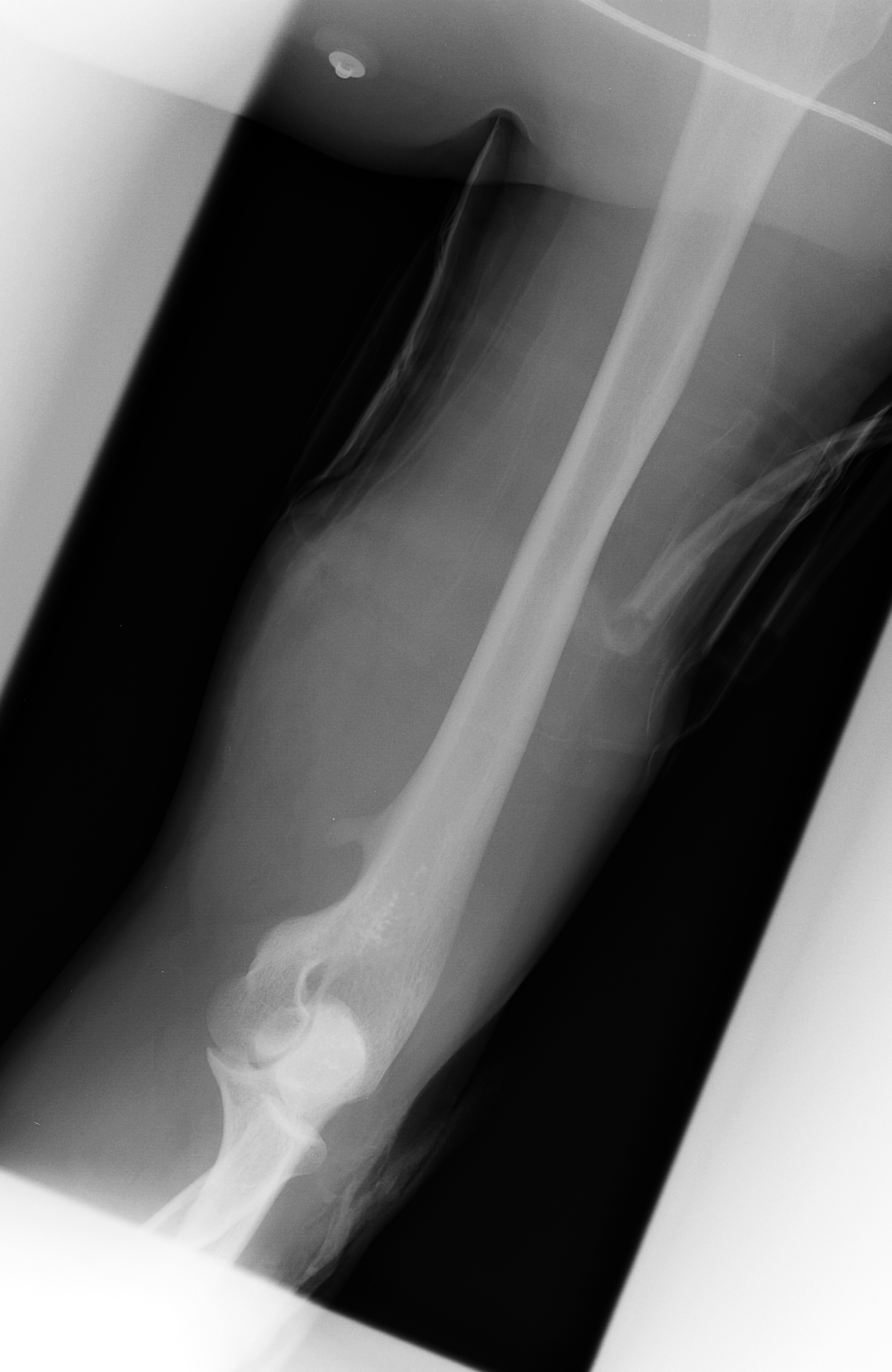
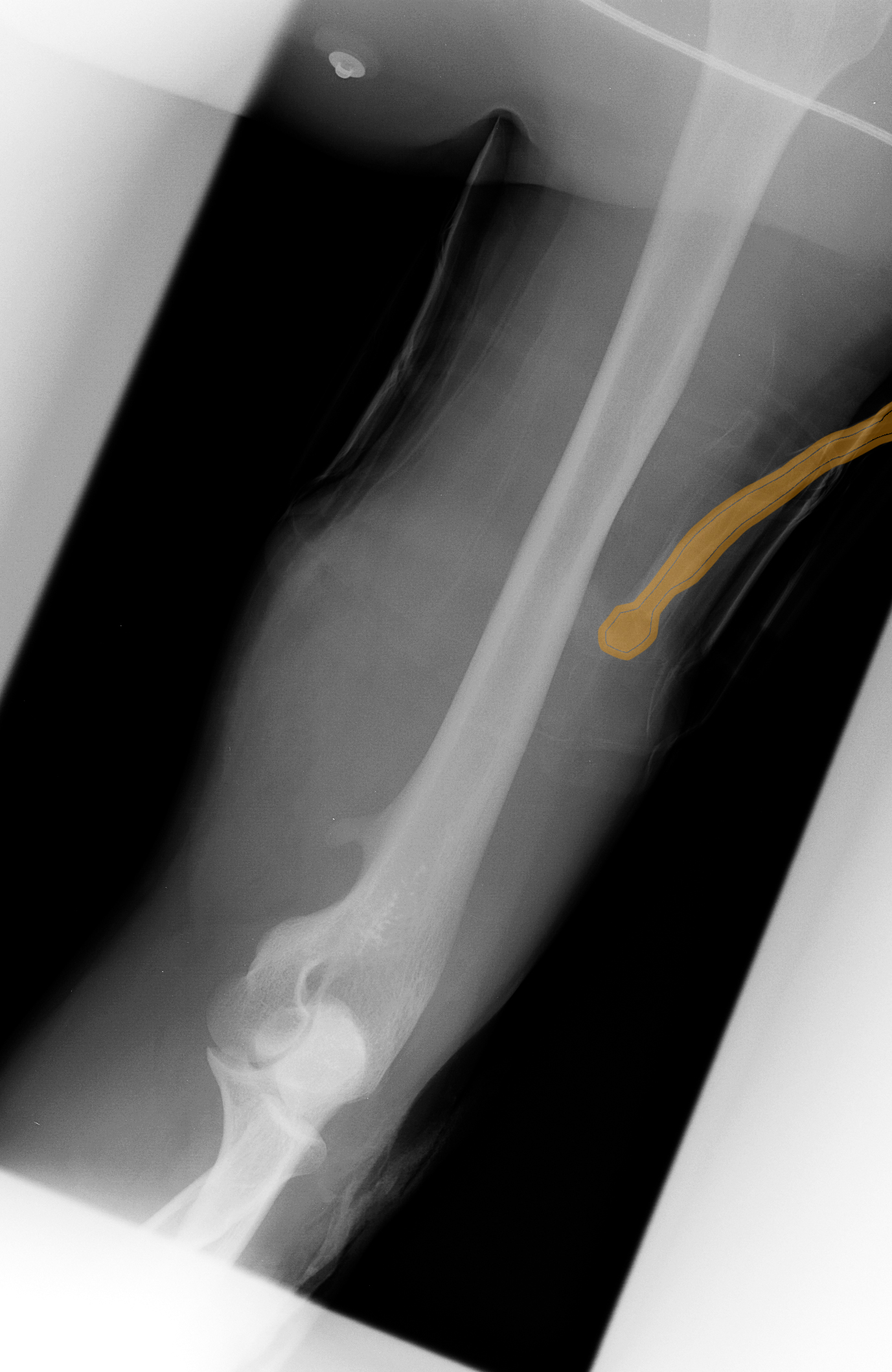
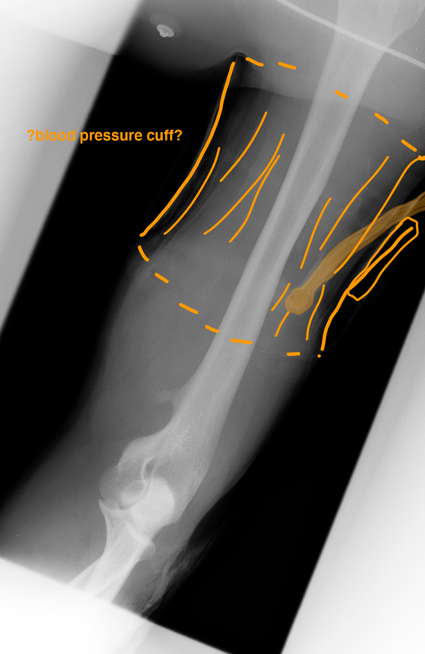
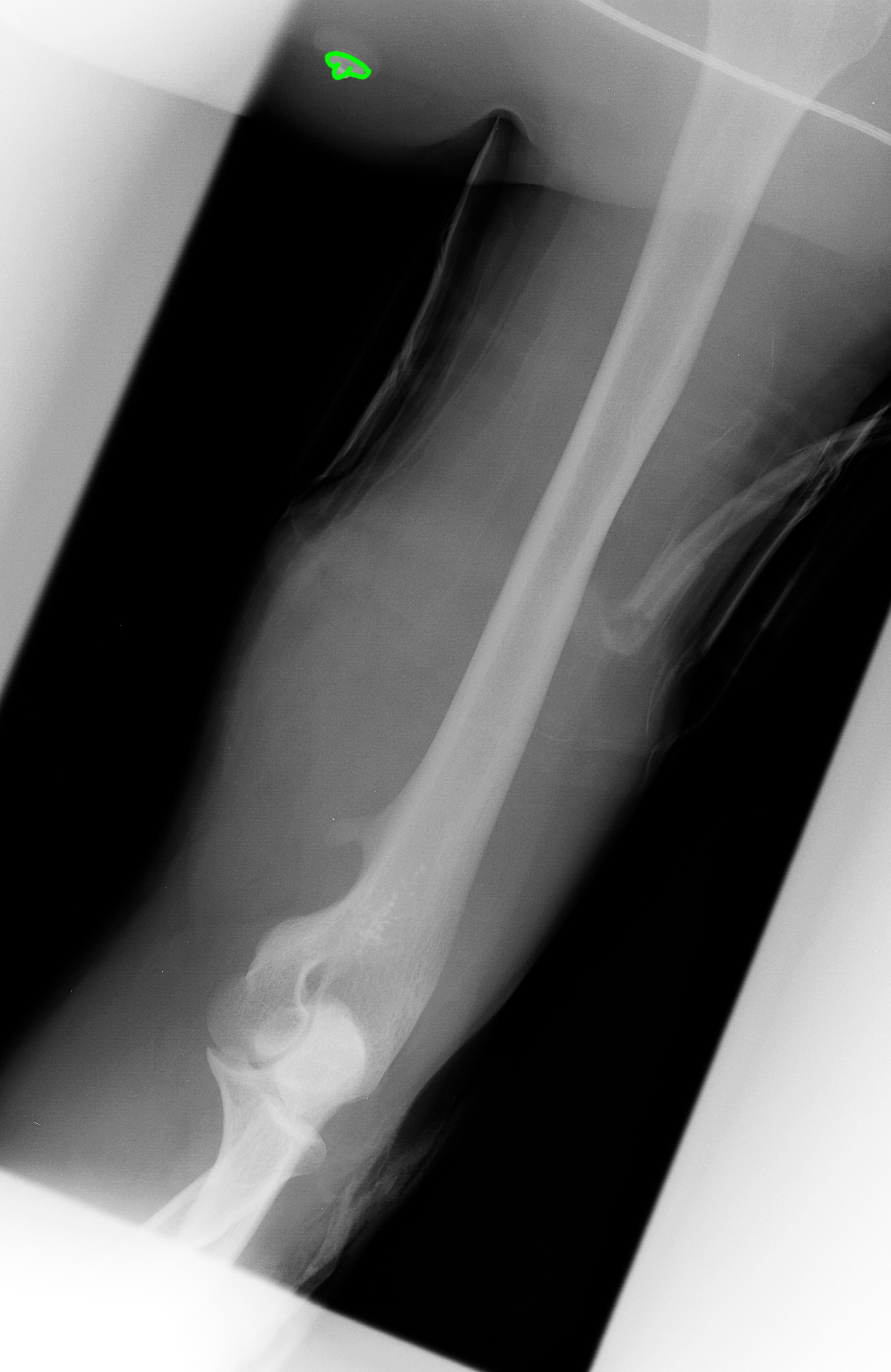
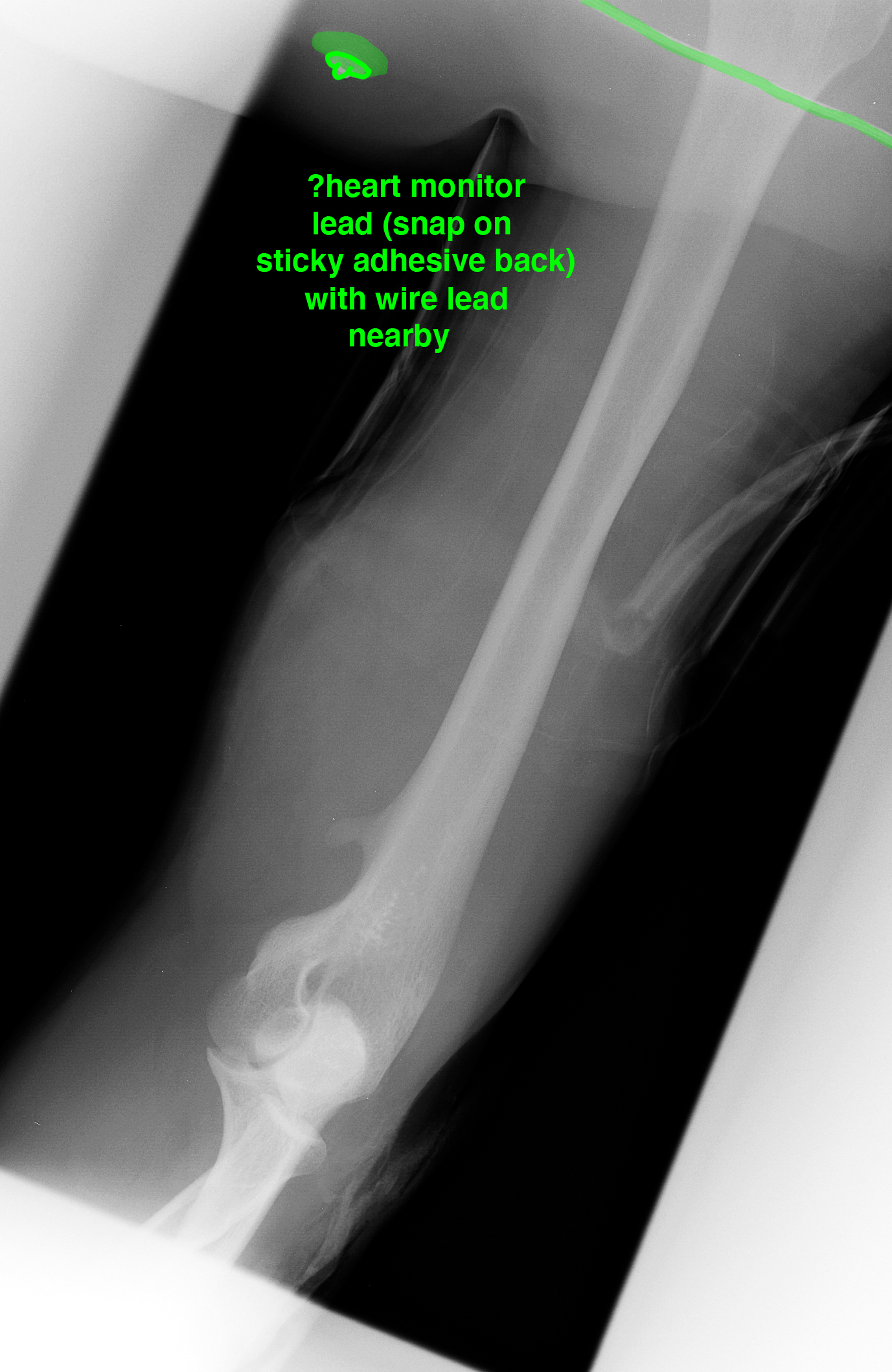
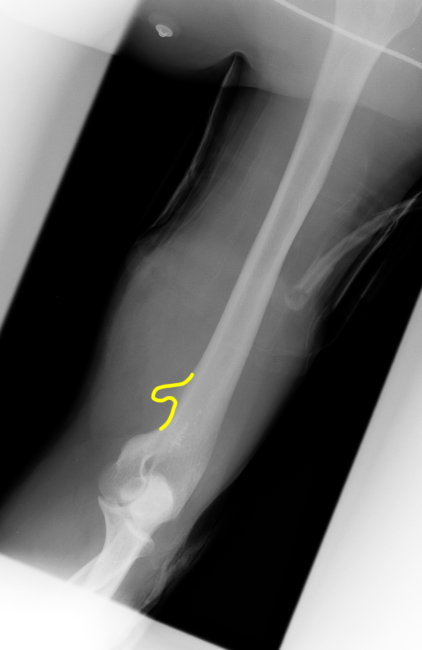
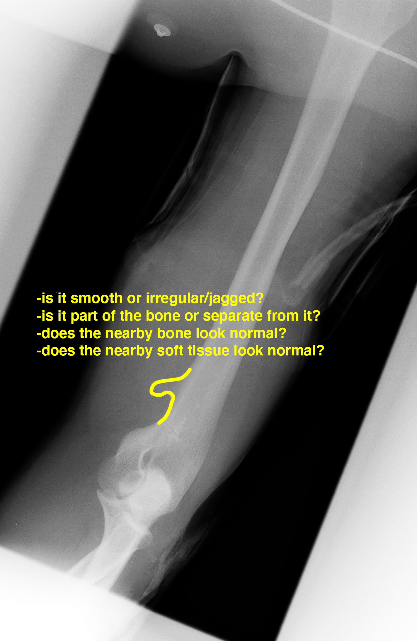
40 year old female with chronic left humerus pain, just proximal to the elbow.
Question 1:
What view do you think this is? For a humerus complaint, do you think you typically need many extra or special views?
This is again likely and AP view (PA is more difficult and has no particular advantage for most limb imaging). It seems, based on the orientation of the elbow, as if it is a lateral view. For simple tubular bones like the humerus, radius, ulna, femur, tibia and fibula, we usually do a frontal (AP from the front) and a lateral view. Do you think a frontal AP will show the abnormality well?
Case 2
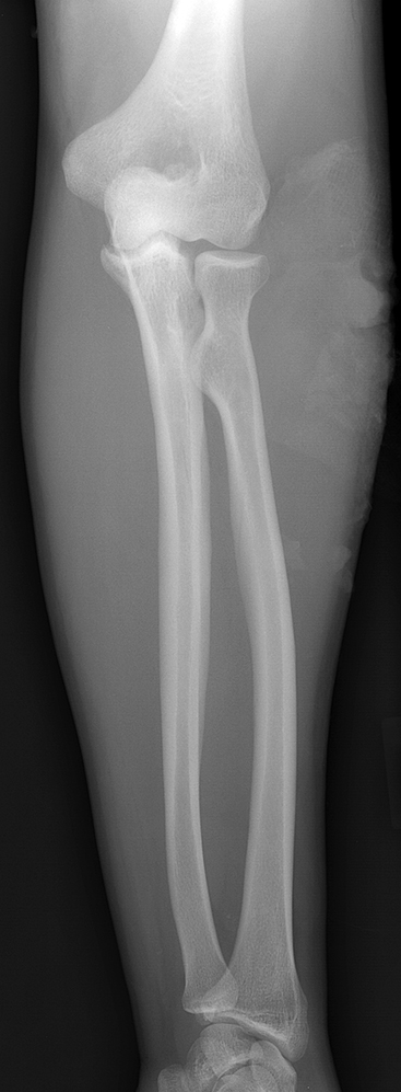
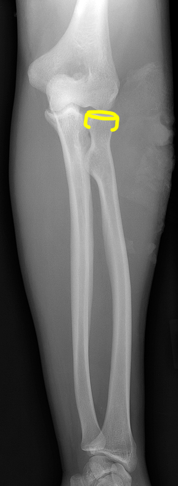
This is another image of the same patient.
Question 2:
Does this image help in assessing this patient's complaint?
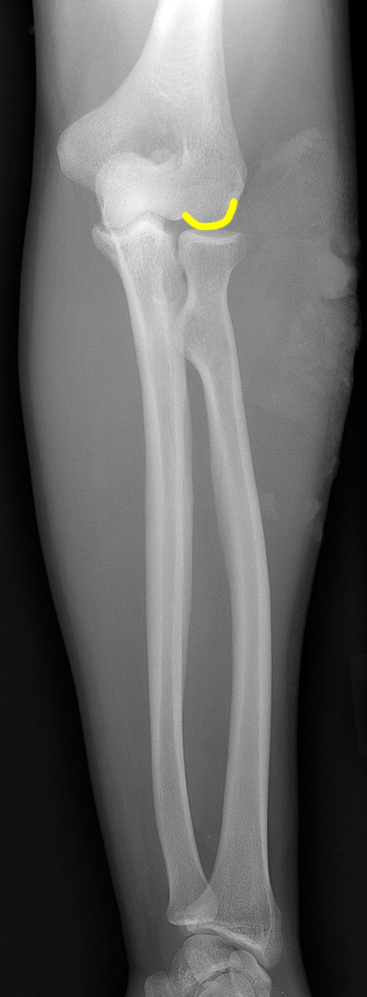
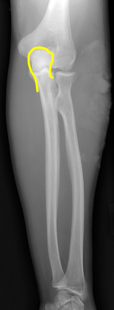
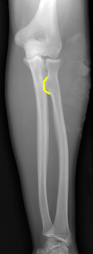
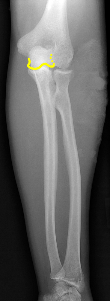
It is really an AP view of the radius and ulna, rather than the humerus. It likely goes just proximal enough to possibly include the finding, but given how it seemed to be projecting out on the lateral view, it would be unlikely to show up very well on an AP view--it would be overlapped by the rest of the distal humerus. This gives us a chance to review some imaging anatomy of the forearm.
Case 2
This is a study on a different patient with arm pain. This is a normal study.
Question 3:
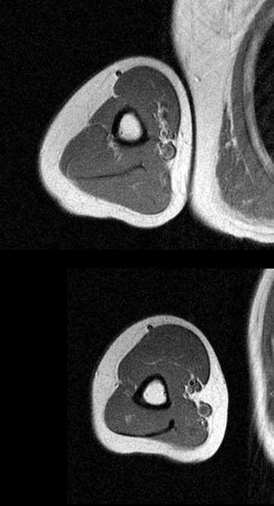
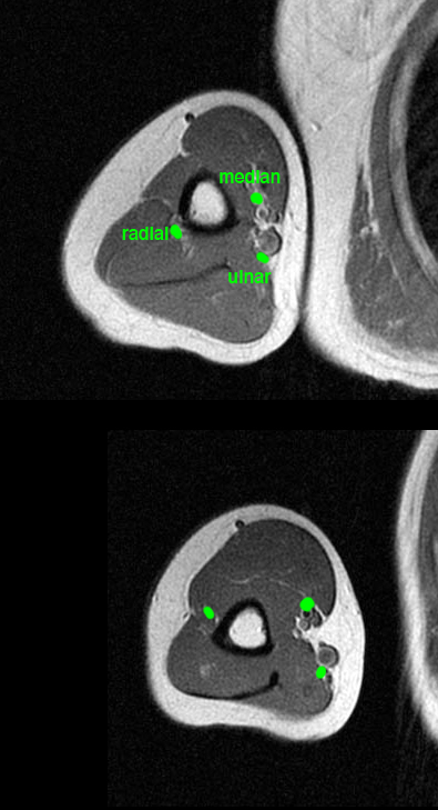
What is this study and what technical aspects can you identify?
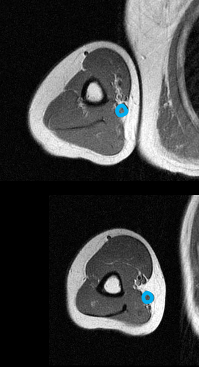
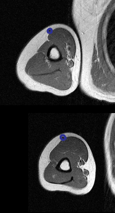
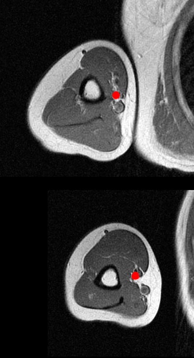
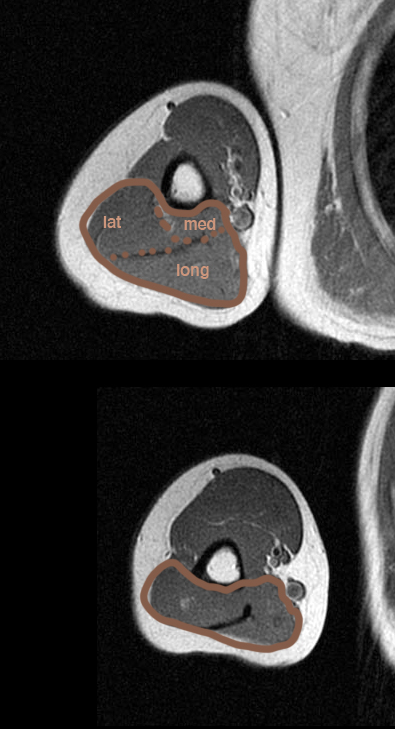
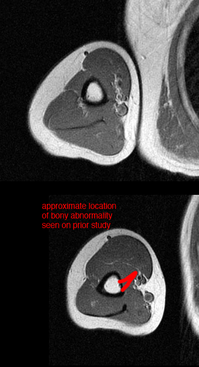
This is an MR of the humerus. The top image is higher than the bottom and both are in the axial plane. You know it is MR because cortical bone of the humerus is DARK. The fat is very high signal so no fat suppression is used. The vessels look more dark than bright. It is hard to tell if it is T1 or T2 weighted as there is no fluid on the image. Some normal anatomy is shown below, as well as an indication of the approximate location of the bony abnormality seen on the prior patient. What nerve would be most likely to be compressed by this bony projection?
Case 2
These are normal views of the humerus in a different patient.
Question 4:
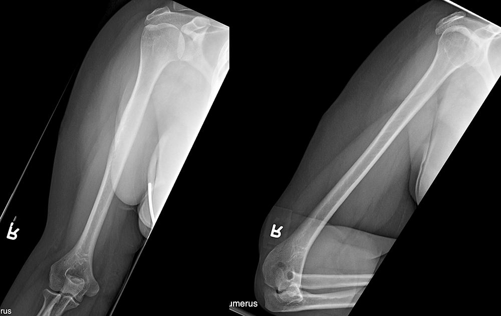
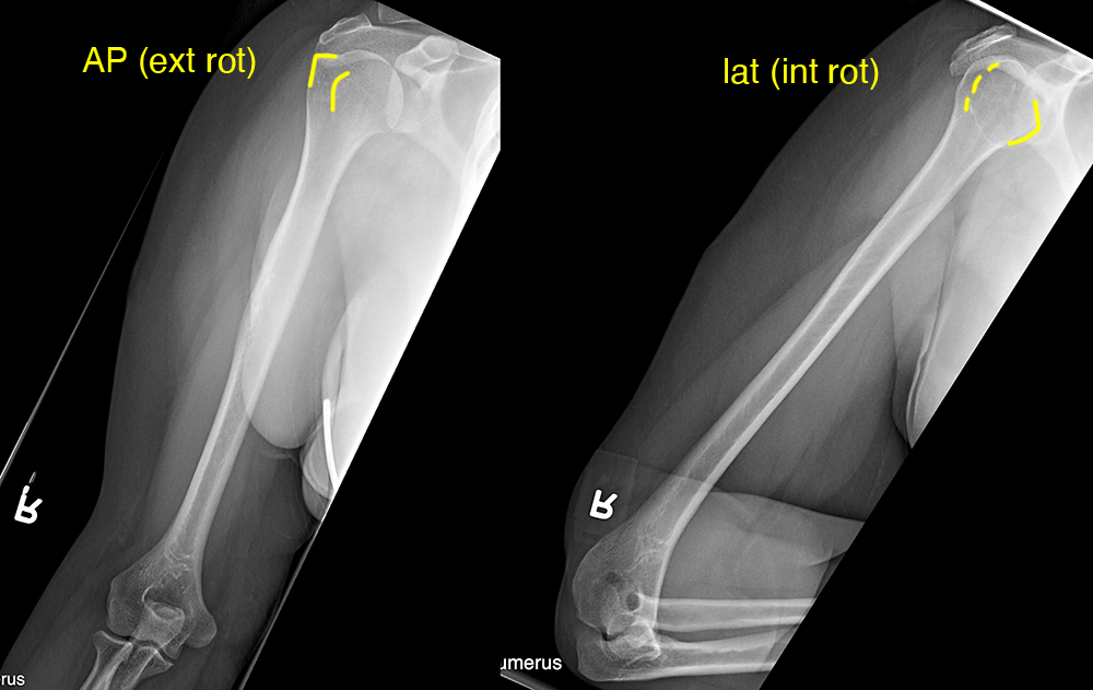
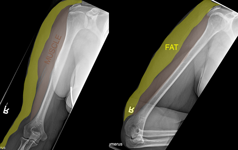
How is the arm positioned in these two views? What do you think of this patient's proportion of muscle to subcutaneous fat?
This is the typical two-view image set that is done for long tubular bones like the humerus: an AP and a lateral. If you look carefully at the humeral head, you can see that this was accomplished by externally and internally rotating the humerus. This patient has a fairly large amount of subcutaneous fat and not very much muscle, which gives some idea of their overall state of physical conditioning and nutrition. It is important when looking at bone radiographs not to forget to look at soft tissues as well.
