

Case 3
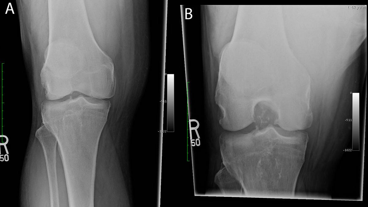
This patient has a painful right knee that appears swollen.
Question 1:
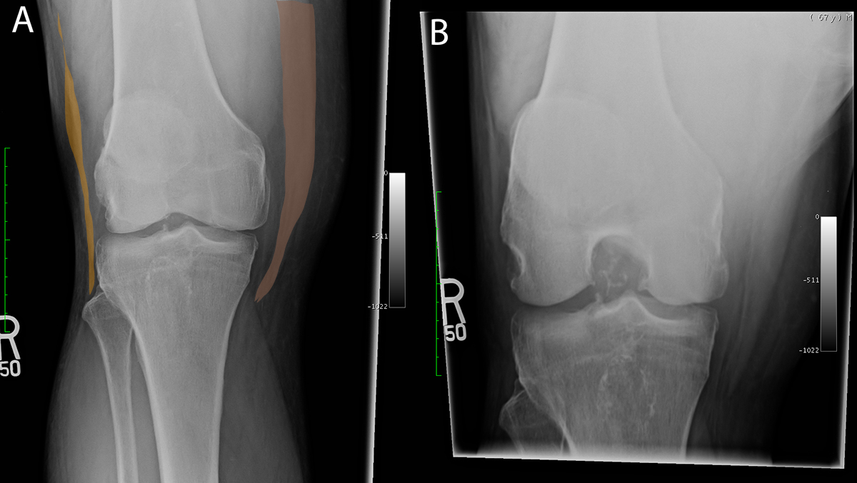
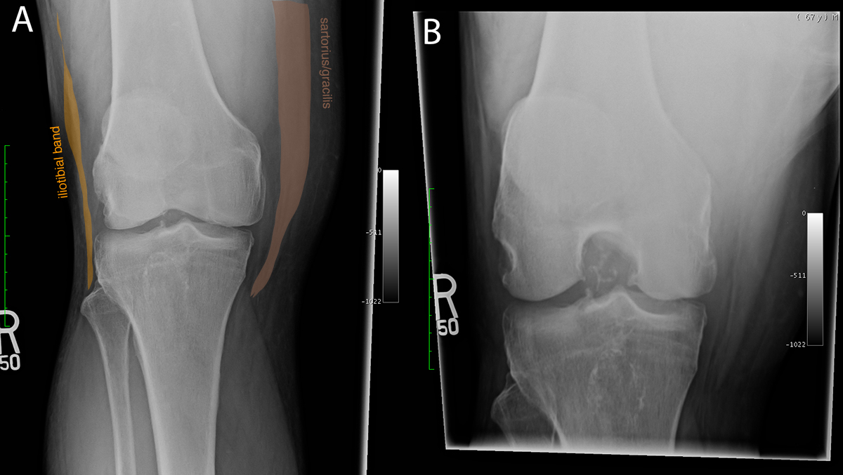
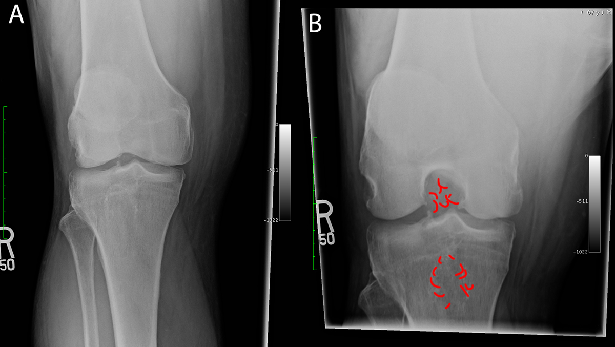
What are these two radiographic views? Do you think most knee radiographs would include more than just and AP and lateral view?
Image A is an AP view but Image B is a special view for the knee, which should not be surprising given how complex this joint is. It is called a 'tunnel' view, done with the knee flexed so that you can see in between the two femoral condyles. Like the shoulder, there are several extra views that are most often done for knee complaints in addition to AP and lateral view. Do you see anything unusual in this patient's tunnel view? Where do you think the abnormality is located--in the joint, in the bone, or elsewhere?
Case 3
This is more imaging on the same patient.
Question 2:
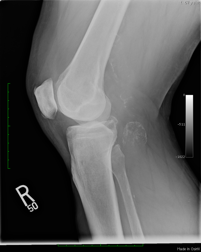
What is this view? Can you see the abnormality and is it where you thought it would be?
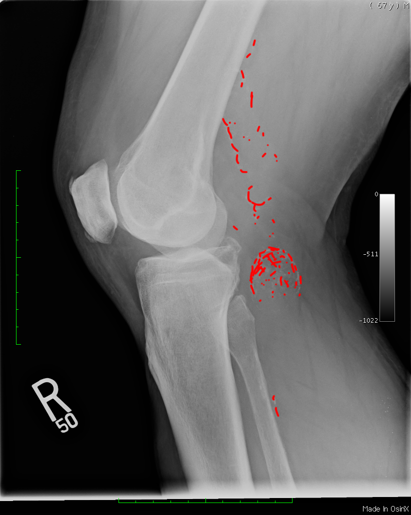
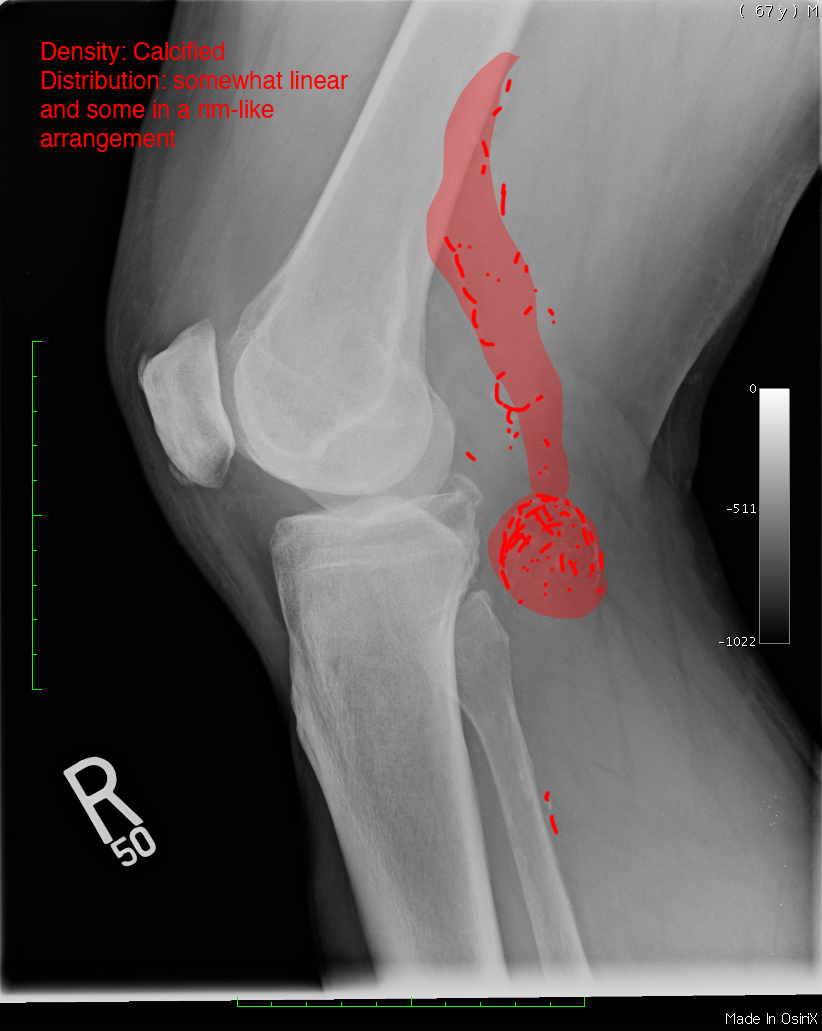
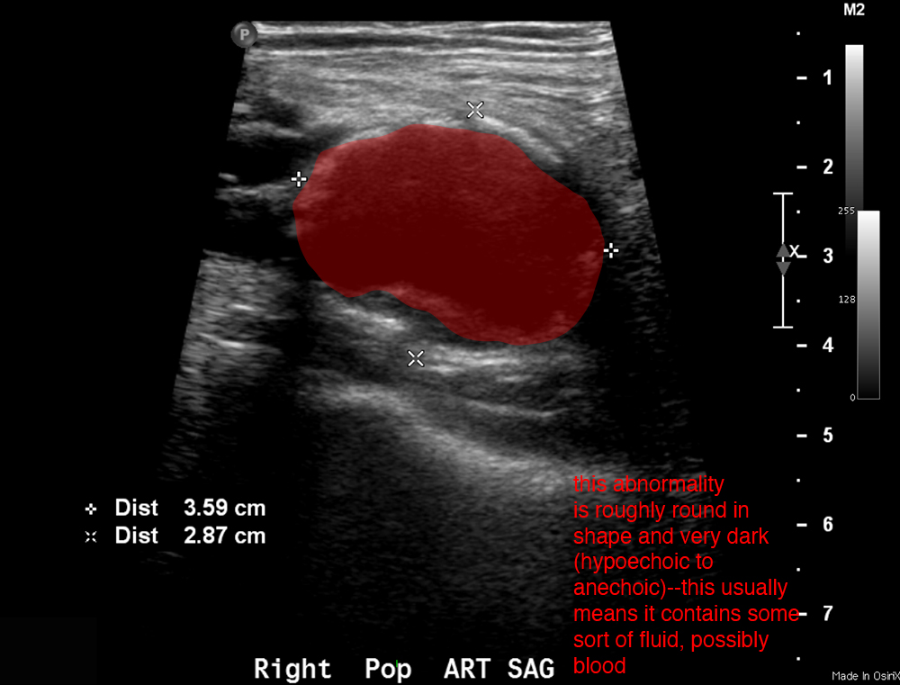
This is a lateral view, performed with the knee slightly flexed. It shows the abnormality much more clearly, and that it is not in the bone or the joint but in the popliteal fossa, which was hard to determine from the AP and tunnel views alone. When you look at the abnormality, think of what adjectives you would use to describe it.
Case 3
This is another study on this same patient.
Question 3:
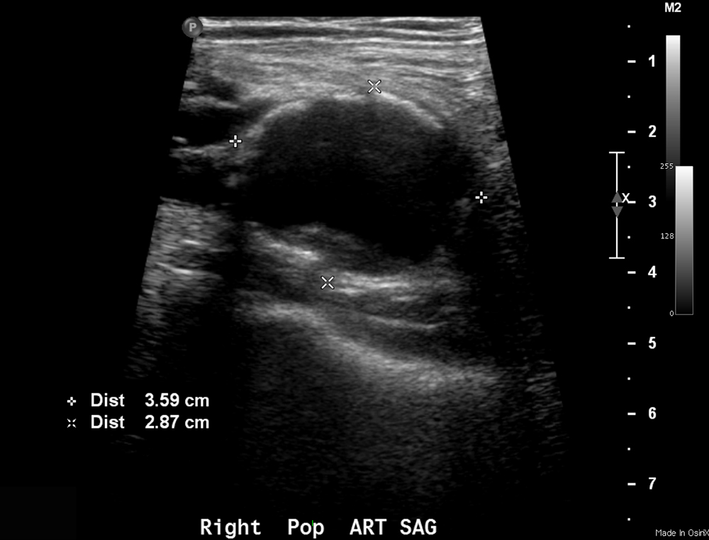
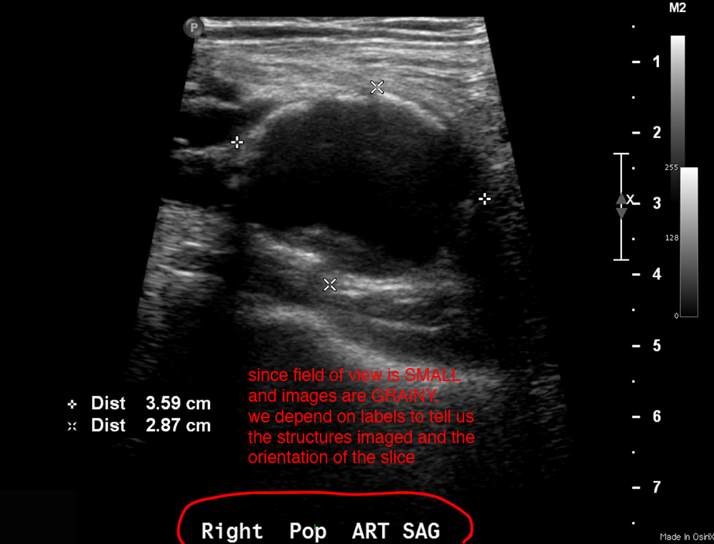
What is this study and does it make sense that this might be a good choice for this patient?
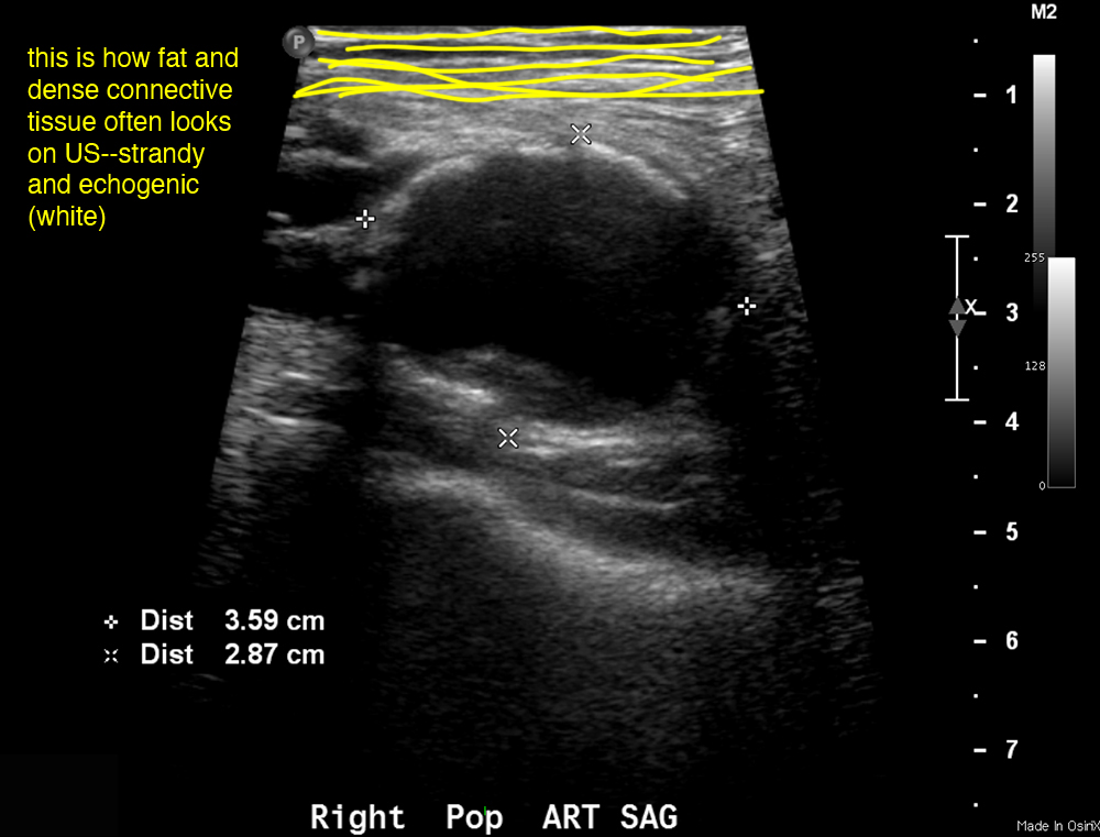
This is an ultrasound. As mentioned, US is great for examining soft tissue structures (not blocked by air or bone) that are near the body surface. The popliteal fossa is a great place for ultrasound. It is particularly good for identifying fluid-filled structures (cysts) and vascular problems.
Case 3
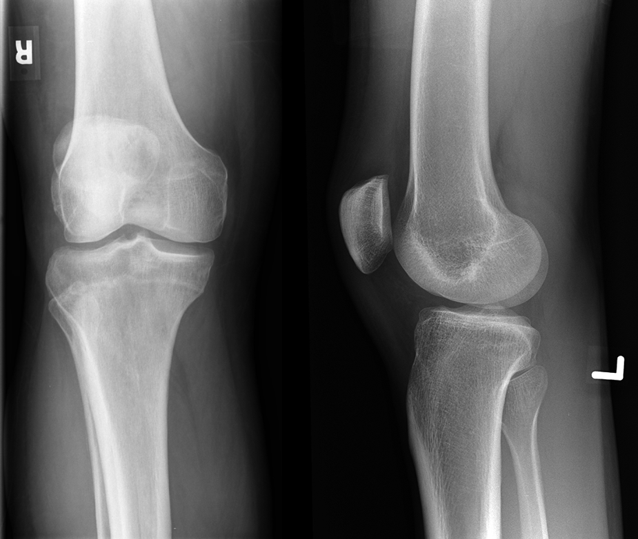
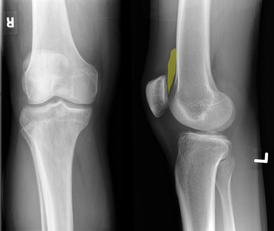
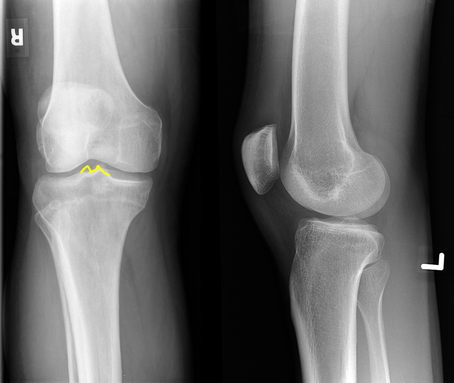
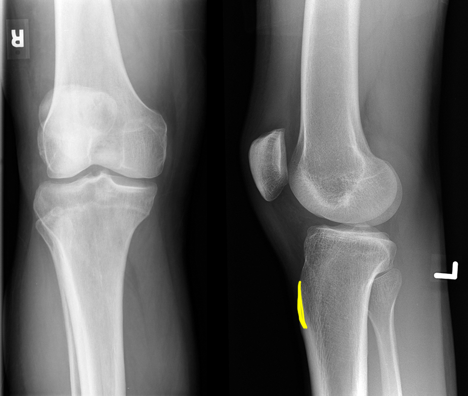
These radiographs are from a normal patient.
Question 4:
If this was being done to check for joint space narrowing (often seen in arthritis), what additional piece of information would you like to have about how the study was done?
These are an AP view of the right knee and a lateral view of the left knee, and to see joint space narrowing, it is often recommended that the views be done with the patient standing rather than seated or lying down. This called 'weight bearing' views and the images would usually be labeled as such.
