


Case 4
Middle-aged male with chronic foot pain.
Question 1:
Just like the hand, the knee, and the shoulder, the foot is a complex shape with many parts. What is this view and what is abnormal?
This is an AP view of the foot. As in the knee, sometimes we do images with weight-bearing to see if this brings out any problems with joints or alignment. Usually the image will say if it was done while the patient was standing. Focus your attention on the lateral part of the mid-foot, the cuboid in particular.


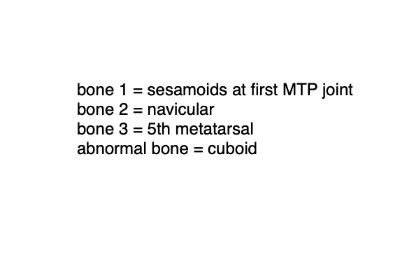
Case 4
This is another image from the same patient.
Question 2:
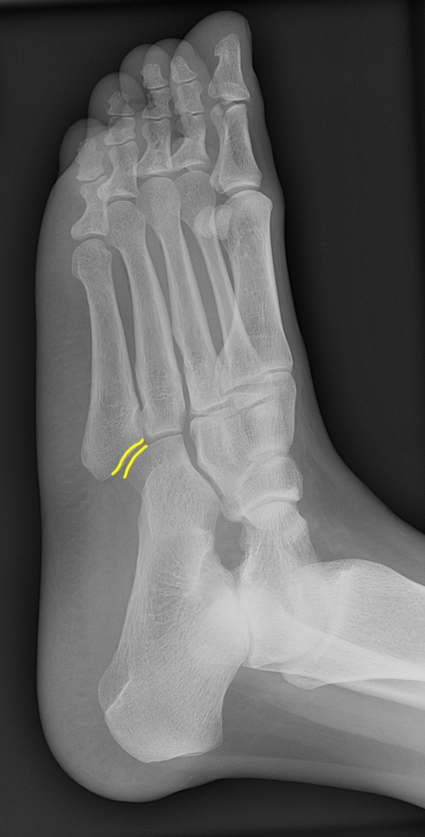
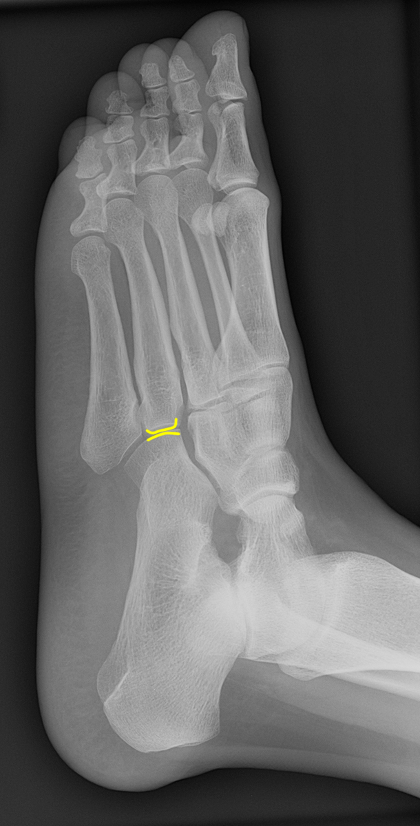
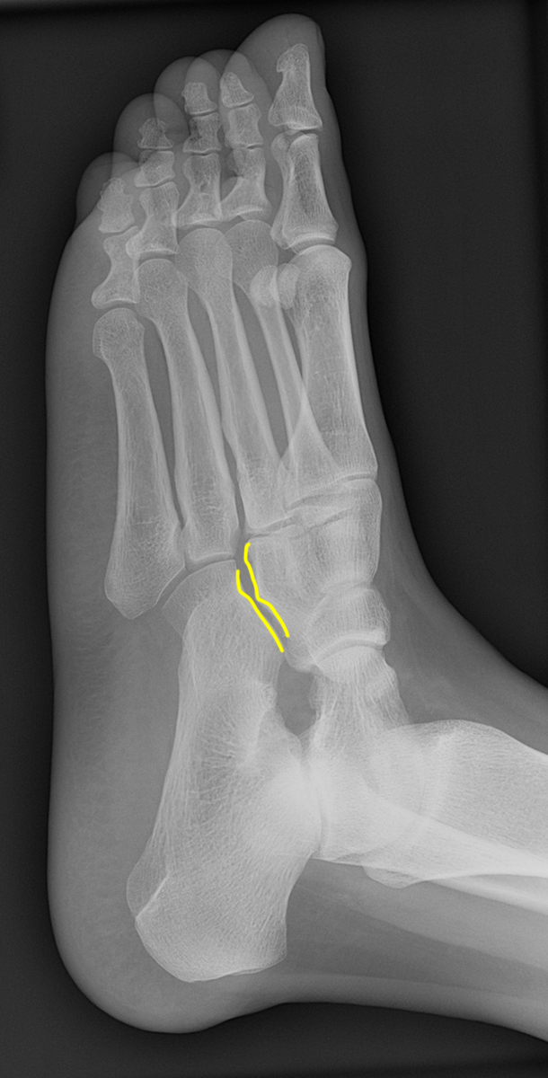
What is this view?
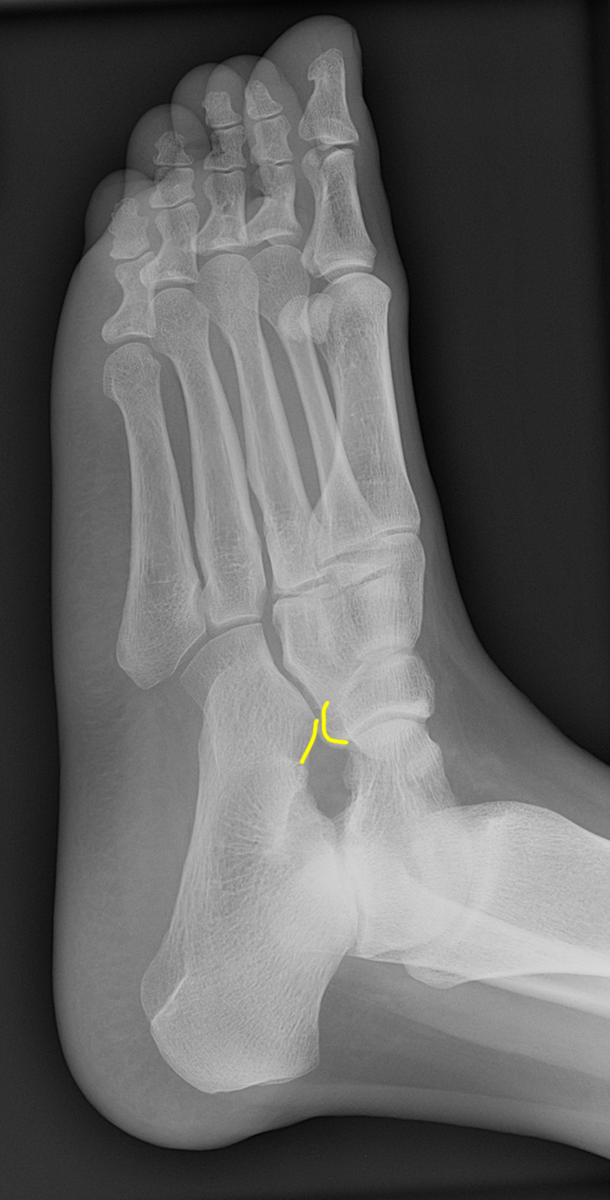
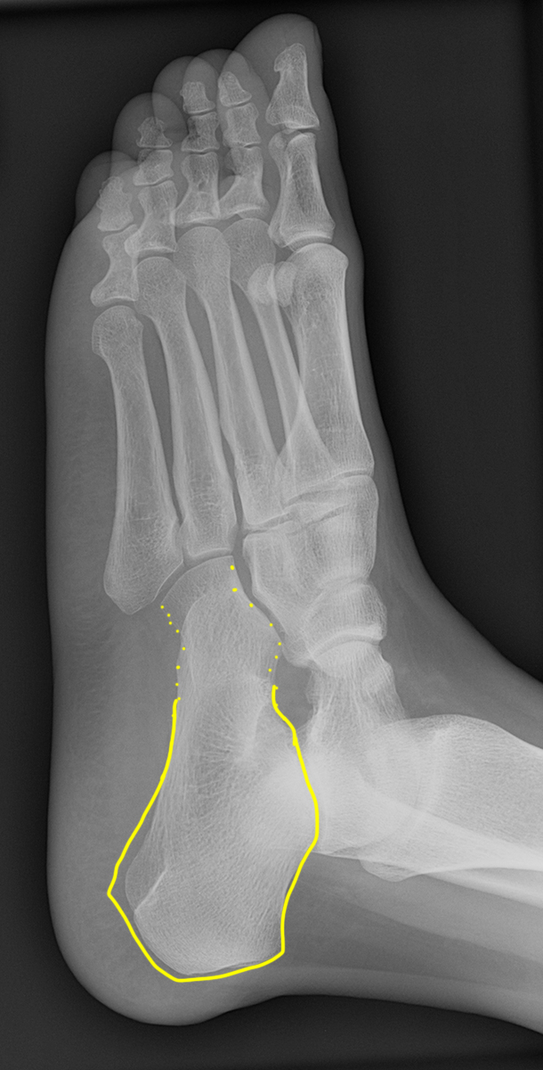
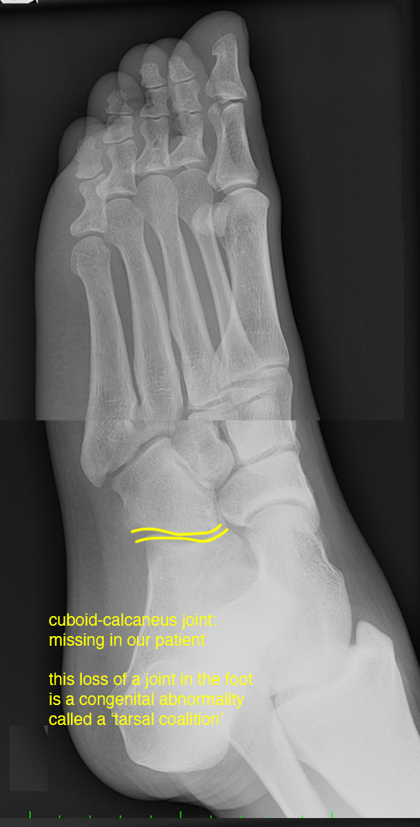
This is a view that is half way between and AP and a lateral. It is called an oblique view and is often done in complicated bony areas like the hand, foot, ankle and wrist. The many joints of the foot are all oriented in slightly different angles, so it is not surprising that an AP and a lateral would not be optimal views for all joints. Look at this oblique radiograph and decide which foot joints are particularly well seen. Decide which tarsal bone(s) appear abnormal.

Case 4
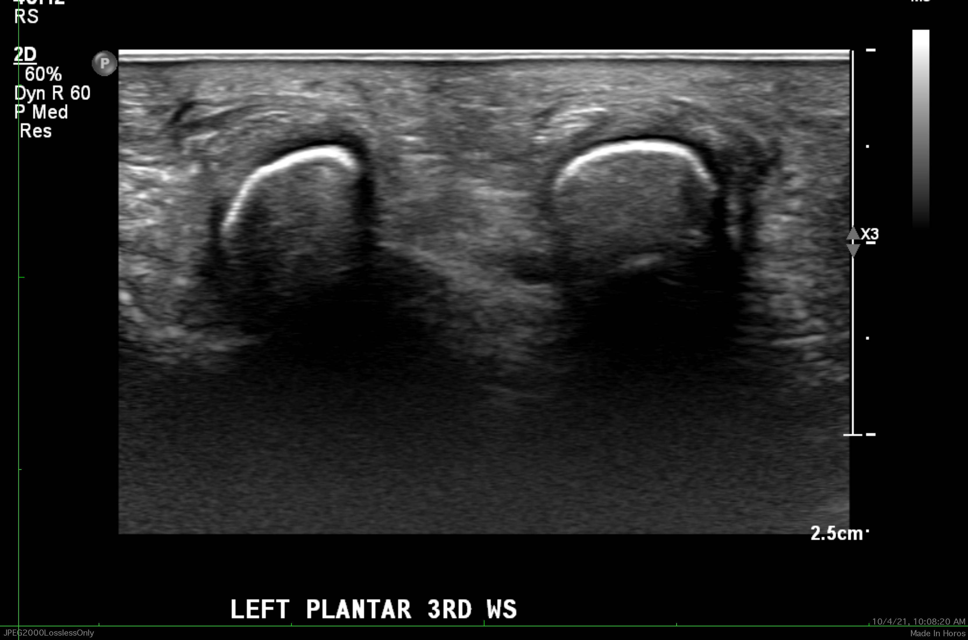
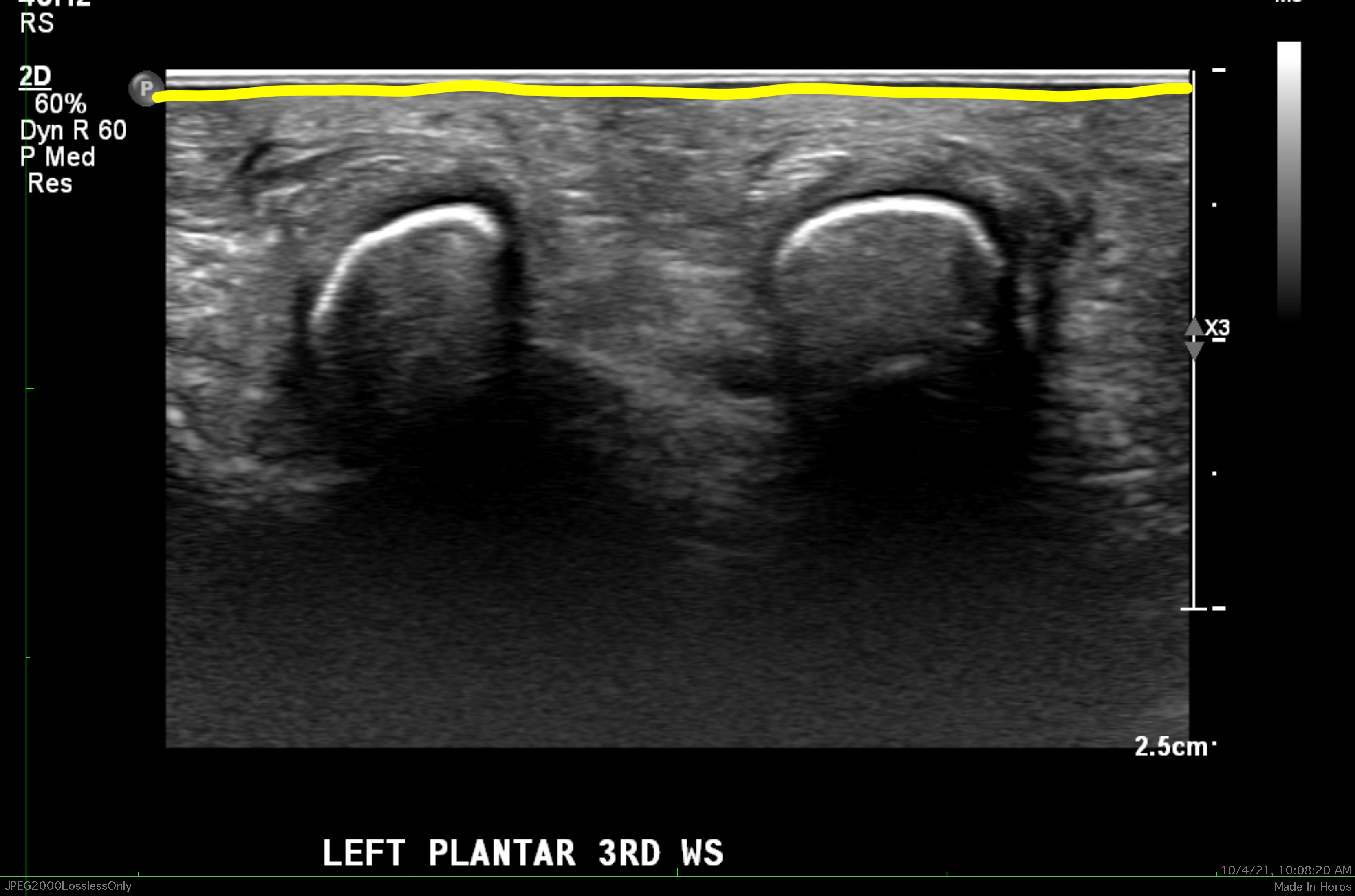
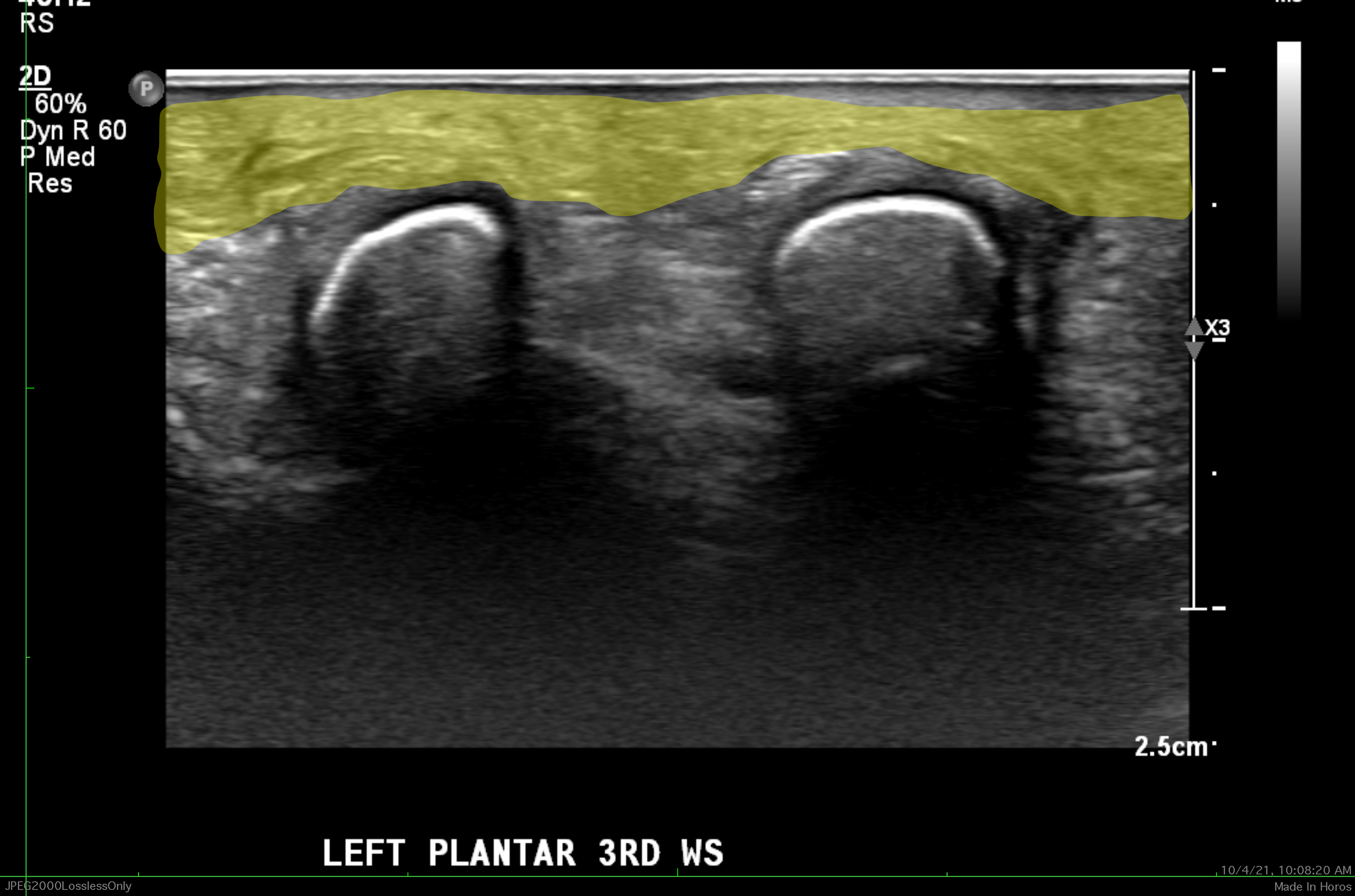
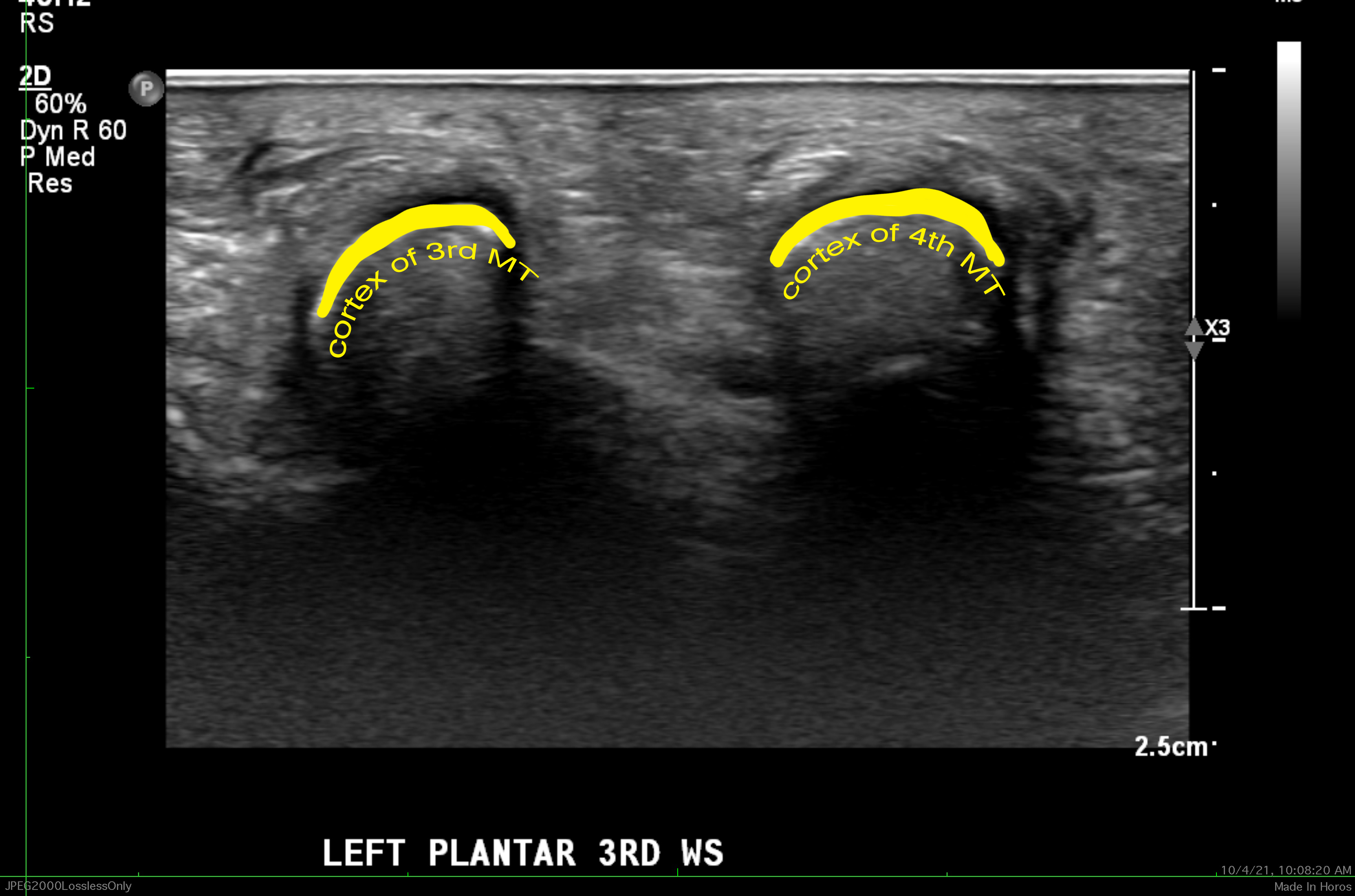
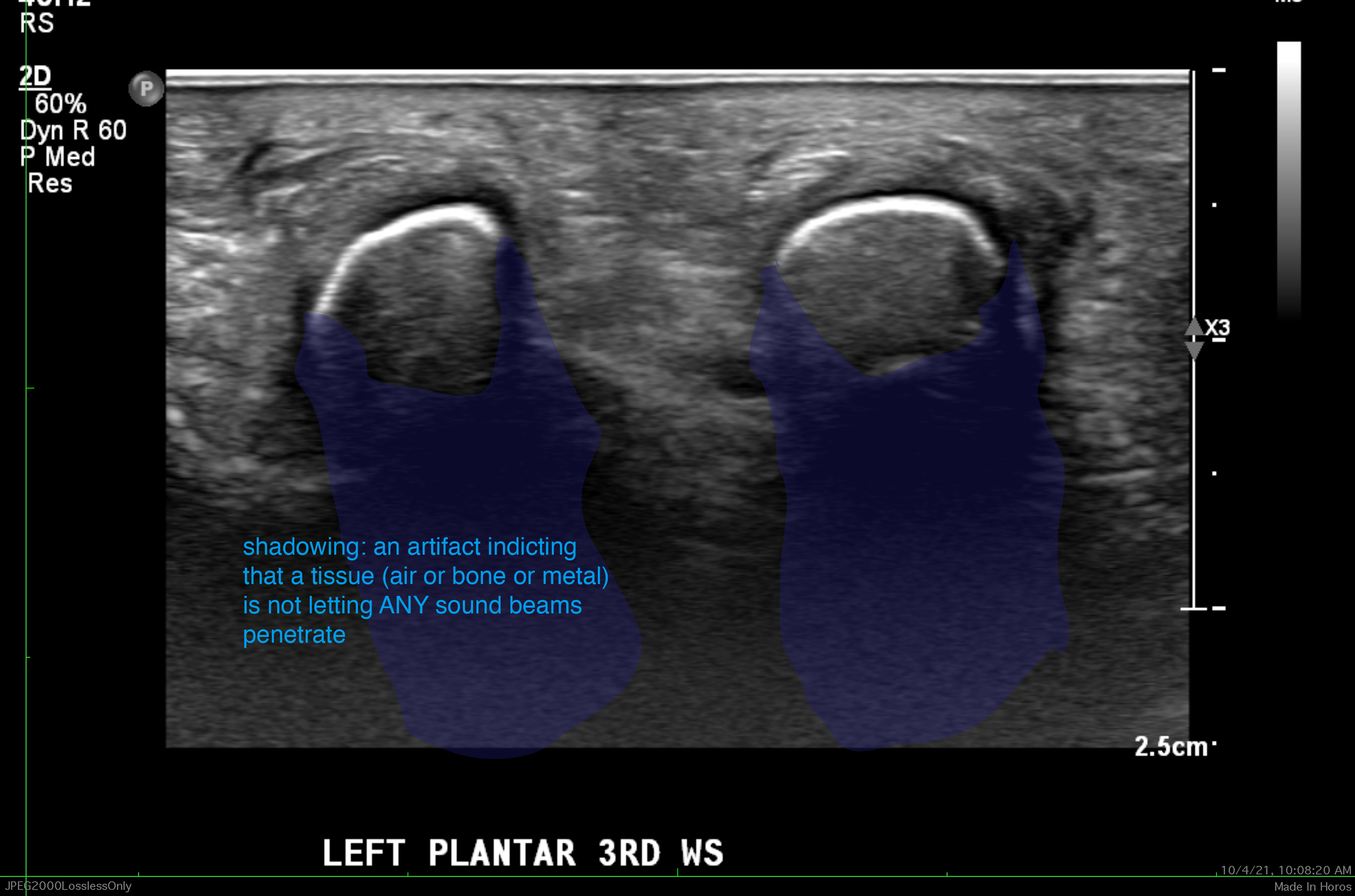
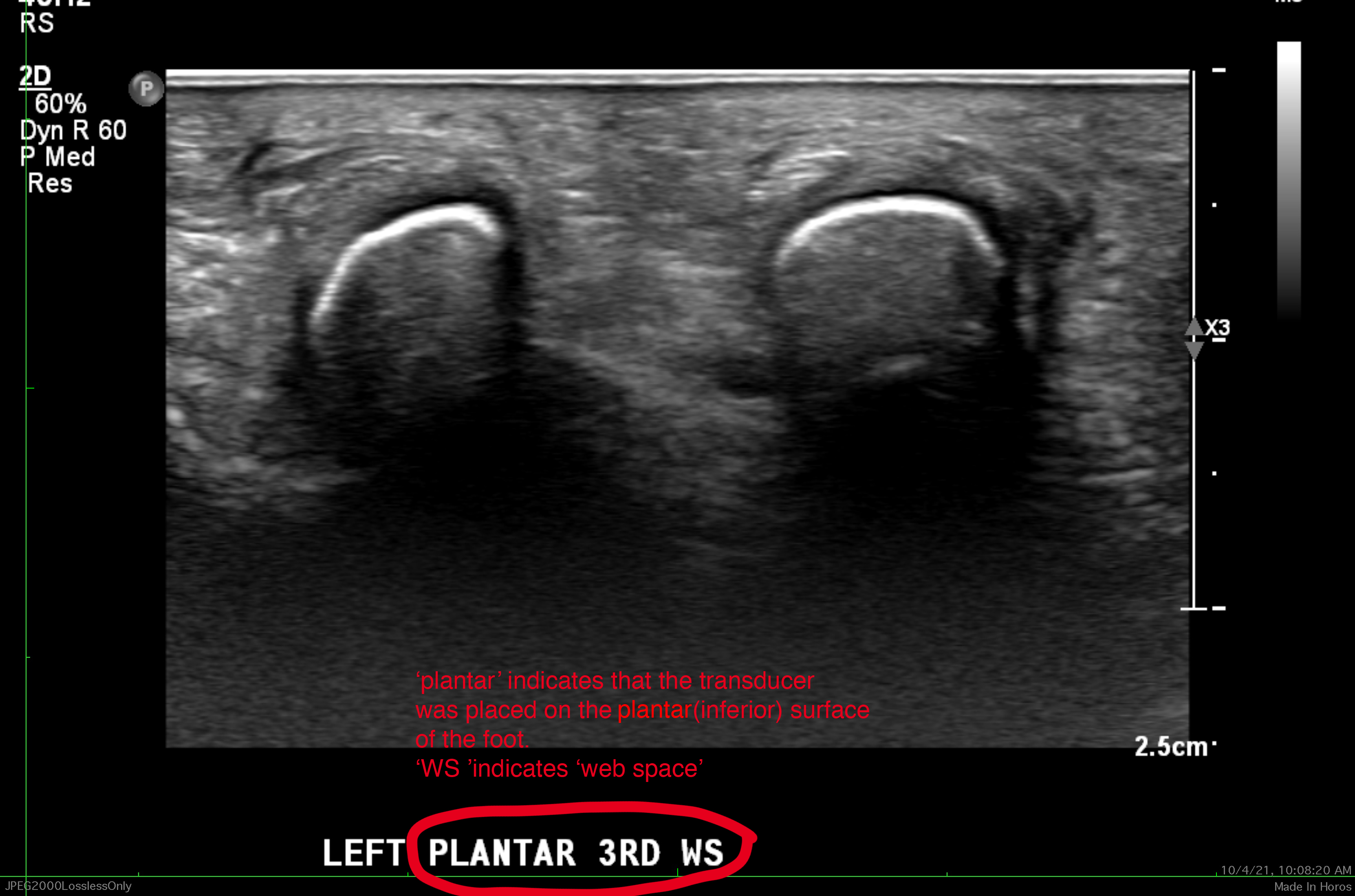
Imaging on a different patient with pain localized to the web space between the 3rd and 4th metatarsals.
Question 3:
What is this image and what do bones look like with this modality?
This is an US of the foot. As previously mentioned, US is a useful tool for body parts that are close to the skin surface, including the most superficial parts of the foot, ankle, and and wrist. The bones have a typical appearance with a bright surface nearest to the transducer and a dark shadow behind this, due to reflection of all of the sound from the first bony surface encountered. This ultrasound is normal.
Case 4
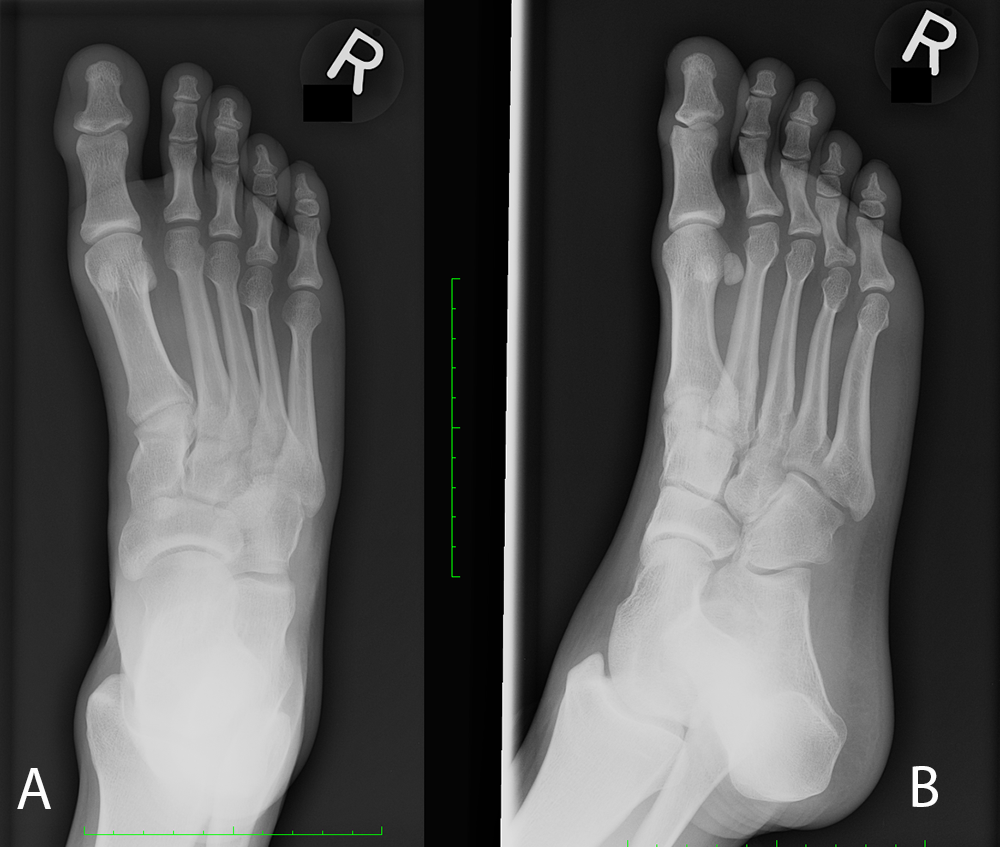
Normal foot images.
Question 4:
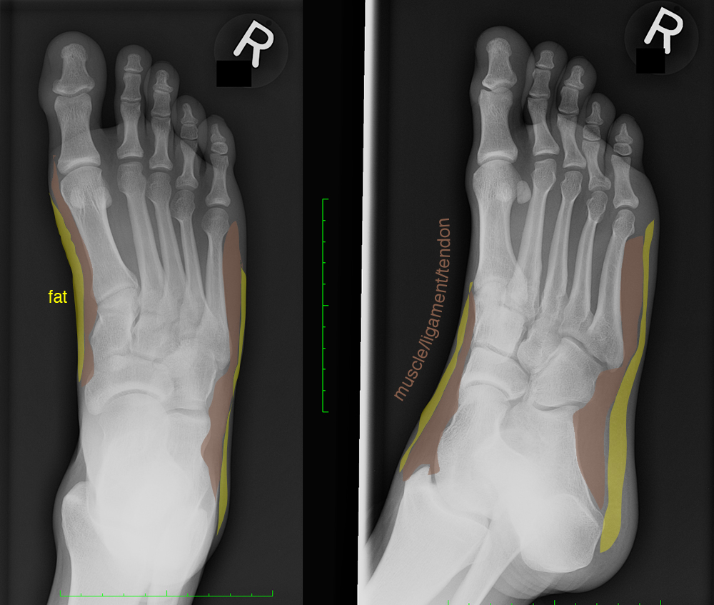
a) What are these two views? Where can you see fat/muscle/ligament interfaces best on these images?
Image A is an AP view and Image B is an oblique view. There is not a lot of subcutaneous fat in the foot, and the intrinsic foot muscles and ligaments are not large, so these interfaces are subtle. You can see a difference between fat and soft tissue (muscle, dense fascia, tendon, ligament) on the inner surface of the mid foot and along the lateral foot margin on the AP view and along the instep and adjacent to the calcaneus on the oblique view. If a patient develops soft tissue swelling due to injury or infection, these interfaces are lost and the thickness of tissue outside of the bone can increase dramatically.
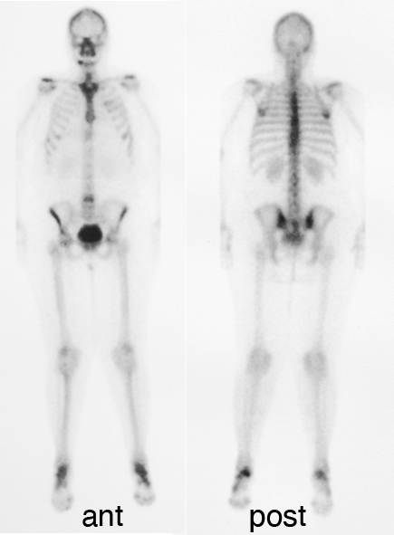
b) What is the other type of imaging shown?
The other imaging shown is an example of a normal bone scan. This type of imaging involves injection of a radio-tracer that homes in on bone, particularly in regions of active bone remodeling. So some of the bony structures are 'hotter' than others. The spine and the sacroiliac joints are often hot, while the long bones of the limbs are barely seen. In this case, the calcaneus on each side appears a bit hot, but symmetric. For this imaging, a large detector (called a gamma camera) is brought close to the body to detect where the radiotracer is coming out. So there are two images shown, one with the detector up against the anterior part of the body, and one with the detector up against the posterior part of the body. How could you identify which view is which without labels?
Case 4
This completes the pre-class cases for your review. These images will be discussed in class, with additional examples. You will get more out of the in-class discussion if you have reviewed these images ahead of time. Please come to class with any questions you have about these cases.
Further Explanation:
THIS is the final page of the preview cases for the Limbs Radiology session
Click the link below to return to other Anatomy Imaging topics-


