

Case 1
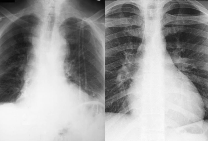
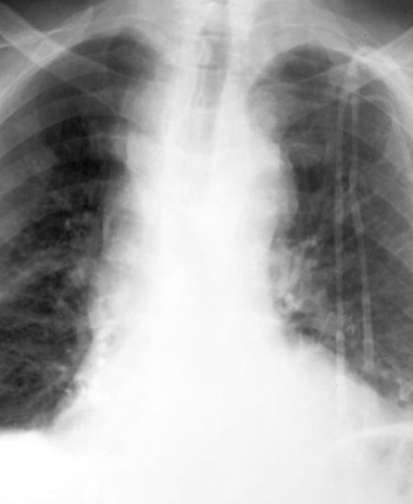
A 62 year old male with history of diabetes required a central line for administration of intravenous antibiotics for a foot infection. You are shown the chest radiograph (chest x-ray, or CXR for short) to check the position of the line.
Question 1:
What is a central line?
A central line is a thin plastic tube that is placed through the skin and into a vein that is close to the surface. The term 'central' means that the tip of the tube is in the region of the superior vena cava, which allows higher volume administration of fluid and medications than is possible with peripheral lines (also called 'IVs' for intravenous lines).
Click 'Reveal' to see the answer to the question. Click 'Next' at the bottom right corner of the screen to move to the next page. Click the pencil icon to enlarge the image and bring up annotation tools to draw on the image, if you are collaborating with other students. Your annotations will not be saved.
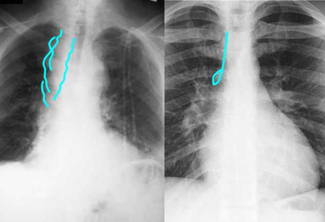
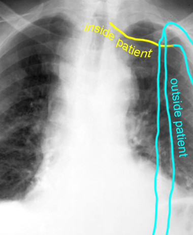
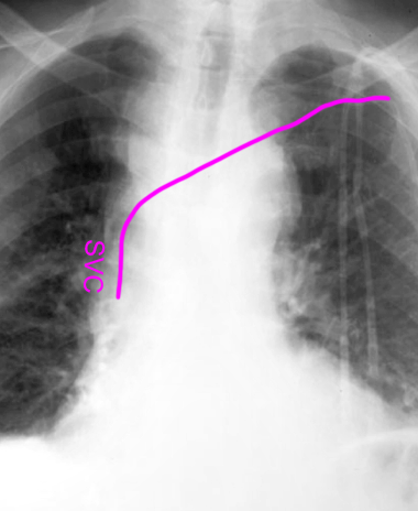
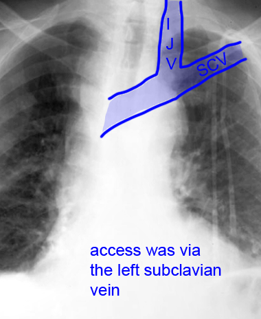
Click the white buttons below to highlight the course of the central line, and other annotations. Because the plastic of the line (also called a catheter) is not very dense, it is often hard to see on CXR. Do you think the tip of this line is in the proper position? What vessel was accessed to put in the line?
Case 1
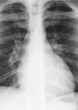
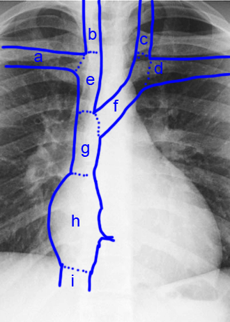
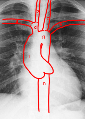
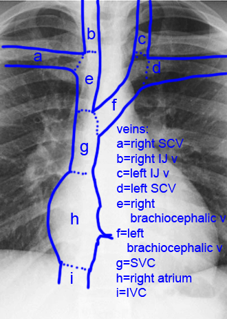
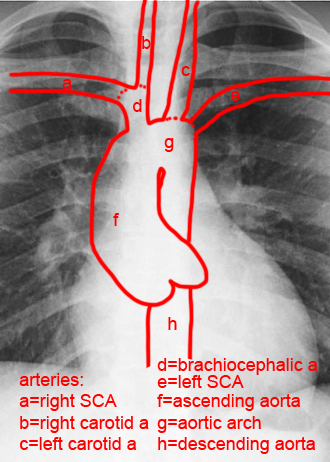
This is a closeup view of a CXR on a normal patient. We will use this image to review the important veins and arteries involved in central line placement.
Question 2:
a) Which vessels are most appropriate for accessing to place a central line?
You want to use vessels that are large, close to the target (the SVC), and close to the surface. The most commonly used vessels are the internal jugular veins and the subclavian veins, and there are advantages and disadvantages to each.
b) Which vessel offers the most direct route to the superior vena cava (SVC)?
The right internal jugular is the most common vessel for placement of a central line because the route from the skin entry point in the neck to the SVC is a straight line.
It is important to realize that the veins are complex, and that a catheter can go into the wrong place relatively easily. It is also possible to accidentally enter an artery rather than a vein, since they are located very close together.
Case 1
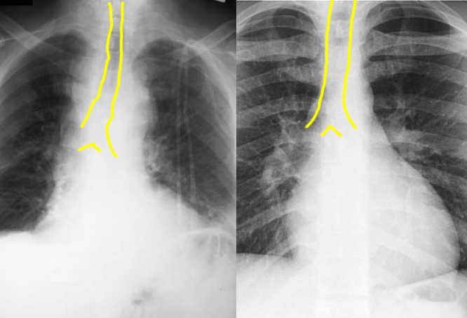
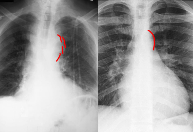
You are shown the patient's CXR side-by-side with the normal image. When looking at imaging after placing a central line, it is important to look for other complications besides abnormal tip location.
Question 3:
a) What looks different between the patient and the normal CXR, shown to the right?
There are several differences, but the most dramatic is the width and contour of the mediastinum. Check the labels below to see where abnormalities are seen.
b) What complication of central line placement is NOT seen on the patient's CXR?
There is no sign of a pneumothorax, which is one of the complications that is more often seen with subclavian line placements.
There are other abnormalities on the patient's CXR, and we will discuss them in class.
