

Case 2
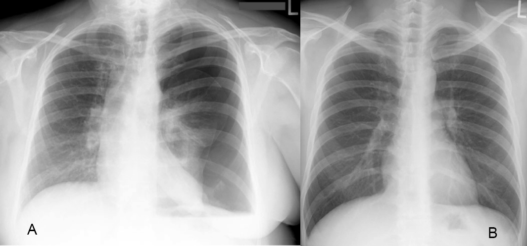
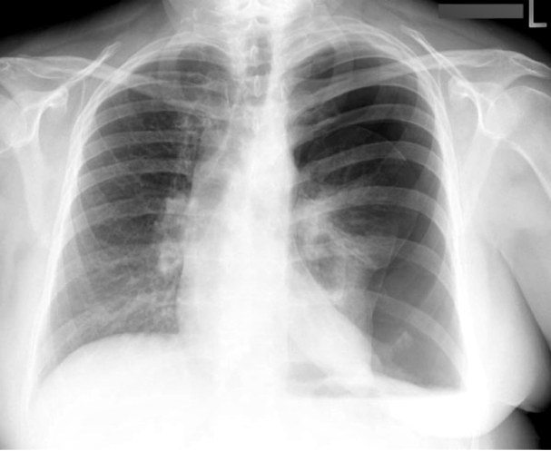
This 55 year old male had new onset of left pleuritic chest pain after an unsuccessful attempt to place a left central line, via a subclavian approach.
Question 1:
a) What does the term 'pleuritic' mean, and what types of pathology produce this type of pain?
As you might imagine, 'pleuritic' pain refers to the pleura, and irritation of the pleura often produces this type of pain. It is sharp and gets worse when the patient coughs or takes a deep breath, causing the pleural surfaces to move past each other. Infections, tumors, effusions, and anything else abnormal involving the pleura can cause this type of pain.
b) What abnormalities in the lungs do you see on this CXR?
The two lungs do not look symmetric, and you can see that there are no lung markings in the periphery on the left. This should make you consider pneumothorax, which as mentioned, is a possible complication of central line placement.
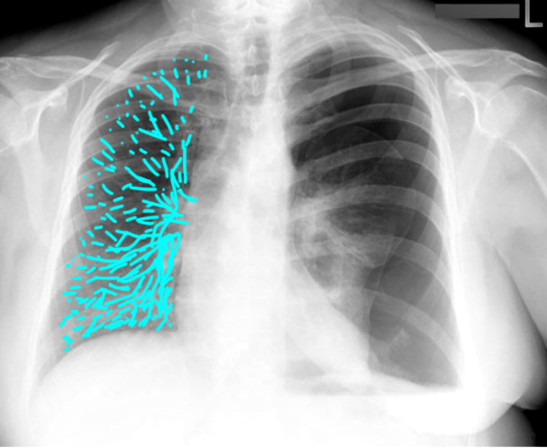
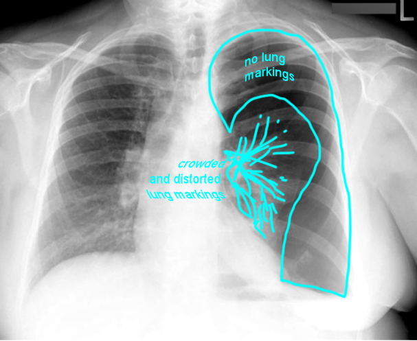
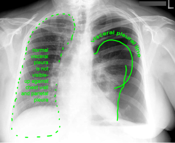
Case 2
When a large pneumothorax is present, as in this case, it is important to be sure not to forget to look at other parts of the chest anatomy.
Question 2:
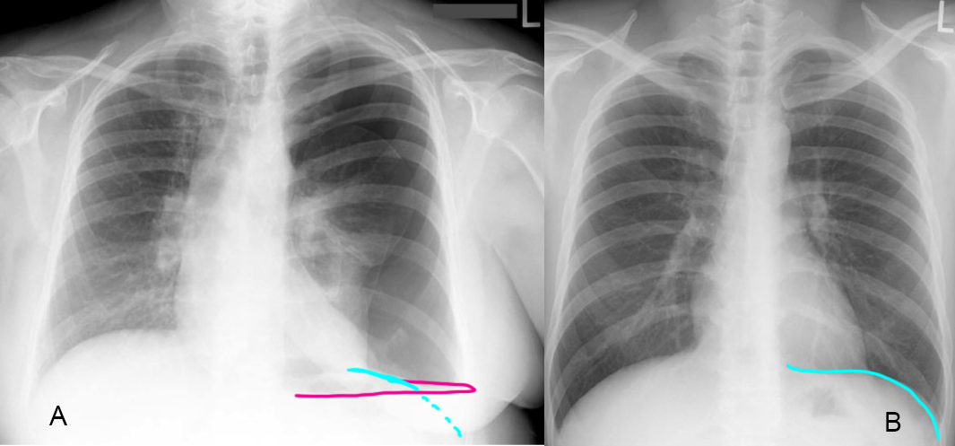
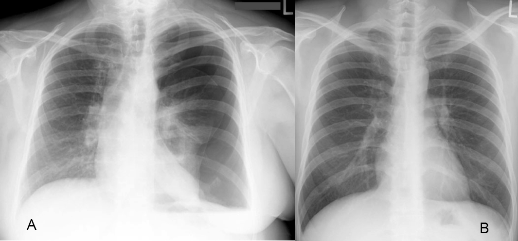
a) What do you think of the heart shape and position in the patient (Image A), compared to a normal CXR in another patient (Image B)?
The heart seems to be moved, or shifted to the patient's right, and is also an abnormal shape. Check the labeled images below to see this illustrated.
b) What kind of problems can occur when this cardiac abnormality is present?
When the heart moves around in the chest, various structures can be twisted or compressed. The vascular structures with the thinnest walls are the IVC and SVC, and if they become compressed, this can be fatal. So this pneumothorax needs to be drained immediately to prevent this complication.
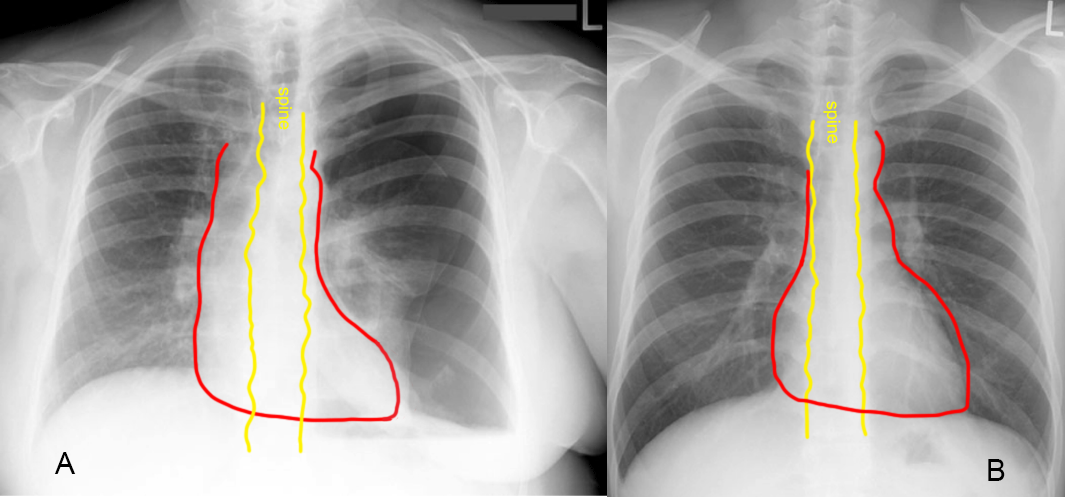
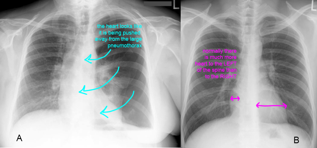
Case 2
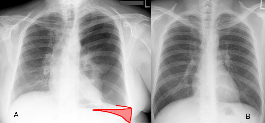
There is another interesting finding related to the pleura that can be seen, particularly if you compare the patient's left diaphragm margin (Image A) to the normal patient (Image B).
Question 3:
a) What do you notice about the shape of the patient's left hemidiaphragm in Image A?
It is not normal in shape, not curved like in the normal patient in Image B.
b) What process could produce this appearance?
This very flat shape is called an air-fluid level, and indicates that the patient was UPRIGHT when the study was done (as is the usual patient position for CXRs), and that there is both air and fluid in the pleural space (which can be called a hydro-pneumothorax). In this situation, with trauma from the attempted line placement, the fluid is very likely blood.
