

Case 3
This case is a 49 year old man who presented with hemoptysis (coughing up blood), and had a CT scan. He was also a smoker, so there was concern for lung malignancy. Enlarged nodes were seen in his mediastinum on the CT, so a biopsy was performed.
Question 1:
a) The procedure that was used to biopsy the nodes was mediastinoscopy. What is this procedure?
As the name implies, mediastinoscopy uses an endoscope (a metal tube through which cameras and biopsy devices can be inserted into the body) to access the mediastinum. This can be done through an incision at the jugular notch, or via a small para-sternal incision between anterior ribs near the costal junction, depending on where in the mediastinum the target is located.
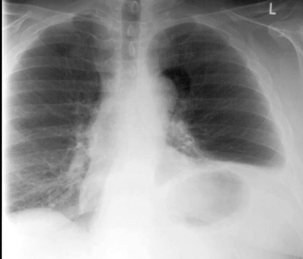
b) What looks abnormal on this CXR?
Several things look abnormal. The left hemidiaphragm is much higher than the right. They should be similar in location, or the left may be a bit lower than the right. There is also a rounded lucency projecting in the upper medial left chest, near the aortic arch. See the labeled images below.
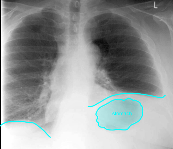
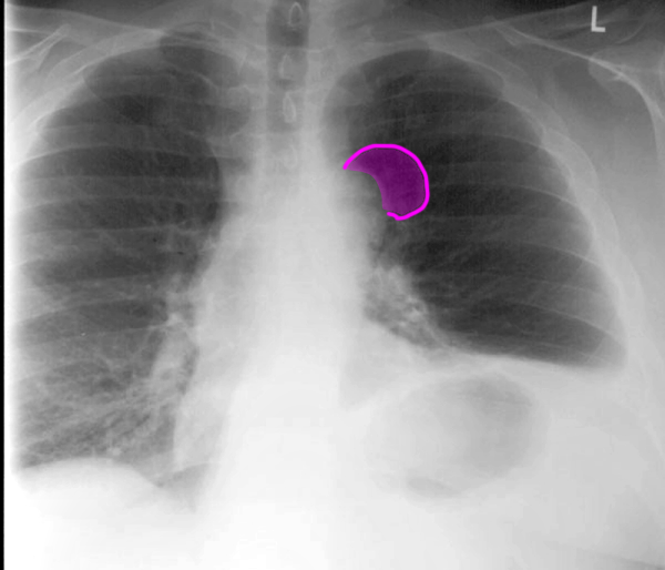
Case 3
You are shown a normal chest CT to help figure out anatomically how to tie the two abnormalities together, that were seen in our patient after anterior mediastinoscopy.
Question 2:
a) What is the imaging plane for this CT (what is the anatomic plane of section)?
These images are in the horizontal (also called transverse or axial) plane, slicing the body like a salami.
b) The arrows on the annotated CT images show approximately where the incision is made and the endoscope inserted during anterior mediastinoscopy. The structure labeled in blue is an important nerve. What nerve is this?
The structure indicated in the annotated CT in blue is the phrenic nerve, which provides innervation to the diaphragm. It is very close to the area where anterior mediastinoscopy is performed. It is rare for this nerve to be injured, but that is what happened in our patient. When the diaphragm loses innervation, it moves UPWARD due to pressure from abdominal contents.
Case 3
These images review the concept of the density spectrum of the body, which is important to understand when looking at CT images.
Question 3:
a) What major advantage does CT offer compared to radiography?
Because the CT images are slices, the density seen on the images is a better representation of the actual density of the tissue that is present. On a radiograph, the density seen on the image is also related to tissue density, but in a much more complex manner, since each pixel is a sum of all the overlapping tissues that part of the beam encountered while passing through the body. CT gives us a much better way to discriminate between tissues based on their density, even for tissues that are quite similar in density.
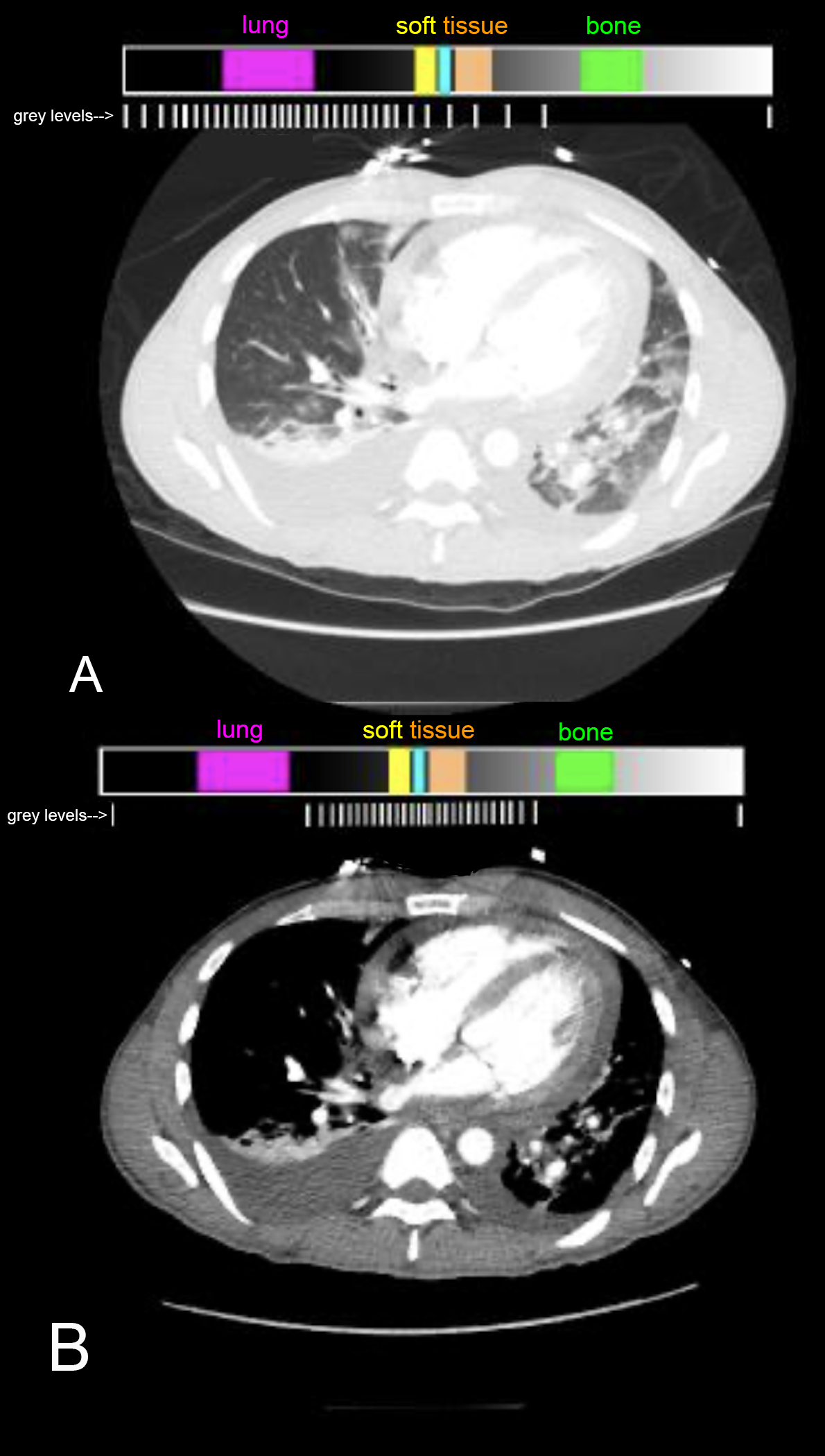
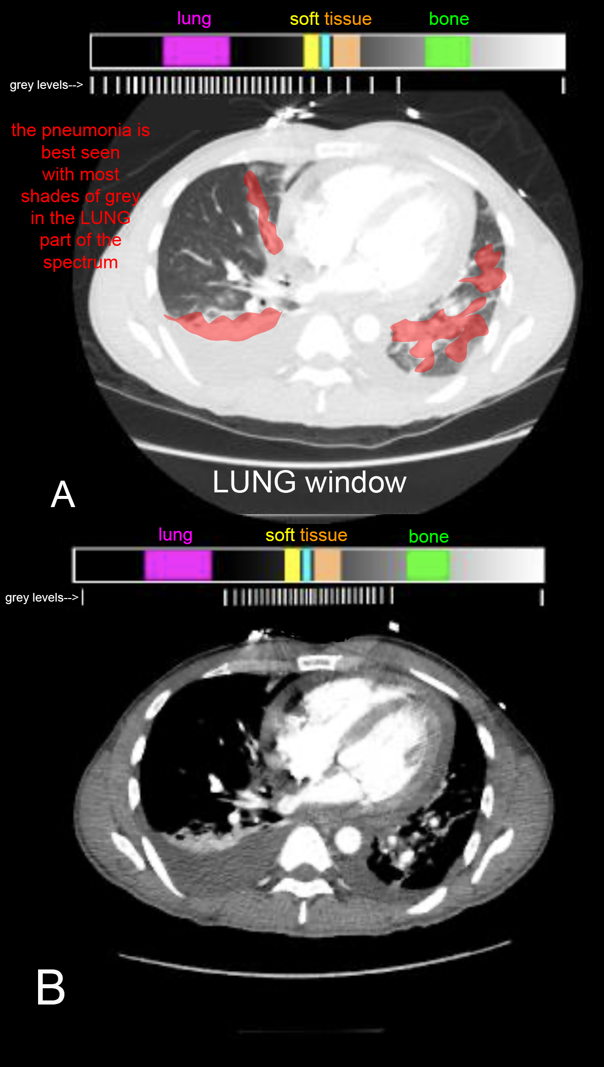
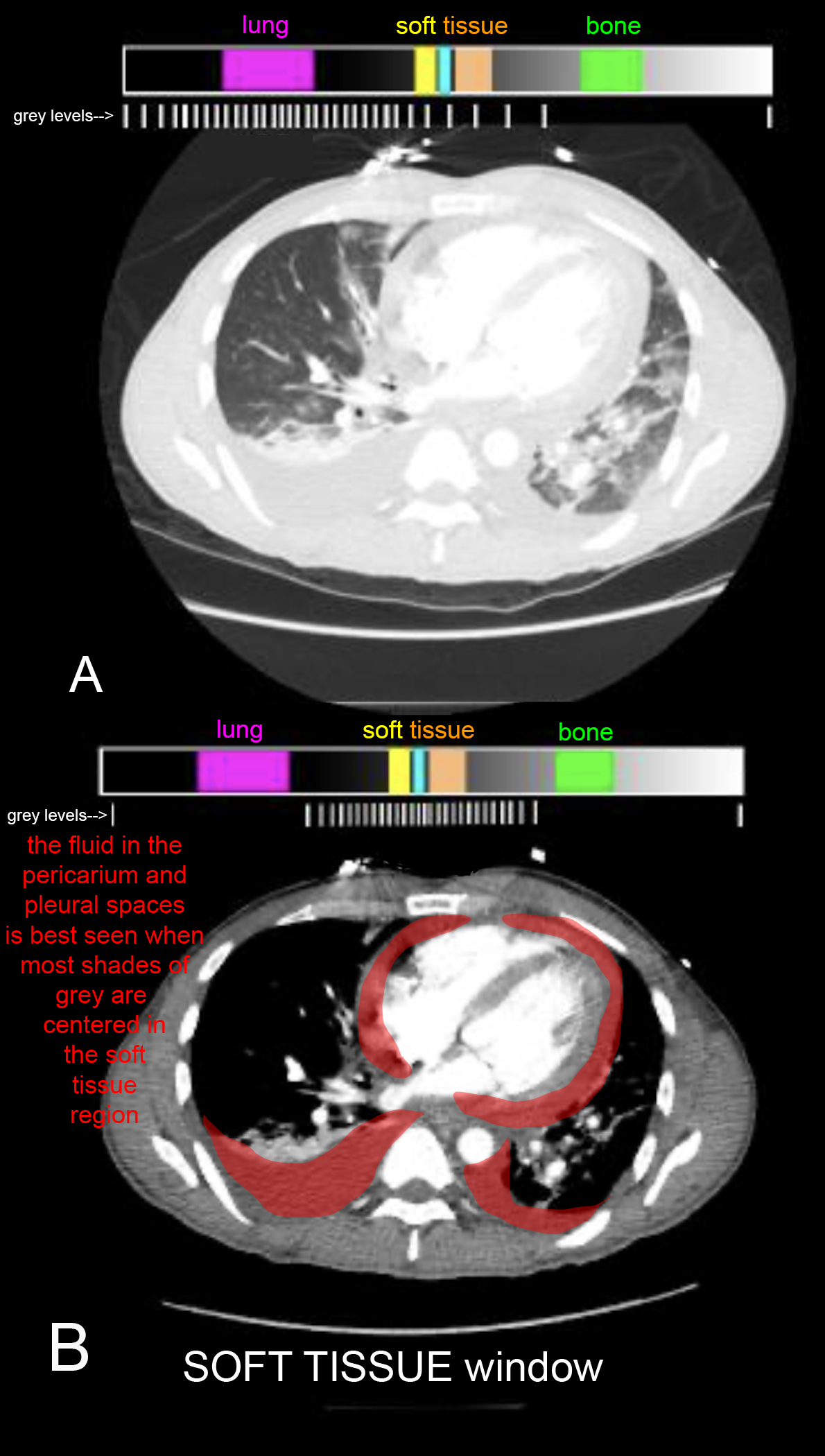
b) You are shown two axial CT images on a different patient, who has pericardial and pleural fluid as well as bilateral pneumonia. How are the two images different?
Image A is a lung window image, meaning that the brightness and contrast settings are optimized for lungs. Most of the shades of grey we can display are set around the density of lung. This is not a good image for soft tissues or bones. To see the patient's pneumonia, you need to be looking at a lung window image. Image B is a soft tissue window. Now, most of our shades of grey are centered in the middle of the density spectrum, where soft tissues are located. This gives us a much better image of the pericardial and pleural fluid that is present.
