


Case 4
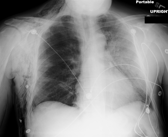
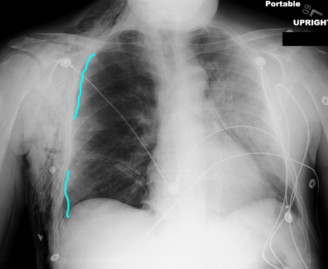
This is the portable chest radiograph (pCXR) of a 25 year old male who was in a high speed motor vehicle collision and had severe shortness of breath due to a large right pneumothorax. A chest tube was placed to evacuate the pleural air.
Question 1:
a) What looks abnormal on his pCXR? Remember to compare right to left, and to look at the soft tissues.
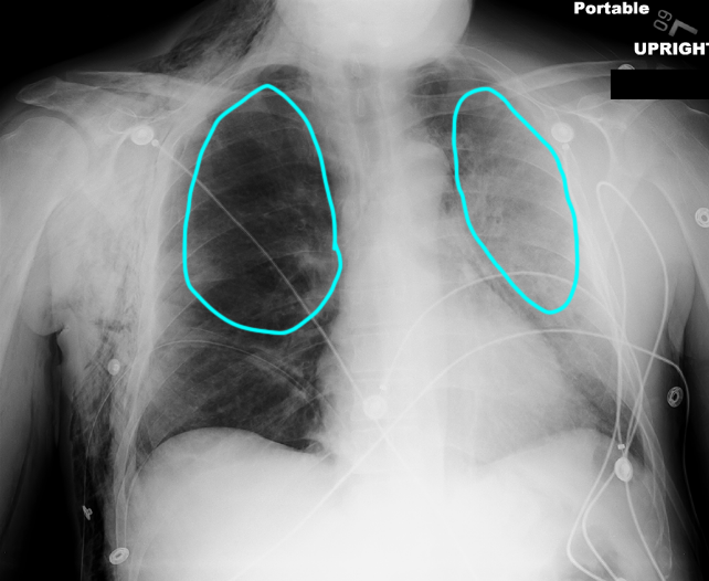
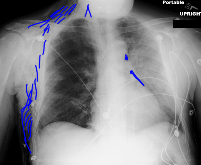
The right lung looks much darker than the left. This could mean that the right lung is too lucent or that the left lung is too dense. If you look along the right chest wall, you will see streaks of black in the soft tissues that look like air. Air in the soft tissues is called subcutaneous emphysema. What do you think the 'mystery' label below indicates?
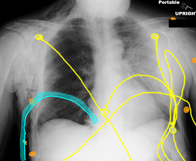
b) What artificial devices do you see, and can you identify any of them?
If you look at the labeled devices image, you will see many wires crossing the chest and ending in round metal plates. These are heart monitor leads. They are very variable in position and appearance and we usually do not comment on them on pCXRs. There are also some metal clips (in orange) that are likely on the patient's hospital gown. There is also a tube (in blue) that is the chest tube, within the pleural space on the right.
Case 4
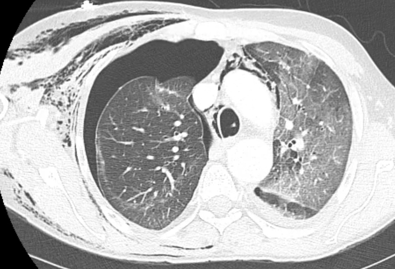
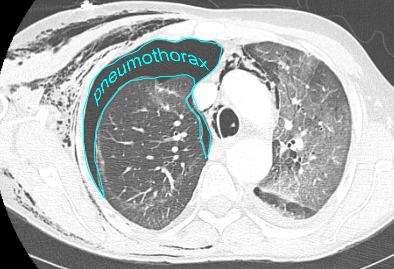
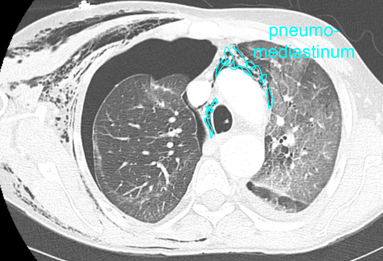
As noted on the prior image, there is a lot of abnormal air in the subcutaneous tissues and a small amount in the mediastinum in this patient. A CT scan was performed to further evaluate these findings.
Question 2:
a) What was the mystery indicated on the pCXR?
The white lines along the lateral right pCXR were visceral pleural lines, indicating that despite the presence of a chest tube in the pleural space, there was still a small pneumothorax.
b) What is the imaging plane and window used for this CT study?
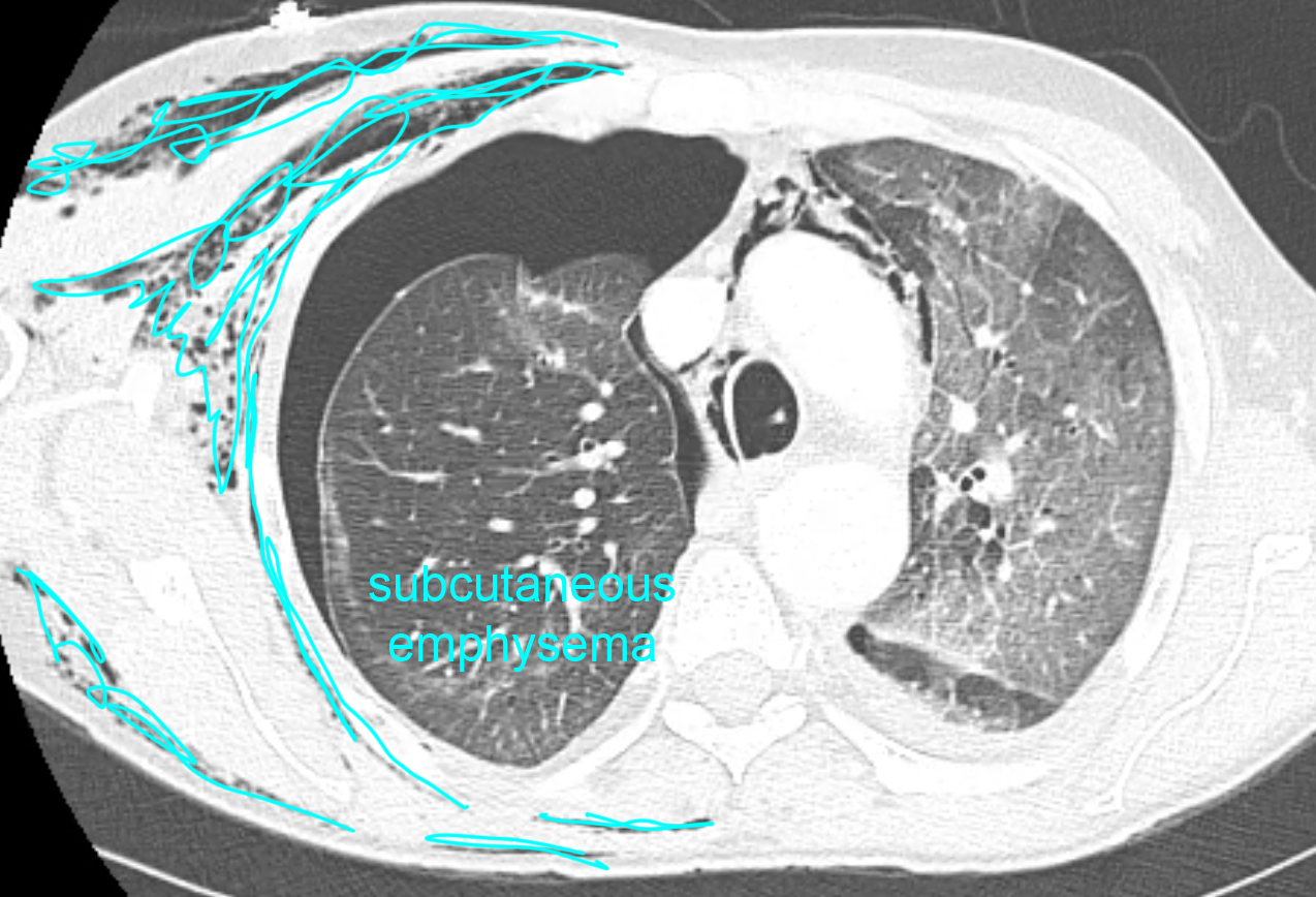
These images are in the axial plane (horizontal or transverse are synonyms). The data is displayed with brightness and contrast settings to optimize the view of the lungs--a LUNG window. It shows the extensive air in the soft tissues (subcutaneous emphysema) very well. It also shows a residual right pneumothorax and air in the mediastinum (called pneumomediastinum). A selected labeled CT image is provided below to highlight these findings.
Case 4
This is another imaging study on the same patient.
Question 3:
What is this study and what technical parameters can you identify?
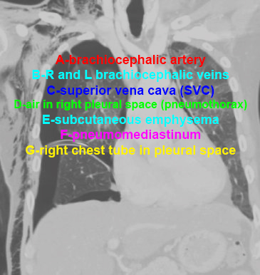
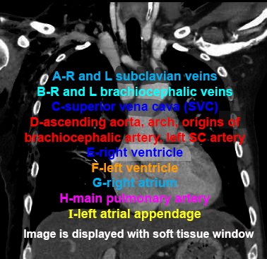
This is also a CT displayed with lung windows, but the data has been reconstructed in the coronal plane. Use the links below to test your knowledge of anatomy on selected images from the study.
Case 4
This completes the pre-class cases for your review. These images will be discussed in class, with additional examples. You will get more out of the in-class discussion if you have reviewed these images ahead of time. Please come to class with any questions you have about these cases.
Further Explanation:
THIS is the final page of the preview cases for the Thorax Radiology session
Click the link below to return to other Anatomy Imaging topics-


