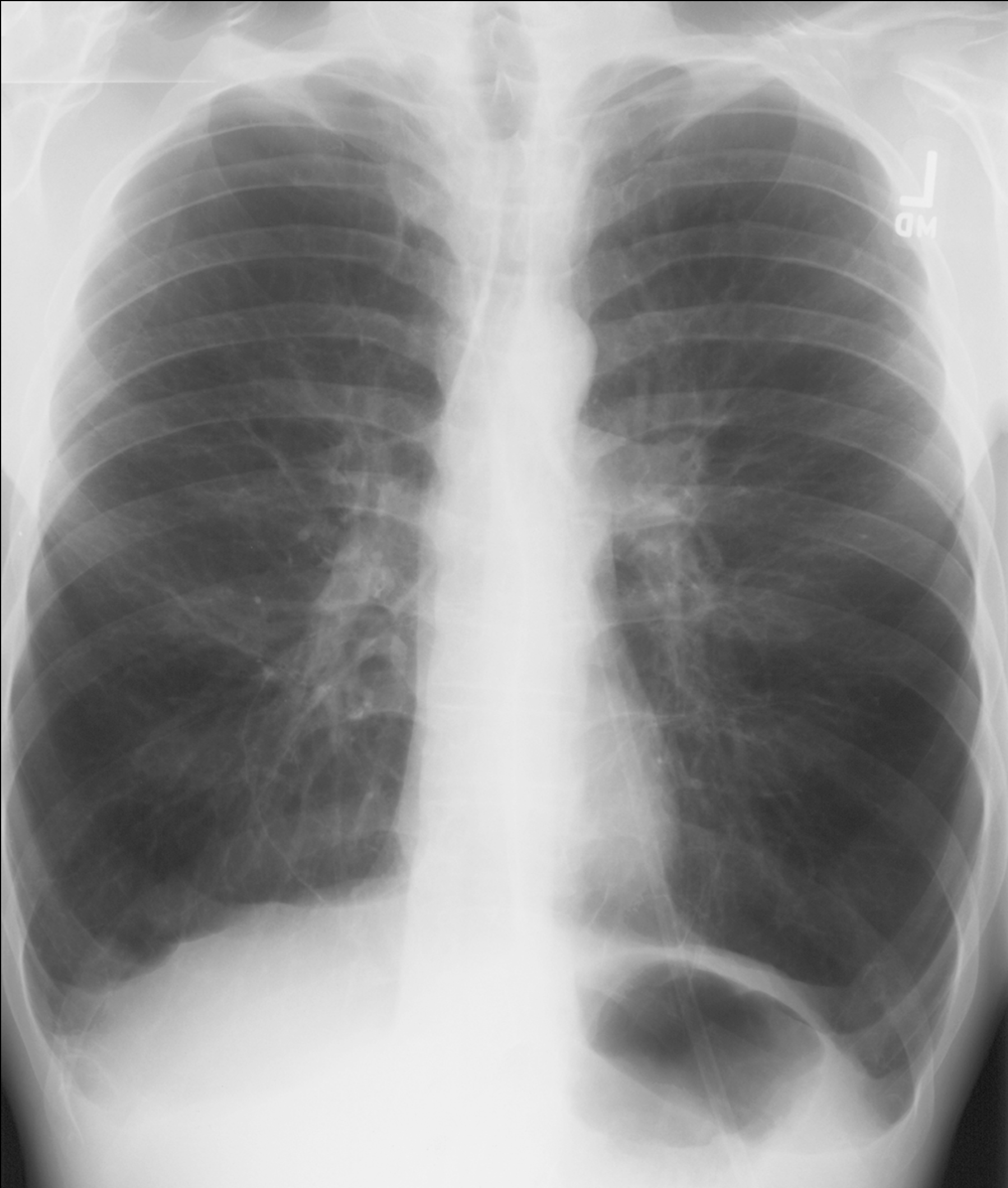


Case 1-chest radiography
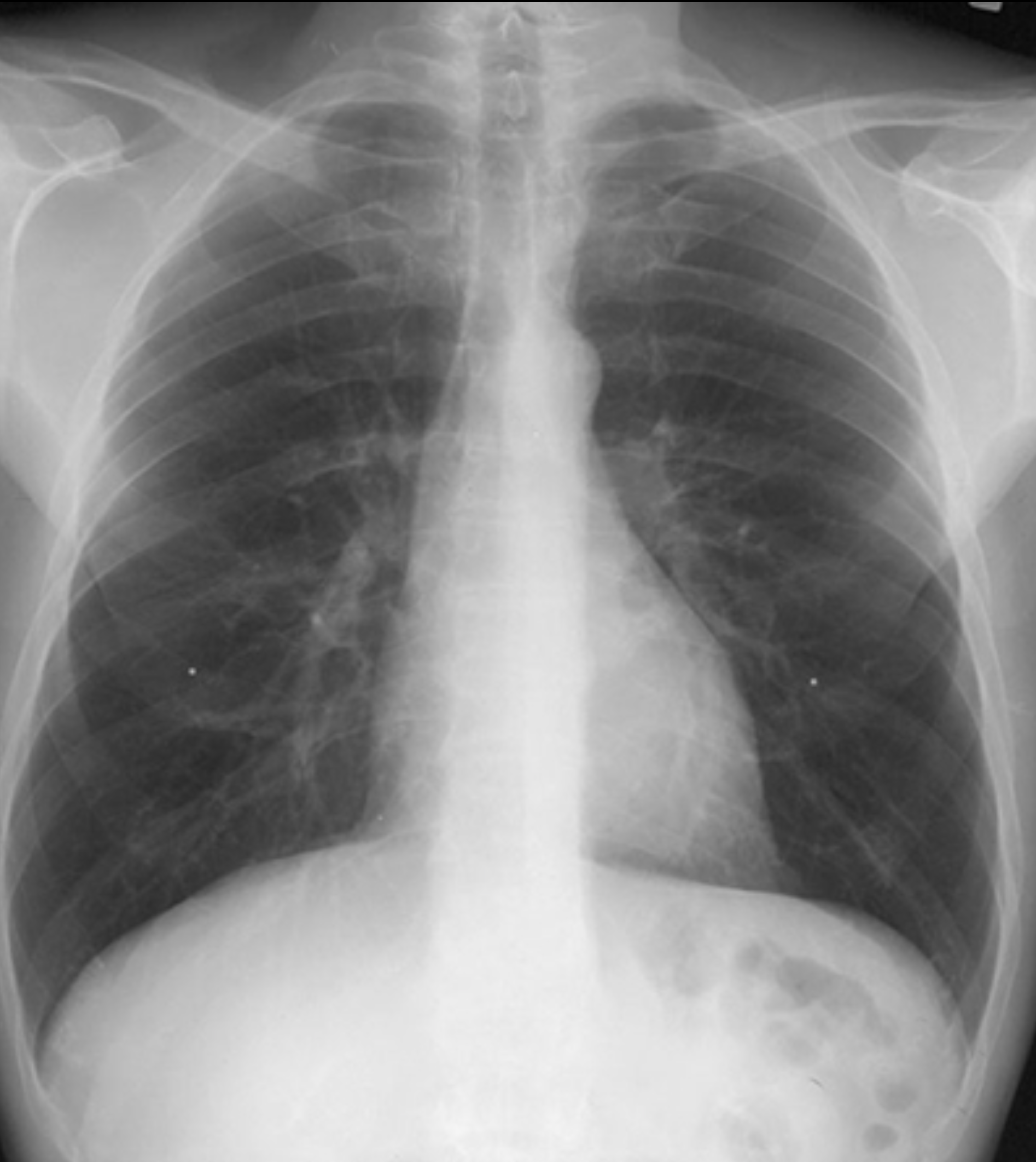
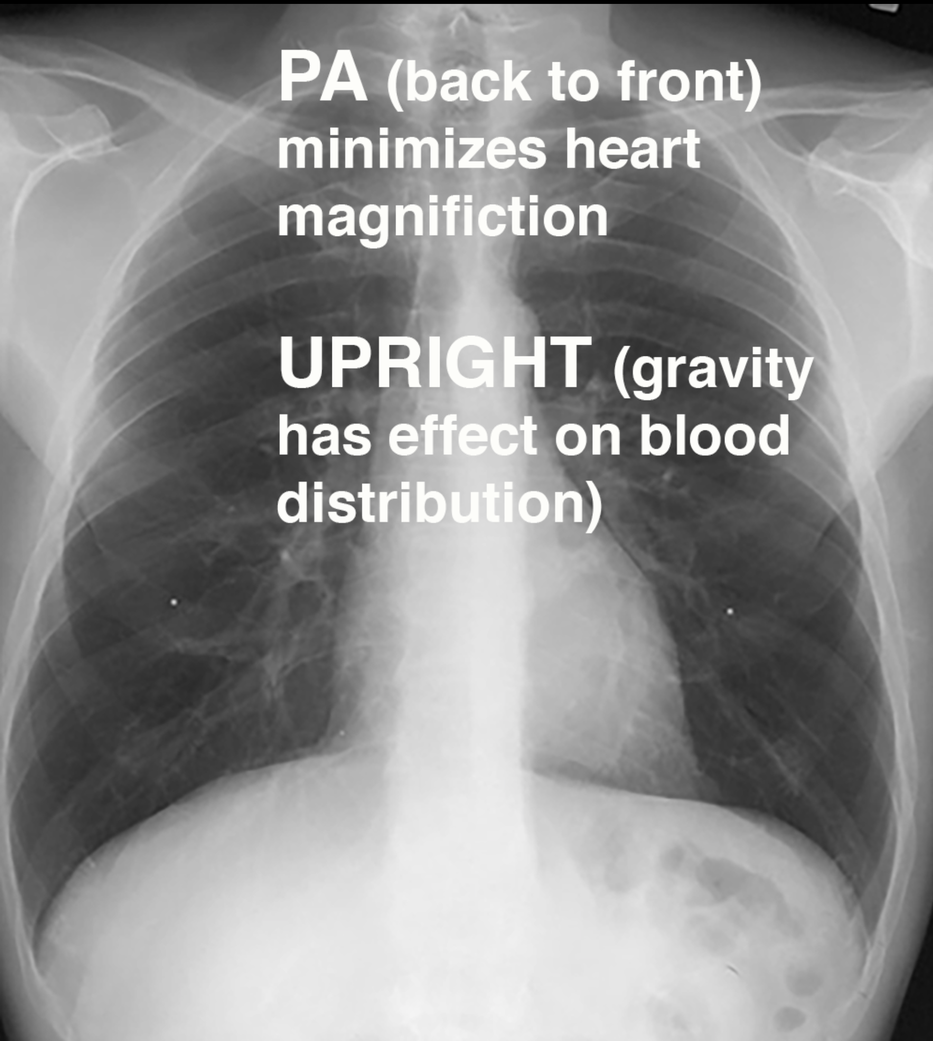
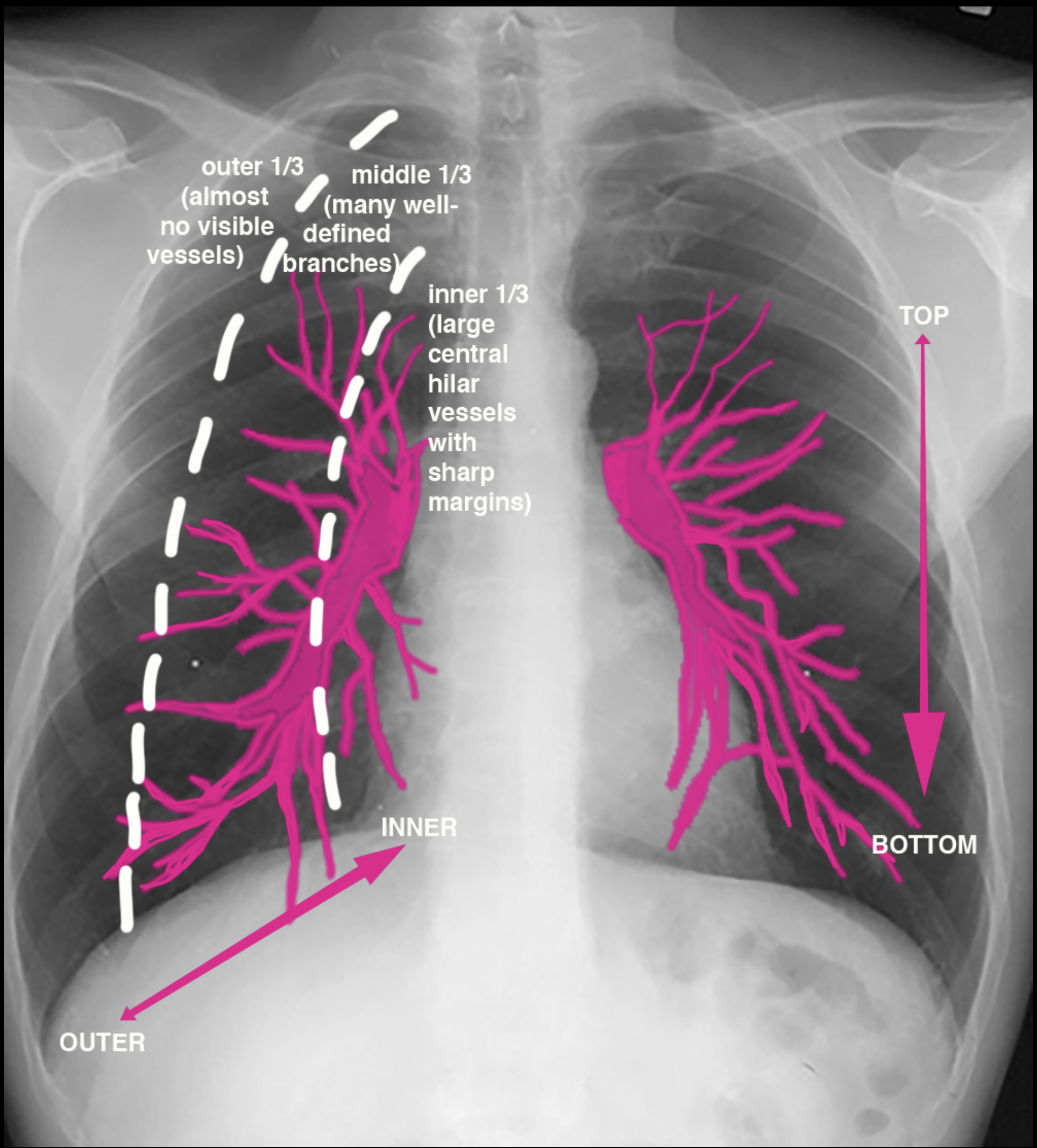
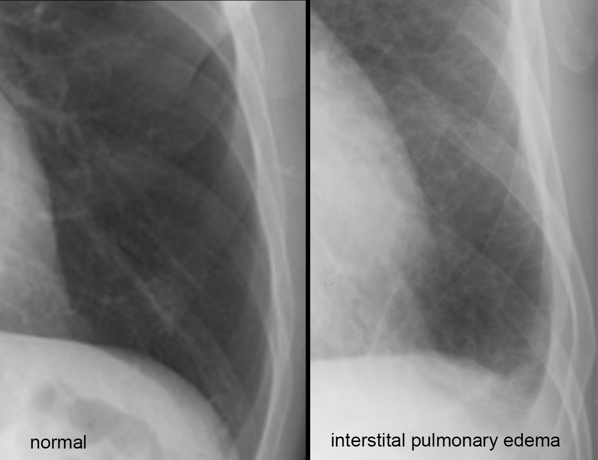
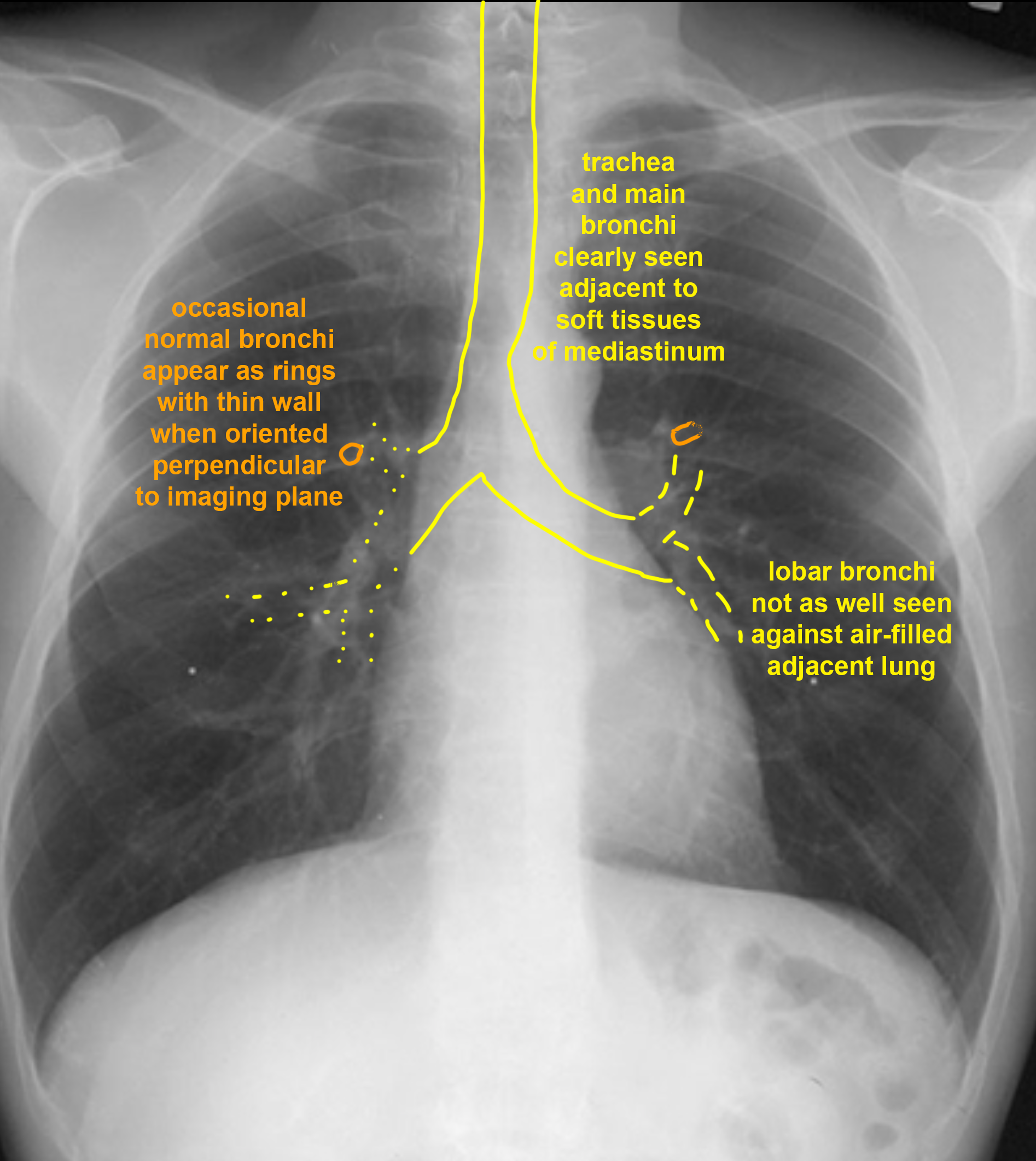
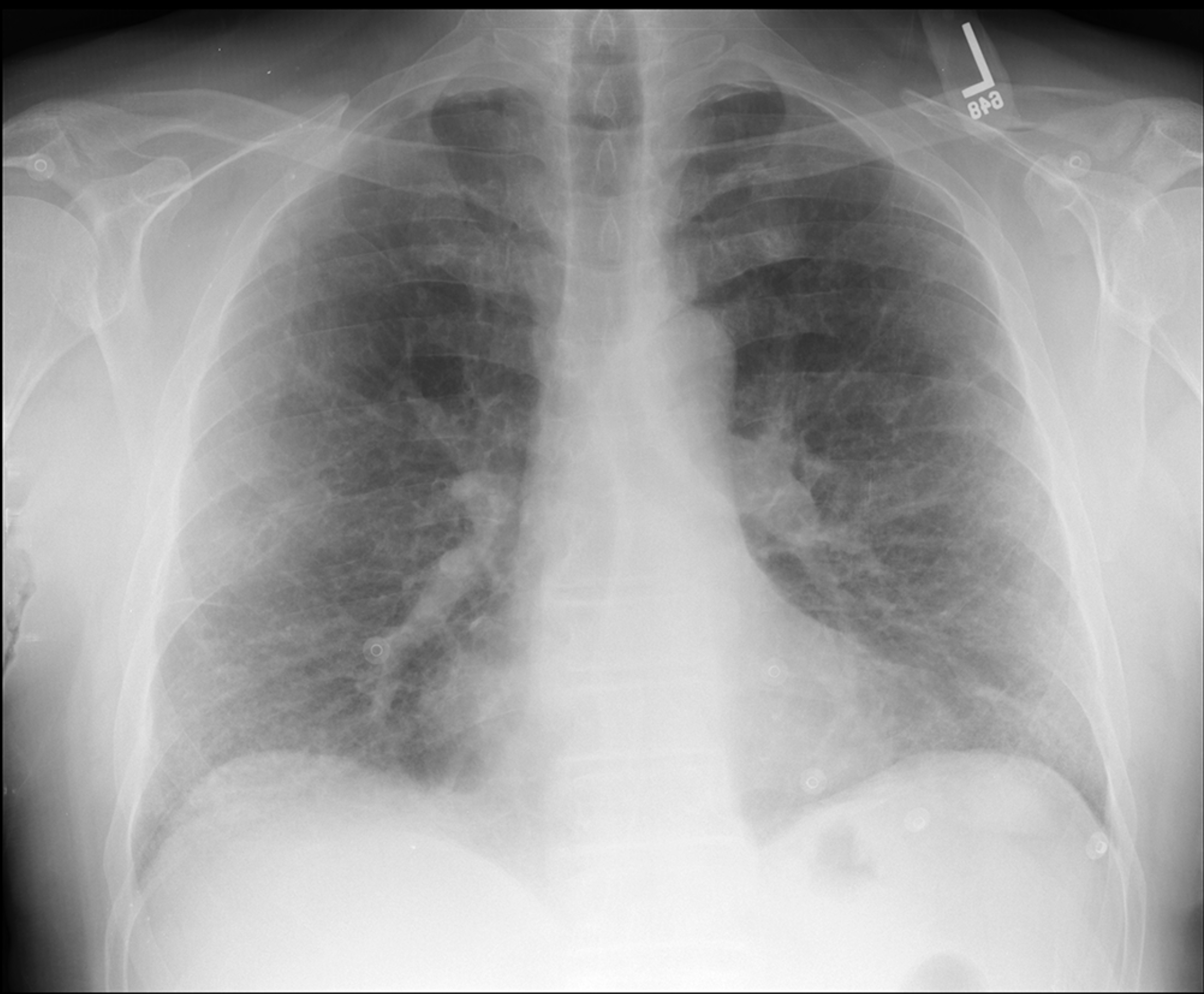
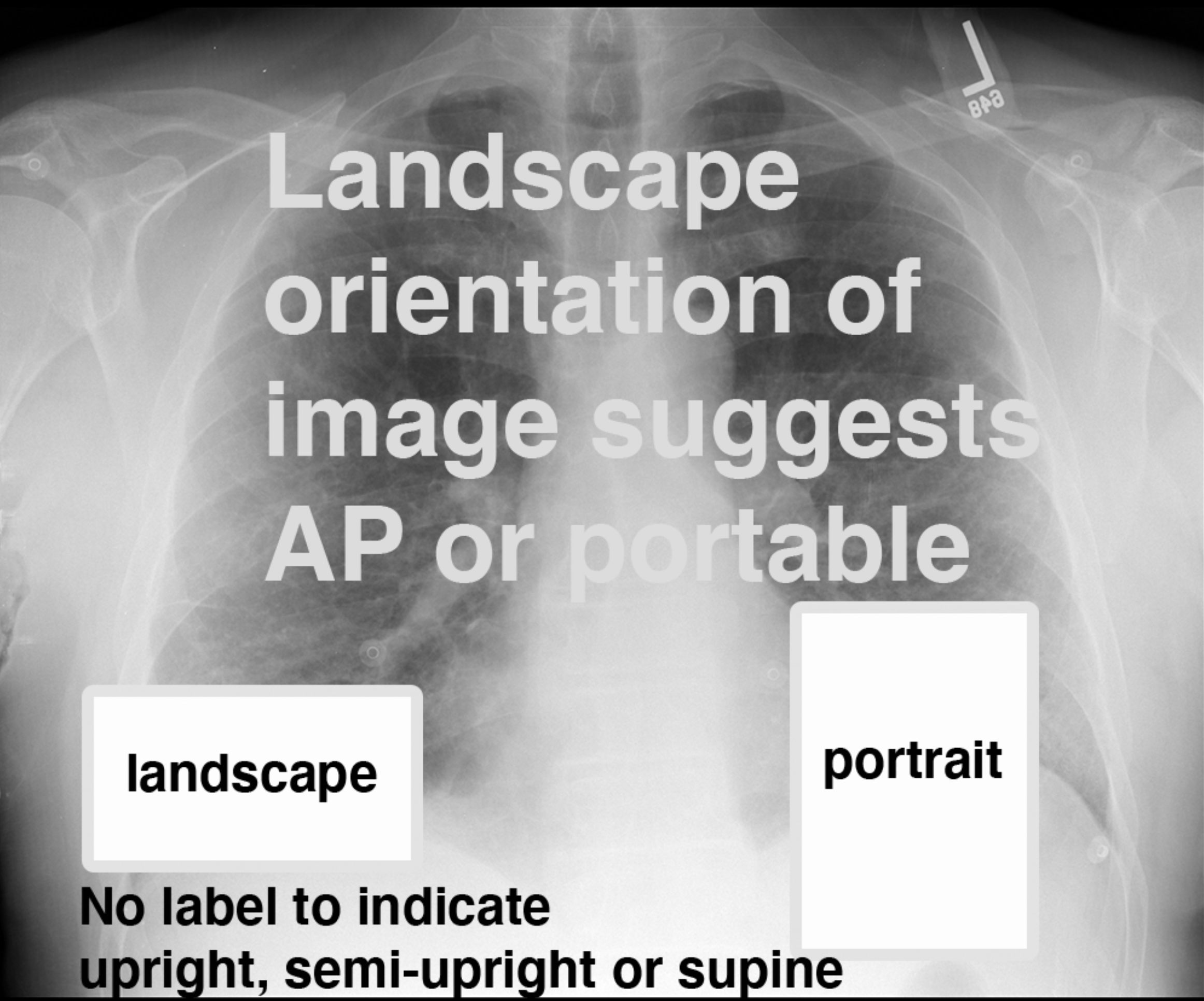
On this page, we focus on the frontal view of the chest--particularly what we can learn about vessels, bronchi, interstitium of the lung, overall level of lung inflation, and pleura. For many of these findings, it is important to know how the image was done. The routine view for the frontal chest is PA in the upright position.
Further Explanation:
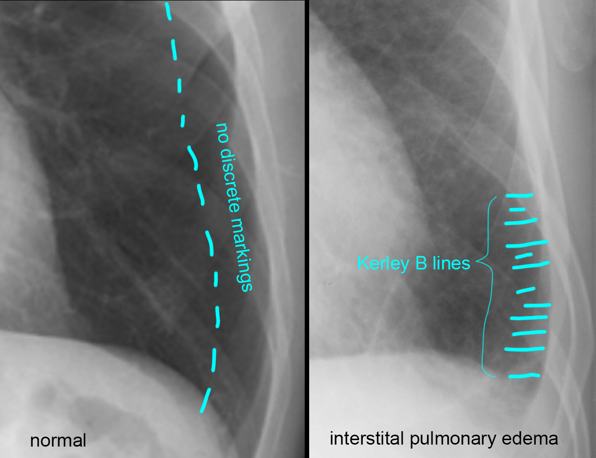
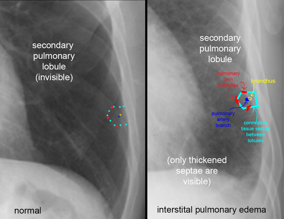
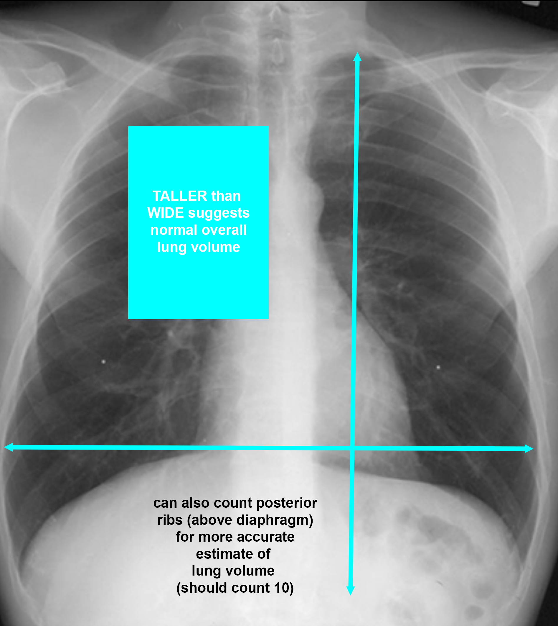
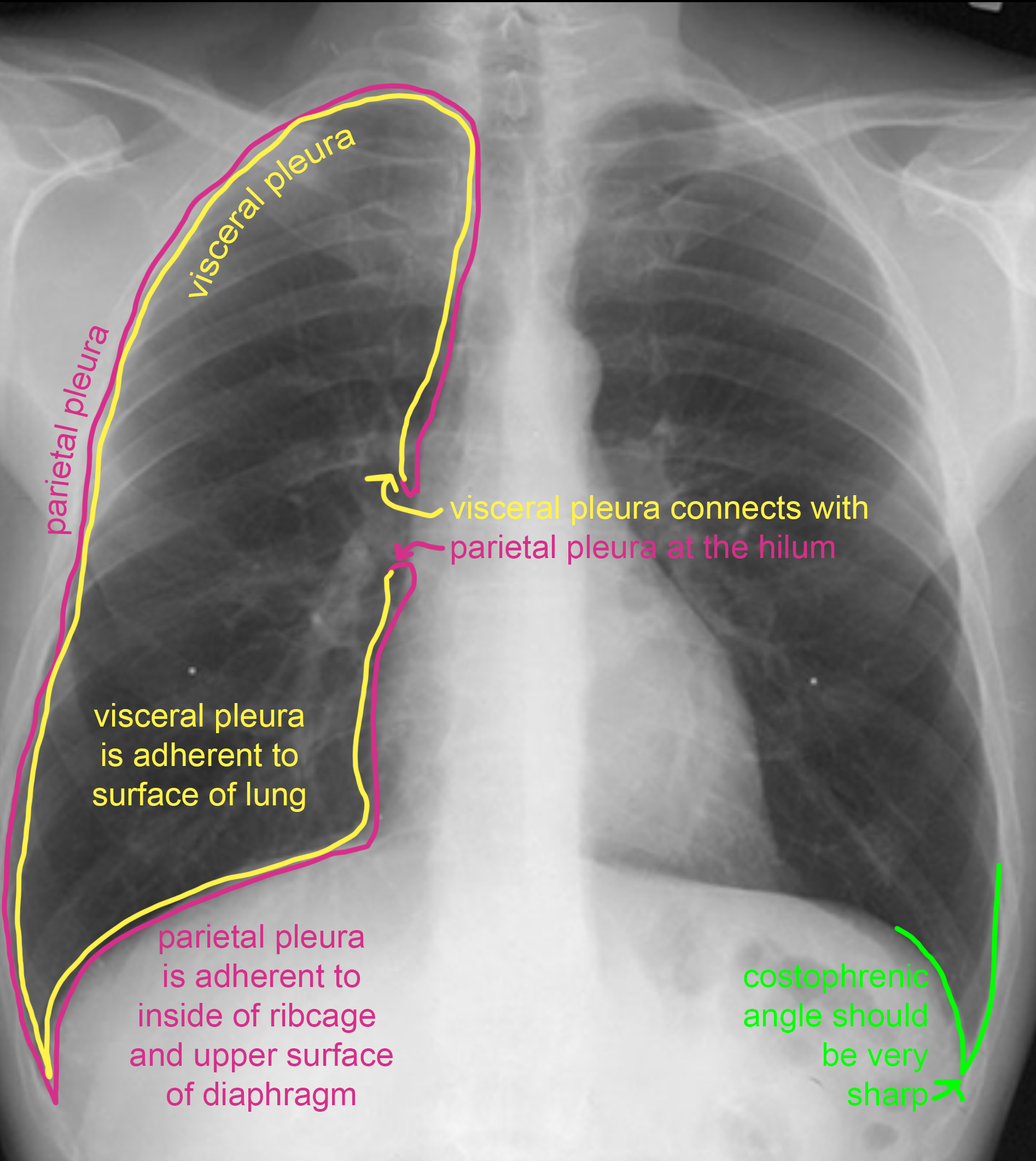
Case 1-chest radiography
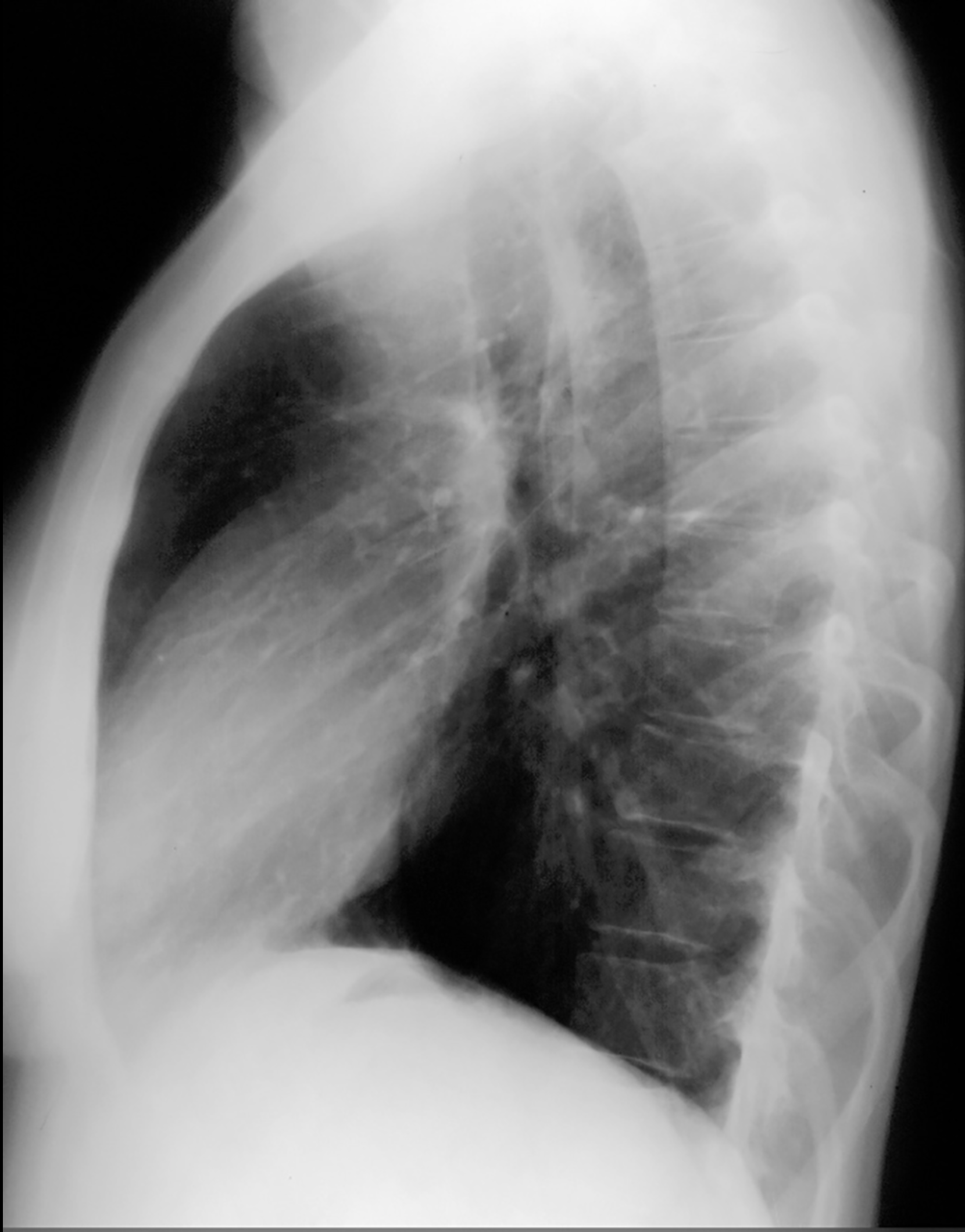
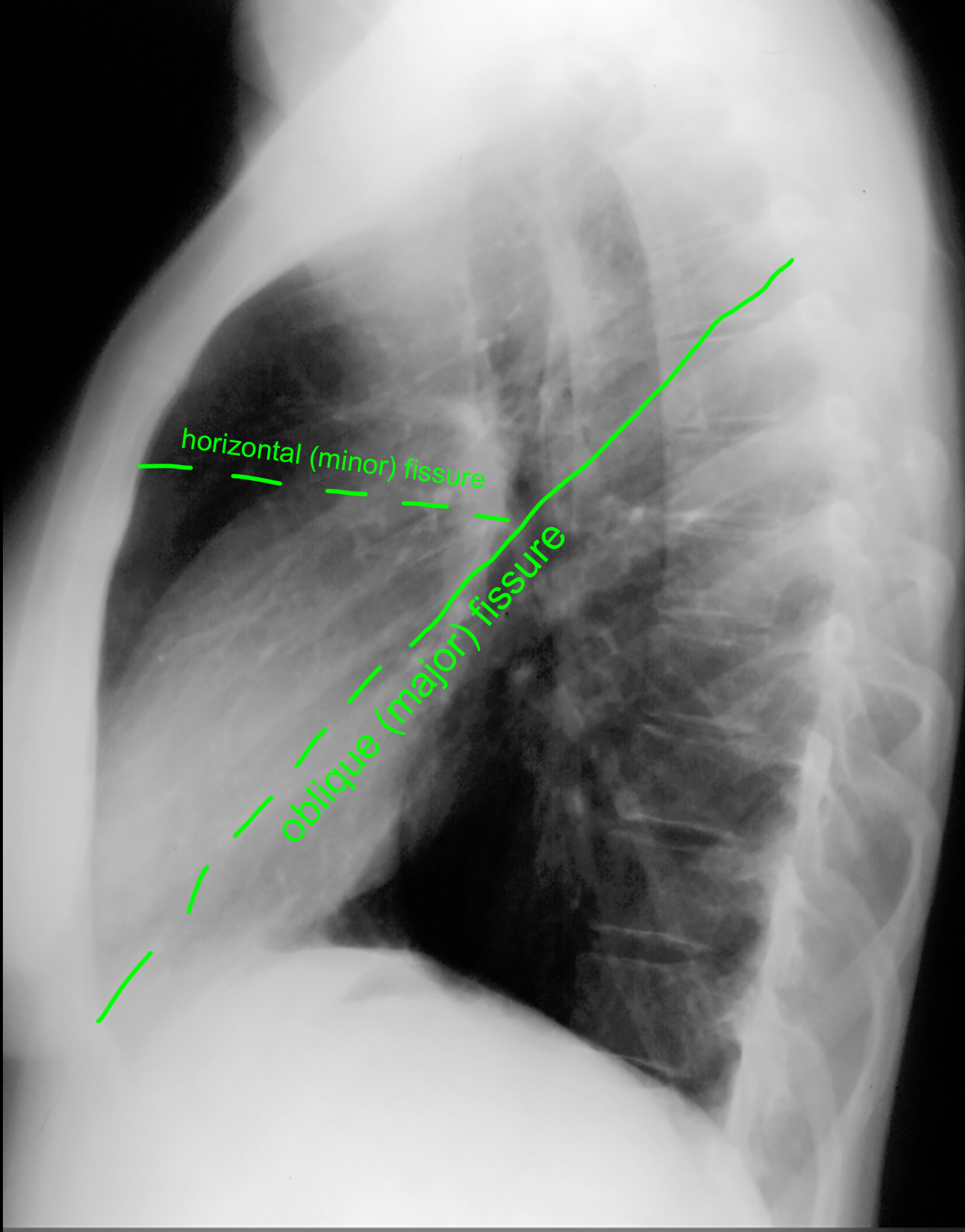
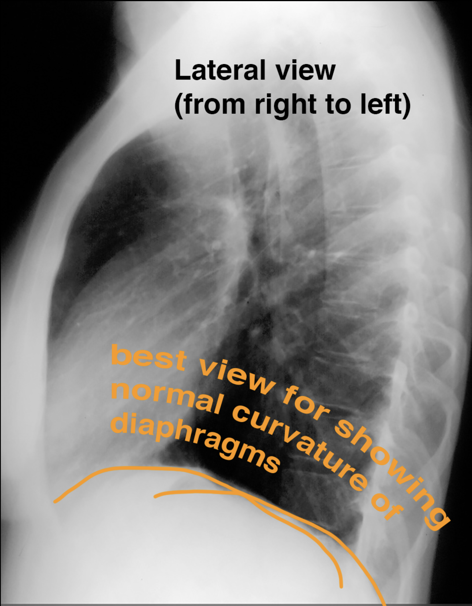
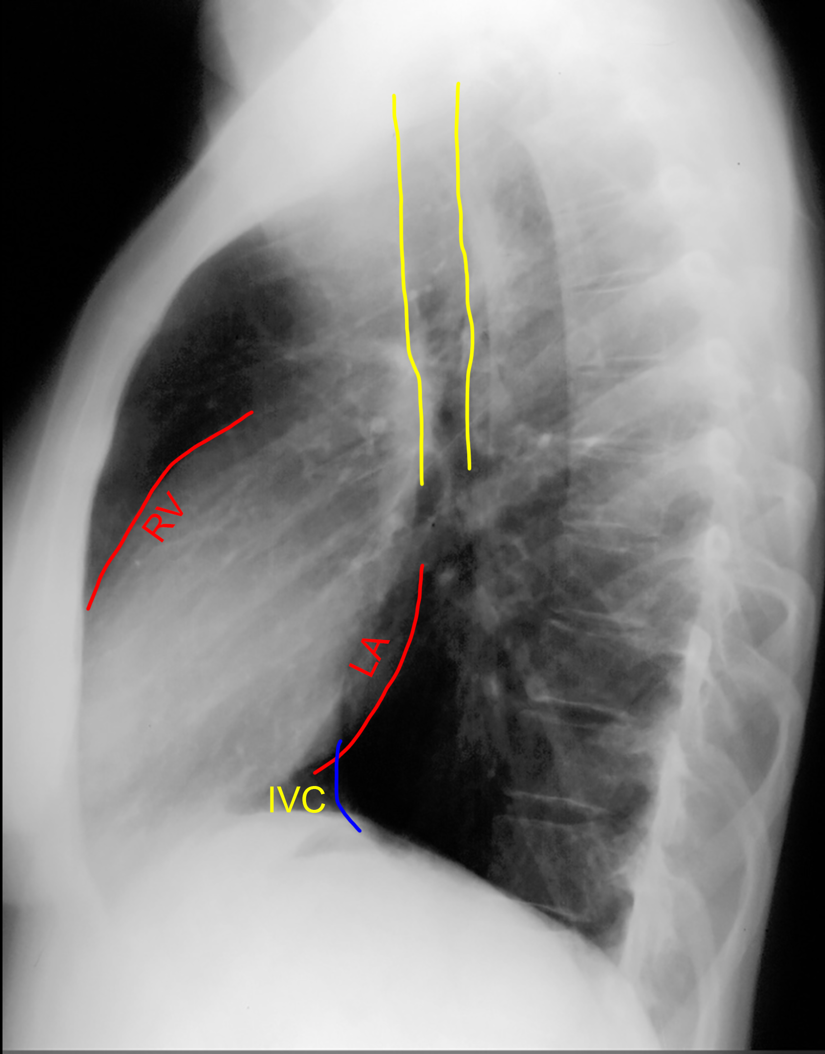
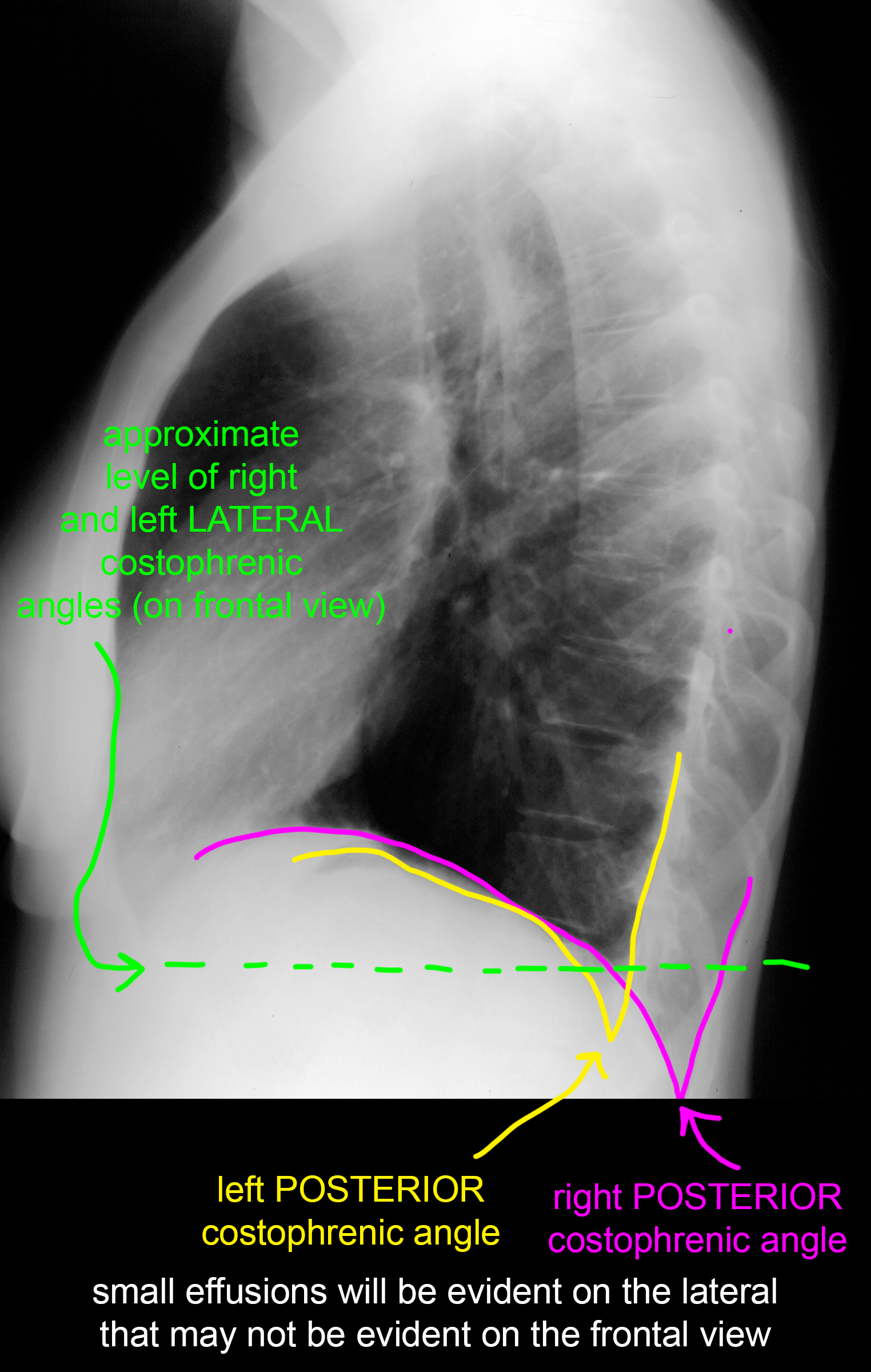
For some portions of the chest, the lateral view may be more revealing than the frontal view. The standard way to obtain this view is upright, with beam passing from the patient's RIGHT side to the LEFT side (which is closest to the receptor).
Further Explanation:
Case 1-chest radiography
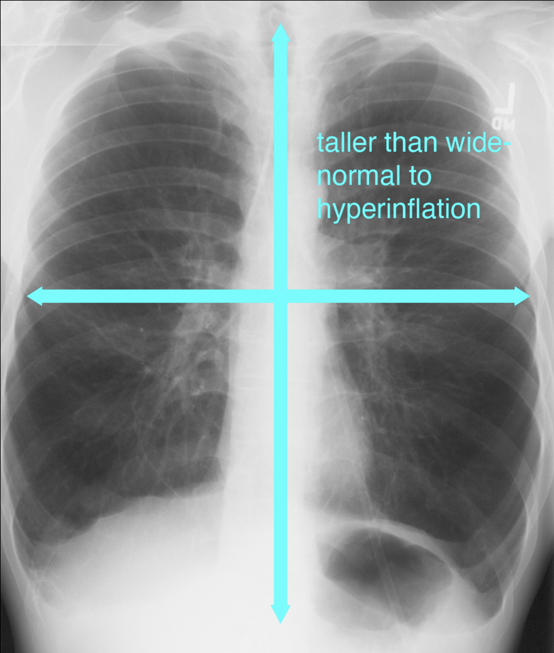
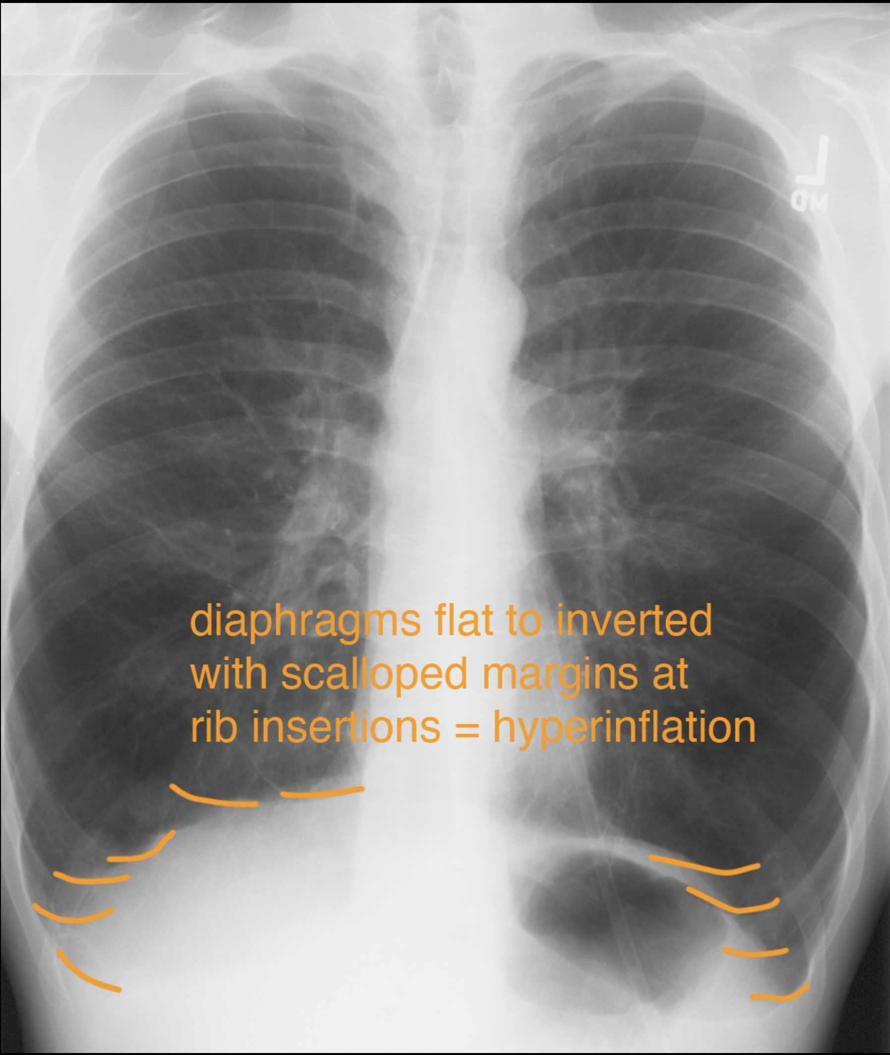
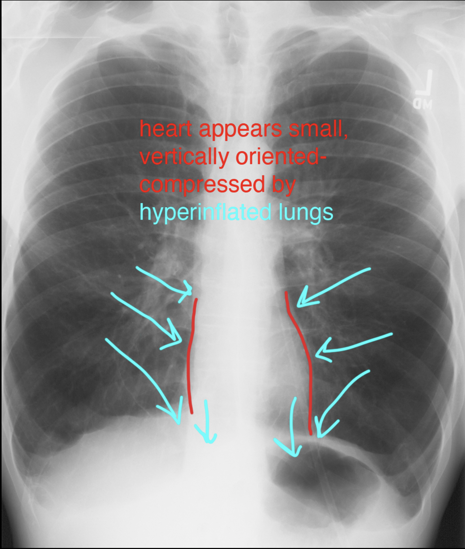
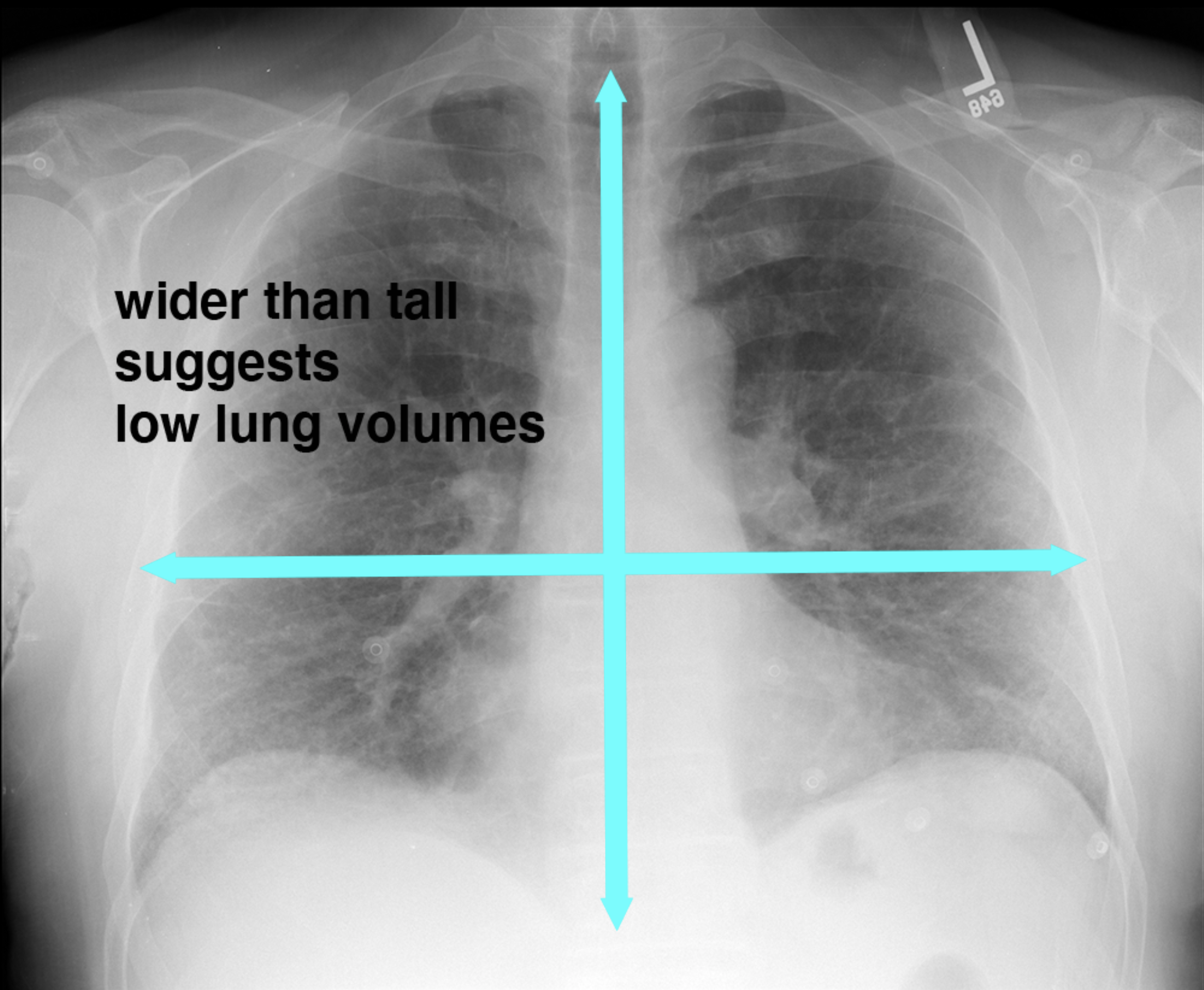
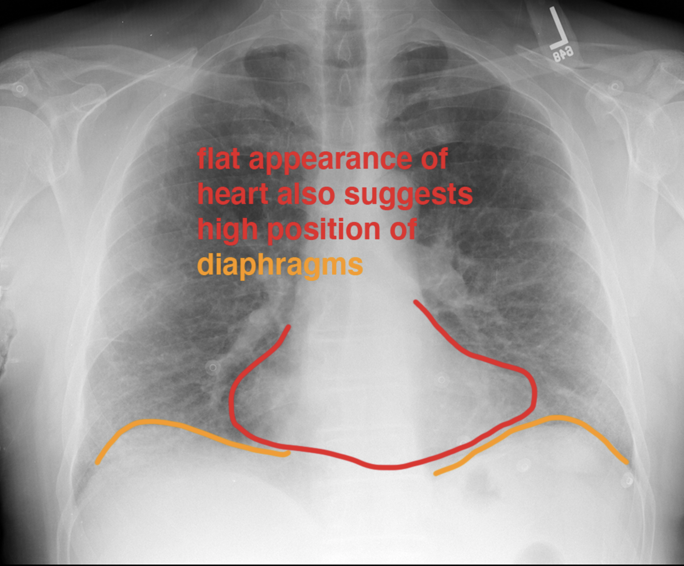
Consider how this patient's frontal radiograph differs from the normal case shown previously.
Further Explanation:
Case 1-chest radiography
Consider how this patient's frontal radiograph differs from the normal example previously shown.
Further Explanation:
